A 13 year old previously healthy female with depression was found unresponsive at home after having overdosed on bupropion and baclofen. Paramedics found her in respiratory arrest, bradycardic, and with a Glasgow Coma Scale score of three. Chest compressions were required and she was intubated. During transport she was hypothermic with decerebrate posturing, and she had a one-minute, self-terminating generalized tonic clonic seizure. In the pediatric intensive care unit, she received intermittent doses of lorazepam and fentanyl for sedation. She remained comatose and continuous video-EEG monitoring was initiated to evaluate for non-convulsive seizures.
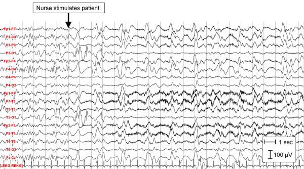
Her electroencephalogram (EEG) demonstrated diffuse polymorphic 5–7 Hz theta activity without organization or normal sleep architecture, as well as waxing and waning spontaneous runs of regular, high amplitude bi-frontally predominant sharp waves. Using American Clinical Neurophysiology Continuous EEG Monitoring Research Terminology [1], these were consistent with abundant (50–90% of record), very prolonged (more than one hour), 1–2 Hz bifrontally predominant generalized periodic epileptiform discharges. By 14 hours post-ingestion the spontaneous periodic discharges had resolved. However, morphologically similar, higher amplitude generalized periodic epileptiform discharges reappeared when the patient was stimulated, for example with sternal rub, pupil examinations, or repositioning, as observed on video-EEG. These bursts demonstrated more pronounced periodicity with longer interburst intervals (1–3 seconds) and more discrete periods of suppression between bursts. Most runs lasted about 30 seconds, although some persisted up to three minutes. None of these evolved into electrographic seizures and none had any associated clinical correlate. These recurred with stimulation until approximately 35 hours post ingestion, when her EEG began to show improved organization with a posterior dominant rhythm of 5–6 Hz theta activity and a well-developed frequency-amplitude gradient. EEG monitoring was stopped at approximately 40 hours post-ingestion when she was extubated and sedative medications were discontinued. Her brain MRI was normal. The patient was quickly transferred out of the intensive care unit and entered rehabilitation three days later. A week later she transferred to an outside psychiatric facility for continued suicidality. She was felt to be neurologically normal.
Stimulus-induced rhythmic, periodic, or ictal discharges (SIRPIDs) were first described in 2004 by Hirsch et al.[2] They were defined as rhythmic, periodic or ictal-appearing discharges, that were consistently induced by alerting stimuli in critically ill patients. These could be focal or generalized and were categorized into three different EEG patterns: periodic epileptiform discharges, frontal rhythmic delta activity, and ictal-appearing discharges. SIRPIDs were found in 22% of 150 consecutive critically ill adults (average patient age = 57 years). Only two patients were under age 18 (17 years and 4 years). Seventeen of the 33 patients with SIRPIDs had more than one SIRPID pattern. Half of the patients had clinical and/or subclinical seizures in addition to their SIRPIDs. Of the 11 patients with subclinical seizures, half arose from the same region as their SIRPIDs. Only one patient demonstrated a clinical correlate with the SIRPIDs (irregular semirhythmic arm jerking during stimulation induced generalized periodic epileptiform discharges. Since the initial description, additional case series have been published, including one series describing three severely ill neonates with stimulus induced seizures among a cohort of 26 neonates undergoing continuous video-EEG monitoring in a neonatal intensive care unit.[3–5] We describe SIRPIDs in a non-neonatal pediatric patient. The pathophysiology of SIRPIDs is not known, but one theory is that sensory stimuli lead to activation of the brainstem arousal circuits, resulting in cortical disturbances via the thalamus. The clinical significance of SIRPID occurrence in critically ill patients is not well characterized, but our case indicates SIRPIDS may be a benign pattern in some critically ill patients.
Figure.
High amplitude 1–2 Hz bifrontally predominant generalized periodic epileptiform discharges with stimulation (arrow). Sensitivity = 7 uV/mm with the exception of O1 which was decreased to 1000uV to minimize electrode artifact; High Filter = 35 Hz; Low Filter = 1.0 Hz.
Acknowledgements
We would like to thank Dennis Dlugos MD for his help in preparing this manuscript.
Drs. Abend and Kessler receive NIH funding (Neurological Sciences Academic Development Award (NSADA): K12 NS049453).
Footnotes
Publisher's Disclaimer: This is a PDF file of an unedited manuscript that has been accepted for publication. As a service to our customers we are providing this early version of the manuscript. The manuscript will undergo copyediting, typesetting, and review of the resulting proof before it is published in its final citable form. Please note that during the production process errors may be discovered which could affect the content, and all legal disclaimers that apply to the journal pertain.
References
- [1].Hirsch LJ, Brenner RP, Drislane FW, So E, Kaplan PW, Jordan KG, Herman ST, LaRoche SM, Young B, Bleck TP, Scheuer ML, Emerson RG. The ACNS subcommittee on research terminology for continuous EEG monitoring: proposed standardized terminology for rhythmic and periodic EEG patterns encountered in critically ill patients. J Clin Neurophysiol. 2005;22:128–35. doi: 10.1097/01.wnp.0000158701.89576.4c. [DOI] [PubMed] [Google Scholar]
- [2].Hirsch LJ, Claassen J, Mayer SA, Emerson RG. Stimulus-induced Rhythmic, Periodic, or Ictal Discharges (SIRPIDs): a common EEG phenomenon in the critically ill. Epilepsia. 2004;45:109–23. doi: 10.1111/j.0013-9580.2004.38103.x. [DOI] [PubMed] [Google Scholar]
- [3].Murphy MM. SIRPIDs: a case study. Am J Electrodiagnostic Technol. 2006;46:a59–64. [Google Scholar]
- [4].Fluri F, Balestra G, Christ M, Marsch S, Fuhr P, Rüegg S. Stimulus-induced rhythmic, periodic or ictal discharges (SIRPIDs) elicited by stimulating exclusively the ophthalmic nerve. Clinical Neurophysiology. 2008;119:1934–38. doi: 10.1016/j.clinph.2008.03.028. [DOI] [PubMed] [Google Scholar]
- [5].Takenouchi T, Yapn VL, Engel M, Perlman JM. Stimulus-induced seizure in sick neonates—Novel observations with potential clinical implications. Epilepsia. 2010;51:308–11. doi: 10.1111/j.1528-1167.2009.02288.x. [DOI] [PubMed] [Google Scholar]


