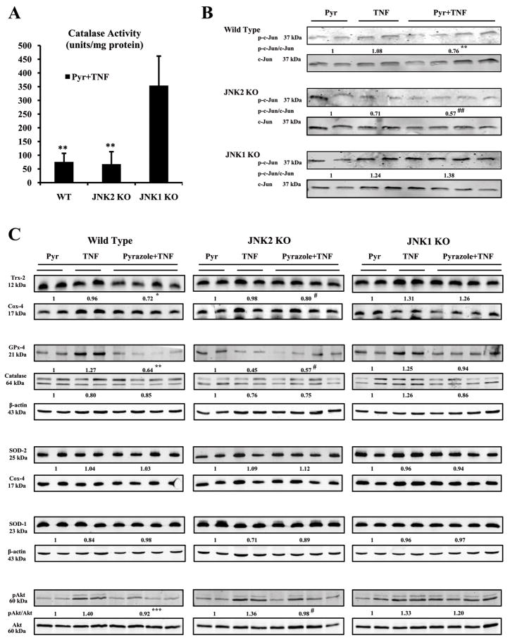
Fig.5.
Antioxidant levels and JNK kinase activity. (A) Catalase activity. ** p<0.05 compared to JNK1 KO. (B) JNK kinase activity. Levels of the phosphorylated c-Jun fusion protein were detected after western blotting using a phospho-c-Jun antibody. ** p<0.05 and ## p<0.05 compared to the pyrazole plus TNF-α-treated JNK1 KO mice. (C) Levels of Trx-2, PHGPx-4, Catalase, pAkt/Akt, SOD-1 or SOD-2 protein in 20–100 μg of protein samples from freshly prepared cytosol, mitochondria, or homogenate fractions were determined by Western blot analysis using the Odyssey Imaging System. All specific bands were quantified with the Automated Digitizing System.β-actin and cox-4 were assayed by Western blot and results expressed as the catalase or GPx-4 or SOD-1/actin ratio or Trx-2 or SOD-2/cox-4 ratio or pAkt/Akt ratio below the specific blots. * p<0.05, ** p<0.01, *** p<0.001 and # p<0.05 compared to the pyrazole plus TNF-α-treated JNK1 KO mice.

