Abstract
A mere forty years ago it was unclear what motor molecules exist in cells that could be responsible for the variety of nonmuscle cell movements, including the “saltatory cytoplasmic particle movements” apparent by light microscopy. One wondered whether nonmuscle cells might have a myosin-like molecule, well known to investigators of muscle. Now we know that there are more than a hundred different molecular motors in eukaryotic cells that drive numerous biological processes and organize the cell's dynamic city plan. Furthermore, in vitro motility assays, taken to the single-molecule level using techniques of physics, have allowed detailed characterization of the processes by which motor molecules transduce the chemical energy of ATP hydrolysis into mechanical movement. Molecular motor research is now at an exciting threshold of being able to enter into the realm of clinical applications.
It is an honor to be sharing the E. B. Wilson Medal with two friends and outstanding scientists, Dick McIntosh and Gary Borisy. We were all introduced to cytoskeletal research at about the same time. In my case, I was introduced to actin and myosin as a postdoctoral fellow at the MRC Laboratory of Molecular Biology in Cambridge, England, working with Hugh Huxley, on whose shoulders all molecular motor researchers stand. Hugh shared the 1983 E. B. Wilson Medal with Joseph Gall. My work with Hugh in 1969 and 1970 on the actin-tropomyosin-troponin-myosin muscle complex led us to postulate the steric blocking mechanism of calcium regulation of skeletal muscle (Spudich et al., 1972) , more fully developed by Hugh elsewhere (Huxley, 1973).

After leaving Cambridge in 1971, I started my laboratory at the University of California, San Francisco, where I spent my first 6 years as a faculty member. In 1977, I moved to Stanford University. Over the years, I have had the privilege of working with the very best students, postdoctoral fellows, research associates, sabbatical visitors, and collaborators. This essay is a tribute to their contributions, rather than a minireview of the field. As it is, due to space limitations, I have had to leave out important contributions by many of my talented lab members, but a complete list of their contributions can be found on our laboratory website (http://spudlab.stanford.edu). This is a story of how our research evolved from 1971 to the present day. To put things in perspective, two key questions about molecular motors around that time were: How does myosin transduce the chemical energy of ATP hydrolysis into mechanical movement?, and What kind of molecular motors are in nonmuscle cells? I decided to focus on two goals: first, to develop quantitative in vitro motility assays for myosin movement on actin, essential for understanding energy transduction in this system; and second, to try to unravel the molecular basis of the myriad nonmuscle cell movements made apparent by light microscopy. During the first year of my assistant professorship, we searched for an ideal model system to study cell movements. We grew Neurospora, Saccharomyces, Physarum, Dictyostelium, Nitella, and other organisms totally unfamiliar to me at the time and searched for a myosin-like motor. Dictyostelium proved to be perfect for this purpose. Margaret Clarke, one of my first postdoctoral fellows, identified a Dictyostelium myosin with properties similar to those of conventional muscle myosin (Clarke and Spudich, 1974), and we then developed methods for visualizing the cytoskeleton in this and other nonmuscle cells (Clarke et al., 1975; Brown et al., 1976).
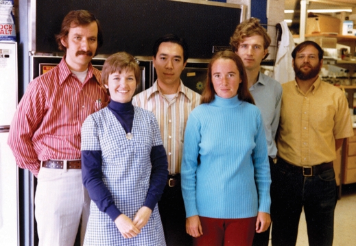
FIGURE 1:
The first Spudich lab group photo, University of California, San Francisco, 1974.
Regarding an in vitro motility assay, Dictyostelium, being a phagocytic organism, offered promise. Actin filaments were known to be in the cortex of nonmuscle cells, with their barbed ends at the cell membrane (Ishikawa et al., 1969; Schroeder, 1973). I fed Dictyostelium small polystyrene beads and isolated the phagocytic vesicles from cell lysates, and found that the vesicles had actin filaments emanating from the membrane-coated surfaces. With great excitement, I tried to establish in vitro motility of these vesicles along myosin-coated coverslips, but directed movements were not readily apparent, and a definitive in vitro motility assay would wait another decade.
THE DISCOVERY OF HOMOLOGOUS RECOMBINATION IN DICTYOSTELIUM
In the mid-1980s, I was considering shifting to a new model system, due to the lack of good genetic approaches for Dictyostelium. In collaboration with Leslie Leinwand, my student Arturo De Lozanne cloned the Dictyostelium myosin heavy chain gene. An interesting set of circumstances then led us to discover homologous recombination in this organism. Arturo disrupted the myosin II heavy chain gene (De Lozanne and Spudich, 1987) and Dietmar Manstein then managed its complete knockout (Manstein et al., 1989). These discoveries added a pivotal dimension to our research for the next couple of decades. We showed that the Dictyostelium nonmuscle myosin II is essential for cytokinesis, but not for cell migration. The latter was a surprise and a very important observation, since the prevailing dogma was that myosin II drove the forward movement of cells.
FIGURE 2:
A recent Spudich lab group photo, Stanford University.
Over the following years, Hans Warrick, Tom Egelhoff, Jihong Zang, Sheri Moores, James Sabry, Wen Liang, Doug Robinson, and others in the lab characterized the structure–function relationship of Dictyostelium myosin II in cell division (e.g., Egelhoff et al., 1993; Sabry et al., 1997; Zang and Spudich, 1998; Robinson and Spudich, 2000). Importantly, having a null myosin II strain, Kathy Ruppel, Taro Uyeda, William Shih, Bruce Patterson, Coleen Murphy, and others were able to use specific mutations in the myosin II heavy chain gene to elucidate how this molecular motor works to transduce the chemical energy of ATP hydrolysis into mechanical movement (Uyeda et al., 1994, 1996; Patterson and Spudich, 1996; Ruppel and Spudich, 1996; Patterson et al., 1997; Shih et al., 2000; Murphy et al., 2001). Cathy Berlot, in collaboration with Peter Devreotes, explored the role of myosin II in Dictyostelium chemotaxis (Berlot et al., 1987), and Meg Titus and Holly Goodson discovered a number of other myosin isoforms in Dictyostelium and Saccharomyces and explored the roles of those isoforms in the biology of those cells (Titus et al., 1989; Goodson et al., 1996).
IN VITRO MOTILITY ASSAYS TAKEN TO THE SINGLE-MOLECULE LEVEL
To understand how myosin transduces the chemical energy of ATP hydrolysis into mechanical movement required us to achieve our second goal—develop quantitative in vitro motility assays for myosin movement on actin. In 1979, Susan Brown managed to grow actin filaments on the surface of polystyrene beads with pointed ends out (Brown and Spudich, 1979); these were cleaner than the Dictyostelium phagocytic vesicles I had explored earlier, but we still failed to see convincing movement of these actin-coated beads in a myosin-coated surface assay. With Alan Weeds, a sabbatical visitor in 1982, we tried the reverse assay, to orient actin filaments on a surface by flow and watch myosin-coated beads move along them. Actin filaments were attached to a streptavidin-coated surface via barbed-end, bound, biotinylated severin. The filaments were oriented by flow and myosin-coated beads were expected to move upstream. Results were encouraging, but again not robust, probably due to poor orientation of the actin filaments. The lack of actin filament orientation was overcome in 1983, now with sabbatical visitor Mike Sheetz, when we used the well-ordered actin cables in Nitella to observe robust movement of myosin-coated beads, the first quantitative in vitro motility assay (Sheetz and Spudich, 1983). This assay allowed Tom Hynes, Steve Block, and Brian White to show that the N-terminal half of the myosin II motor was all that was needed for motility, ruling out some models of contraction (Hynes et al., 1987). The results from the Nitella assay led me and my new graduate student Steve Kron to push harder to get good orientation of actin filaments in the biotinylated severin filament assay, and in 1985 we published the first demonstration that purified actin and ATP are sufficient to support movement of myosin at rates consistent with the speeds of muscle contraction and other forms of cell motility (Spudich et al., 1985).
An even simpler in vitro motility assay harked back to the use of myosin-coated surfaces, but this time using Toshio Yanagida's pivotal observation in Fumio Oosawa's lab in 1984: individual actin filaments labeled with rhodamine-phalloidin are visible by fluorescence microscopy, and they show increased thermal bending motion of actin filaments in suspension in the presence of myosin and ATP (Yanagida et al., 1984). In 1985, Steve Kron showed robust directional gliding of individual rhodamine-phalloidin–labeled actin filaments along myosin-coated surfaces at velocities similar to those observed in muscle (Kron and Spudich, 1985, 1986). Shortly thereafter, Yoko Toyoshima and others in the lab established that the globular head, or subfragment 1 (S1) of the myosin molecule, is the motor domain (Toyoshima et al., 1987). Thereafter, all research on how the myosin family of molecular motors transduce the chemical energy of ATP hydrolysis into mechanical motion focused on the S1 head.
Fundamental questions remained, primarily focused on the step size that the myosin takes for each ATP hydrolysis. Various experiments from my lab suggested a step size of ∼10 nm, while similar experiments from Yanagida's lab reported values in excess of 50 nm (for review, see Altman and Spudich, 2005). A step size considerably larger than 10 nm would compel a model for how the actin-myosin system works different from the prevailing swinging lever arm hypothesis put forward by Hugh Huxley in 1969 (Huxley, 1969). The large range in reported step-size values probably reflected the difficulty in determining how many myosin molecules were acting on a single actin filament in the Kron in vitro motility assay. So, it became critical to watch a single myosin molecule go through one cycle of its ATP hydrolysis and measure the step size directly. Thus, the Kron assay was refined to the single-molecule level by graduate student Jeff Finer and sabbatical visitor Bob Simmons using a dual-beam laser trap they built in collaboration with Steve Chu (Simmons et al., 1993). Using the dual-beam laser trap allowed a single actin filament to be lowered onto a single myosin II molecule on the surface for direct observation of the step size (∼10 nm) and the force (∼5 pN) produced during a single cycle of ATP hydrolysis (Finer et al., 1994).
NONMUSCLE PROCESSIVE MYOSINS DRIVE HOME THE SWINGING LEVER ARM HYPOTHESIS
The dual-beam laser-trap experiments led to a host of studies in our lab and others on nonmuscle myosins that are processive, meaning they undergo numerous steps along actin before completely dissociating from the filament (for reviews, see Sellers and Veigel, 2006; Sweeney and Houdusse, 2007, 2010; Trybus, 2008; Spudich and Sivaramakrishnan, 2010). Some of the important experiments from my laboratory using myosin V included the direct demonstration of myosin V processivity by Amit Mehta, Ron Rock, and Matthias Rief, in collaboration with Mark Mooseker and Richard Cheney (Mehta et al., 1999; Rief et al., 2000); the demonstration of single-molecule, high-resolution colocalization of two fluorescent probes (or SHREC) by Stirling Churchman (Churchman et al., 2005); and the observation of the behavior of the leading head as it searches for its actin binding site, ∼36 nm in front of the rear head, by Alex Dunn (Dunn and Spudich, 2007). Other experiments by Ron Rock, Sarah Rice, and Tom Purcell, in collaboration with Lee Sweeney's lab, showed processivity of myosin VI (Rock et al., 2001). This study resulted in the biggest challenge to the lever arm hypothesis; the step size was much too large for the presumed structure of the myosin VI dimer. The resolution of this dilemma came partially from experiments by Zev Bryant and David Altman, who showed that the myosin VI lever arm swings a full ∼180 degrees during its powerstroke (Bryant et al., 2007), and by Ben Spink and Sivaraj Sivaramakrishnan, who showed that the central part of the myosin VI tail is not a coiled-coil, but rather is a stable, relatively rigid, single α-helix, which could allow the dimer to straddle 36 nm along the actin filament (Spink et al., 2008). The processivity and large step size taken by these two myosin motors allowed detailed characterizations of how they work, and drove home the swinging lever arm hypothesis proposed by Hugh Huxley (Huxley, 1969).
WHERE DO WE GO FROM HERE?
There is still much to do in molecular motor research. For example, dynein is considerably more complex than kinesin and myosin, and its molecular basis of energy transduction is just beginning to be elucidated. With more than 50 kinesin types and 40 myosin types in most mammalian cells, the protein complexes that organize and regulate how these motors distribute the various cargoes in the cell into the detailed arrangements of the cell's dynamic city plan remain mostly a mystery. My graduate students Dina Finan and Mandi Hartman recently identified a large number of proteins associated with just one of these motors, myosin VI (Finan et al., 2011). The field is also ready for emphasizing translational research involving the cytoskeleton (Malik et al., 2011). My laboratory now returns to muscle, this time cardiac muscle. Our goal is to use the many tools we have developed over the last several decades to understand the molecular basis of hypertrophic and dilated cardiomyopathy mutations that affect one out of 500 people in the general population and lead to heart failure and sudden cardiac death. I am fortunate to have a current laboratory group that is every bit as talented, energetic, and enthusiastic as in the previous years, and we look forward to a productive and stimulating period ahead.
Acknowledgments
I am most thankful to all the talented students, postdoctoral fellows, research associates, and sabbatical visitors, past and present, who have made our journey into the field of molecular motors and the cytoskeleton so rewarding. I am totally immersed in our current studies on hypertrophic and dilated cardiomyopathies being carried out, in collaboration with Leslie Leinwand and her colleagues, by my current highly talented group, including Kathy Ruppel, Shirley Sutton, Ruth Sommese, Mary Elting, Elizabeth Choe, Peiying Chuan, Jongmin Sung, Kim Mortensen, Sadie Bartholomew, John Mercer, Tejas Gupta, and Suman Nag, all of whom I thank for joining this new effort. None of our work could have been done without the long-standing and generous support by the National Institutes of Health, as well as by grants from the Human Frontiers Science Program and the many agencies that provided fellowships to my students and postdoctoral fellows along the way.
Abbreviations used:
- S1
subfragment 1
Footnotes
James A. Spudich is corecipient of the 2011 E. B. Wilson Medal awarded by the American Society for Cell Biology.
REFERENCES
- Altman D, Spudich JA. Single-molecule optical trap studies and the myosin family of motors. In: Greco RS, Prinz FB, Smith RL, editors. Nanoscale Technology in Biological Systems. Boca Raton: CRC Press; 2005. pp. 175–212. [Google Scholar]
- Berlot CH, Devreotes PN, Spudich JA. Chemoattractant-elicited increases in Dictyostelium myosin phosphorylation are due to changes in myosin localization and increases in kinase activity. J Biol Chem. 1987;262:3918–3926. [PubMed] [Google Scholar]
- Brown S, Levinson W, Spudich JA. Cytoskeletal elements of chick embryo fibroblasts revealed by detergent extraction. J Supramol Struct. 1976;5:119–130. doi: 10.1002/jss.400050203. [DOI] [PubMed] [Google Scholar]
- Brown SS, Spudich JA. Nucleation of polar actin filament assembly by a positively-charged surface. J Cell Biol. 1979;80:499–504. doi: 10.1083/jcb.80.2.499. [DOI] [PMC free article] [PubMed] [Google Scholar]
- Bryant Z, Altman D, Spudich JA. The power stroke of myosin VI and the basis of reverse directionality. Proc Natl Acad Sci USA. 2007;104:772–777. doi: 10.1073/pnas.0610144104. [DOI] [PMC free article] [PubMed] [Google Scholar]
- Churchman LS, Ökten Z, Rock RS, Dawson JF, Spudich JA. Single molecule high-resolution colocalization of Cy3 and Cy5 attached to macromolecules measures intramolecular distances through time. Proc Natl Acad Sci USA. 2005;102:1419–1423. doi: 10.1073/pnas.0409487102. [DOI] [PMC free article] [PubMed] [Google Scholar]
- Clarke M, Schatten G, Mazia D, Spudich JA. Visualization of actin fibers associated with the cell membrane in amoebae of Dictyostelium discoideum. Proc Natl Acad Sci USA. 1975;72:1758–1762. doi: 10.1073/pnas.72.5.1758. [DOI] [PMC free article] [PubMed] [Google Scholar]
- Clarke M, Spudich JA. Biochemical and structural studies of actomyosin-like proteins from nonmuscle cells. I. Isolation and characterization of myosin from amoebae of Dictyostelium discoideum. J Mol Biol. 1974;86:209–222. doi: 10.1016/0022-2836(74)90013-8. [DOI] [PubMed] [Google Scholar]
- De Lozanne A, Spudich JA. Disruption of the Dictyostelium myosin heavy chain gene by homologous recombination. Science. 1987;236:1086–1091. doi: 10.1126/science.3576222. [DOI] [PubMed] [Google Scholar]
- Dunn A, Spudich JA. Dynamics of the unbound head during myosin V processive translocation. Nat Struct Mol Biol. 2007;14:246–248. doi: 10.1038/nsmb1206. [DOI] [PubMed] [Google Scholar]
- Egelhoff TT, Lee RJ, Spudich JA. Dictyostelium myosin heavy chain phosphorylation sites regulate myosin filament assembly and localization in vivo. Cell. 1993;75:363–371. doi: 10.1016/0092-8674(93)80077-r. [DOI] [PubMed] [Google Scholar]
- Finan D, Hartman MA, Spudich JA. Proteomics approach to study the functions of Drosophila myosin VI through identification of multiple cargo-binding proteins. Proc Natl Acad Sci USA. 2011;108:5566–5571. doi: 10.1073/pnas.1101415108. [DOI] [PMC free article] [PubMed] [Google Scholar]
- Finer JT, Simmons RM, Spudich JA. Single myosin molecule mechanics: piconewton forces and nanometre steps. Nature. 1994;368:113–119. doi: 10.1038/368113a0. [DOI] [PubMed] [Google Scholar]
- Goodson HV, Anderson BL, Warrick HM, Pon LA, Spudich JA. Synthetic lethality screen identifies a novel yeast myosin I gene (MY05): myosin I proteins are required for polarization of the actin cytoskeleton. J Cell Biol. 1996;133:1277–1291. doi: 10.1083/jcb.133.6.1277. [DOI] [PMC free article] [PubMed] [Google Scholar]
- Huxley HE. The mechanism of muscle contraction. Science. 1969;164:1356–1365. [Google Scholar]
- Huxley HE. Structural changes in the actin- and myosin-containing filaments during contraction. Cold Spring Harbor Symp Quant Biol. 1973;37:361–376. [Google Scholar]
- Hynes TR, Block SM, White BT, Spudich JA. Movement of myosin fragments in vitro: Domains involved in force production. Cell. 1987;48:953–963. doi: 10.1016/0092-8674(87)90704-5. [DOI] [PubMed] [Google Scholar]
- Ishikawa H, Bischoff R, Holtzer H. Formation of arrowhead complexes with heavy meromyosin in a variety of cell types. J Cell Biol. 1969;43:312–328. [PMC free article] [PubMed] [Google Scholar]
- Kron SJ, Spudich JA. Reconstitution of actin-myosin movement in vitro: fluorescent actin filaments move on myosin fixed to a surface. In: Yanagida T., editor. Actin: Structure and Functions, Proceedings of the 11th Taniguchi Symposium. Osaka, Japan: Osaka University; 1985. [Google Scholar]
- Kron SJ, Spudich JA. Fluorescent actin filaments move on myosin fixed to a glass surface. Proc Natl Acad Sci USA. 1986;83:6272–6276. doi: 10.1073/pnas.83.17.6272. [DOI] [PMC free article] [PubMed] [Google Scholar]
- Malik FI, et al. Cardiac myosin activation: a potential therapeutic approach for systolic heart failure. Science. 2011;331:1439–1443. doi: 10.1126/science.1200113. [DOI] [PMC free article] [PubMed] [Google Scholar]
- Manstein DJ, Titus MA, De Lozanne A, Spudich JA. Gene replacement in Dictyostelium: generation of myosin null mutants. EMBO J. 1989;8:923–932. doi: 10.1002/j.1460-2075.1989.tb03453.x. [DOI] [PMC free article] [PubMed] [Google Scholar]
- Mehta AD, Rock RS, Rief M, Spudich JA, Mooseker MS, Cheney RE. Myosin-V is a processive actin-based motor. Nature. 1999;400:590–593. doi: 10.1038/23072. [DOI] [PubMed] [Google Scholar]
- Murphy CT, Rock RS, Spudich JA. A myosin II mutation uncouples ATPase activity from motility and shortens step size. Nat Cell Biol. 2001;3:311–315. doi: 10.1038/35060110. [DOI] [PubMed] [Google Scholar]
- Patterson B, Ruppel KM, Wu Y, Spudich JA. Cold-sensitive mutants G680V and G691C of Dictyostelium myosin II confer dramatically different biochemical defects. J Biol Chem. 1997;272:27612–27617. doi: 10.1074/jbc.272.44.27612. [DOI] [PubMed] [Google Scholar]
- Patterson B, Spudich JA. Cold-sensitive mutations of Dictyostelium myosin heavy chain highlight functional domains of the myosin motor. Genetics. 1996;143:801–810. doi: 10.1093/genetics/143.2.801. [DOI] [PMC free article] [PubMed] [Google Scholar]
- Rief M, Rock RS, Mehta AD, Mooseker MS, Cheney RE, Spudich JA. Myosin-V stepping kinetics: a molecular model for processivity. Proc Natl Acad Sci USA. 2000;97:9482–9486. doi: 10.1073/pnas.97.17.9482. [DOI] [PMC free article] [PubMed] [Google Scholar]
- Robinson DN, Spudich JA. Dynacortin, a genetic link between equatorial contractility and global shape control discovered by library complementation of a Dictyostelium discoideum cytokinesis mutant. J Cell Biol. 2000;150:823–838. doi: 10.1083/jcb.150.4.823. [DOI] [PMC free article] [PubMed] [Google Scholar]
- Rock RS, Rice SE, Wells AL, Purcell TJ, Spudich JA, Sweeney HL. Myosin VI is a processive motor with a large step size. Proc Natl Acad Sci USA. 2001;98:13655–13659. doi: 10.1073/pnas.191512398. [DOI] [PMC free article] [PubMed] [Google Scholar]
- Ruppel KM, Spudich JA. Structure-function studies of the myosin motor domain: importance of the 50-kDa cleft. Mol Biol Cell. 1996;7:1123–1136. doi: 10.1091/mbc.7.7.1123. [DOI] [PMC free article] [PubMed] [Google Scholar]
- Sabry JH, Moores SL, Ryan S, Zang JH, Spudich JA. Myosin heavy chain phosphorylation sites regulate myosin localization during cytokinesis in live cells. Mol Biol Cell. 1997;8:2605–2615. doi: 10.1091/mbc.8.12.2605. [DOI] [PMC free article] [PubMed] [Google Scholar]
- Schroeder TE. Actin in dividing cells: contractile ring filaments bind heavy meromyosin. Proc Natl Acad Sci USA. 1973;70:1688–1692. doi: 10.1073/pnas.70.6.1688. [DOI] [PMC free article] [PubMed] [Google Scholar]
- Sellers JR, Veigel C. Walking with myosin V. Curr Opin Cell Biol. 2006;18:68–73. doi: 10.1016/j.ceb.2005.12.014. [DOI] [PubMed] [Google Scholar]
- Sheetz MP, Spudich JA. Movement of myosin-coated fluorescent beads on actin cables in vitro. Nature. 1983;303:31–35. doi: 10.1038/303031a0. [DOI] [PubMed] [Google Scholar]
- Shih WM, Gryczynski Z, Lakowicz JR, Spudich JA. A FRET-based sensor reveals large ATP hydrolysis-induced conformational changes and three distinct states of the molecular motor myosin. Cell. 2000;102:683–694. doi: 10.1016/s0092-8674(00)00090-8. [DOI] [PubMed] [Google Scholar]
- Simmons RM, Finer JT, Warrick HM, Kralik B, Chu S, Spudich JA. Force on single actin filaments in a motility assay measured with an optical trap. Adv Exp Med Biol. 1993;332:331–336. doi: 10.1007/978-1-4615-2872-2_32. [DOI] [PubMed] [Google Scholar]
- Spink BJ, Sivaramakrishnan S, Lipfert J, Doniach S, Spudich JA. Long single α-helical tail domains bridge the gap between structure and function of myosin VI. Nat Struct Mol Biol. 2008;15:591–597. doi: 10.1038/nsmb.1429. [DOI] [PMC free article] [PubMed] [Google Scholar]
- Spudich JA, Huxley HE, Finch J. Regulation of skeletal muscle contraction. II. Structural studies of the interaction of the tropomyosin-troponin complex with actin. J Mol Biol. 1972;72:619–632. doi: 10.1016/0022-2836(72)90180-5. [DOI] [PubMed] [Google Scholar]
- Spudich JA, Kron SJ, Sheetz MP. Movement of myosin-coated beads on oriented filaments reconstituted from purified actin. Nature. 1985;315:584–586. doi: 10.1038/315584a0. [DOI] [PubMed] [Google Scholar]
- Spudich JA, Sivaramakrishnan S. Myosin VI: An innovative motor that challenged the swinging lever arm hypothesis. Nat Rev Mol Cell Biol. 2010;11:128–137. doi: 10.1038/nrm2833. [DOI] [PMC free article] [PubMed] [Google Scholar]
- Sweeney HL, Houdusse A. What can myosin VI do in cells? Curr Opin Cell Biol. 2007;19:57–66. doi: 10.1016/j.ceb.2006.12.005. [DOI] [PubMed] [Google Scholar]
- Sweeney HL, Houdusse A. Myosin VI rewrites the rules for myosin motors. Cell. 2010;141:573–582. doi: 10.1016/j.cell.2010.04.028. [DOI] [PubMed] [Google Scholar]
- Titus MA, Warrick HM, Spudich JA. Multiple actin-based motor genes in Dictyostelium. Cell Regulation. 1989;1:55–63. doi: 10.1091/mbc.1.1.55. [DOI] [PMC free article] [PubMed] [Google Scholar]
- Toyoshima YY, Kron SJ, McNally EM, Niebling KR, Toyoshima C, Spudich JA. Myosin subfragment-1 is sufficient to move actin filaments in vitro. Nature. 1987;328:536–539. doi: 10.1038/328536a0. [DOI] [PubMed] [Google Scholar]
- Trybus KM. Myosin V from head to tail. Cell Mol Life Sci. 2008;65:1378–1389. doi: 10.1007/s00018-008-7507-6. [DOI] [PMC free article] [PubMed] [Google Scholar]
- Uyeda TQP, Abramson PD, Spudich JA. The neck region of the myosin motor domain acts as a lever arm to generate movement. Proc Natl Acad Sci USA. 1996;93:4459–4464. doi: 10.1073/pnas.93.9.4459. [DOI] [PMC free article] [PubMed] [Google Scholar]
- Uyeda TQP, Ruppel KM, Spudich JA. Enzymatic activities correlate with chimaeric substitutions at the actin-binding face of myosin. Nature. 1994;368:567–569. doi: 10.1038/368567a0. [DOI] [PubMed] [Google Scholar]
- Yanagida T, Nakase M, Nishiyama K, Oosawa F. Direct observation of motion of single F-actin filaments in the presence of myosin. Nature. 1984;307:58–60. doi: 10.1038/307058a0. [DOI] [PubMed] [Google Scholar]
- Zang JH, Spudich JA. Myosin II localization during cytokinesis occurs by a mechanism that does not require its motor domain. Proc Natl Acad Sci USA. 1998;95:13652–13657. doi: 10.1073/pnas.95.23.13652. [DOI] [PMC free article] [PubMed] [Google Scholar]


