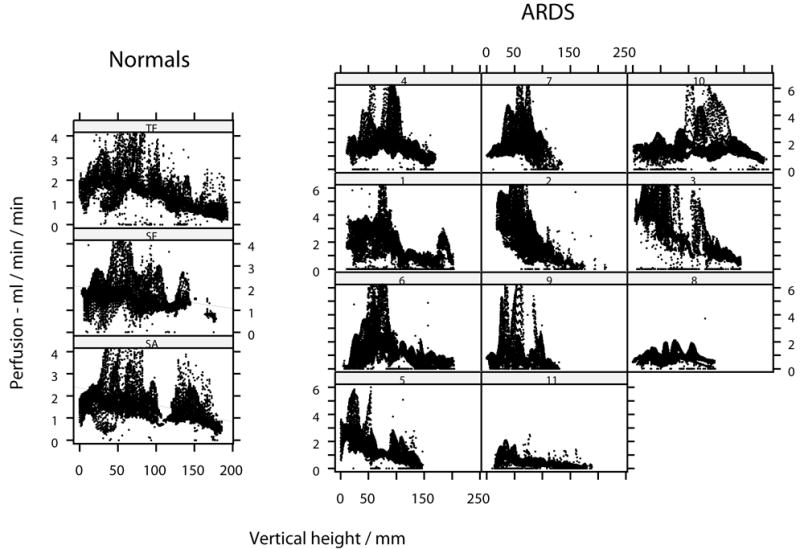
Figure 4.

Distribution of raw perfusion throughout the axial plane for lungs of each normal subject and for patients with ARDS. The right lung only is represented. Location in the axial plane expressed as vertical height above the scanning table. Linear regression lines added for normal subjects.
