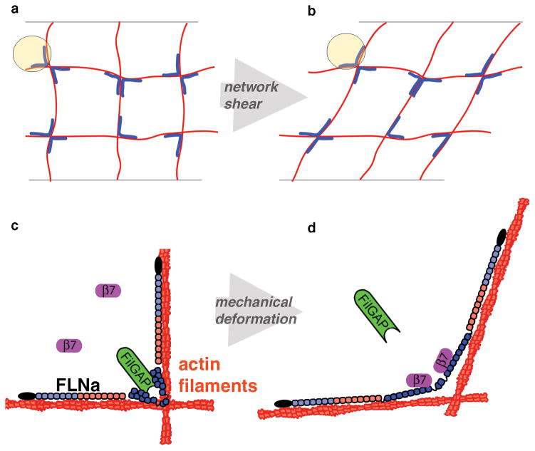
Figure 1. Differential mechanotransduction in FLNa occurs through spatial separation of binding sites and opening cryptic sites.
a) A Filamin (blue) crosslinked actin (red) gel forms an orthogonal network. b) When this network is strained, crosslinks are deformed. c) The actin-binding domain of FLNa is shown in black, followed by repeats 1–7 (light blue) and 8–15 (red), which form the linear rod 1 region. Repeats 16–23 (dark blue) form the compact rod 2 region. FilGAP (green) binds repeats 23 and the cytoplasmic domain of β7 integrin (purple) is unbound. d) When FLNa is mechanically deformed, the cryptic integrin site on repeat 21 is exposed allowing β7 integrin to bind, while repeats 23 are spatially separated, preventing FilGAP from binding both.

