Abstract
Combining ultrasonic modulation and optical phase conjugation allows light to be tightly focused in a scattering medium, providing benefits for studies of photophysical, photochemical and photobiological processes
Developing new methods for the focusing and delivery of light deep into biological tissue is of critical importance for biomedical imaging. The main problem is that tissue strongly scatters light, making both tasks extremely difficult, with high-resolution optical imaging often being limited to just a few hundred micrometres in turbid tissues.
Now, reporting in Nature Photonics, Lihong Wang and co-workers at Washington University in St. Louis, USA, demonstrate a new idea that tackles this problem by combining ultrasound and near-infrared (NIR) light to overcome the negative impact of multiple scattering when imaging biological media1.
The use of NIR light for medical imaging is attractive because it is non-ionizing, low cost, convenient to generate and detect, and sensitive to intrinsic tissue contrast such as haemoglobin concentration and oxygenation state. In the 1930s, Max Cutler used NIR light to transilluminate human breast tissue for cancer detection2. Subsequent studies were conducted in the late 1970s and 1980s to image breast3 and brain4 tissue. Although NIR light penetrates tissue to depths of several centimetres, early methods did not account for distortions caused by refractive-index fluctuations within and between microscopic tissue structures. These fluctuations limit the mean free path lengths of NIR photons to ~20–40 μm, such that multiple scattering dominates light propagation in tissue. Thus, high-resolution imaging methods that require coherent or ‘ballistic’ light, such as confocal microscopy and optical coherence tomography, are restricted to imaging depths of a few hundred micrometres and a few millimetres, respectively.
Over the past twenty years, significant attention has been devoted to developing methods that can overcome multiple light scattering when imaging deep inside tissue. A common approach is to model the propagation of the photons and then adjust the spatially varying optical properties of the tissue until the model’s predictions agree with experimental measurements. This approach bore the field of diffuse optical tomography, which derives its name from the observation that light propagation in a tissue resembles a random walk and can be mathematically described using a diffusion equation5,6. Implementations of diffuse optical tomography have so far included the measurement of either the detected light intensity, the phase of photon density waves produced by an intensity-modulated laser or the time-of-flight of photons from a pulsed source, as well as the removal of longer-path-length photons by time-domain or spatial-frequency gating techniques (for a review see ref. 7). However, the high degree of scattering for such techniques results in either an ill-posed image reconstruction problem — not enough information to reconstruct a unique solution — or data with a low signal-to-noise ratio, making high-resolution images difficult to produce.
The strategy of Wang and co-workers builds on previous hybrid imaging techniques that integrate optics with ultrasound1. These methods attempt to combine molecular sensitivity to light with the high spatial resolution of ultrasound. Such hybrid techniques fall into two main categories: those that use light to generate sound and those that tag light with focused sound waves. Photo-acoustic tomography and microscopy belong to the first category8, relying on pulses of light to cause a small but rapid expansion of the light-absorbing structures in tissue. Ultrasonic transducers detect the resulting pressure wave, and the propagation times and intensities of the detected signals are used to reconstruct images of light absorption.
The work of Wang and co-workers is closely related to ultrasound-modulated optical tomography, which belongs to the second category of such ultrasound–optical hybrid techniques9. In this scheme, the sample is illuminated with coherent light and scanned using a focused ultrasonic wave. By compressing and rarefying the tissue, the ultrasonic wave modulates the displacement of tissue scatterers and the index of refraction, resulting in an intensity modulation in the detected speckles produced by light passing through the ultrasound focus. Measurements of light intensity at the frequency of the ultrasonic wave are sensitive to the optical properties of the sample in the ultrasound focus. For example, high optical absorption at the ultrasound focus will cause a decrease in the modulated signal. The key point is that the spatial resolution is determined by the focused ultrasound beam and is not limited by the diffuse light field. However, this technique has proved to be challenging because the amount of ultrasound-modulated light is small compared with the diffuse background.
To overcome this limitation, Wang and colleagues combine the concept of ultrasonically tagging photons with optical phase conjugation, allowing them to focus light and ultrasound in the same location with a resolution of 0.63 mm (ref. 1). This major advance builds on the work of Yaqoob et al., who were able to refocus a beam of light that had passed through a 0.69 mm slice of chicken breast10. In that experiment, the light transmitted through the chicken breast was combined with a reference beam to record a hologram in an LiNbO3 photorefractive crystal. A second beam was then used to read the hologram and produce the phase-conjugate wave, which retraced the original path through the chicken breast.
The work of Wang and co-workers is significant because the use of ultrasound allows the light to be focused inside the tissue, rather than at the surface, as was done by Yacoob et al. In their experiment, two acousto-optic modulators shift the frequency of the NIR beam before it reaches the specimen. The specimen was a 10-mm-thick scattering slab containing two absorbing objects dyed with black ink, one measuring 1.3 mm × 4.5 mm × 1 mm and the other 1.7 mm × 4.5 mm × 0.6 mm.
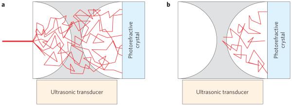
The frequency change of the NIR beam is equal to that of the ultrasonic beam focused inside the sample. The movement of scatterers at the ultrasonic focus causes some of the light to be Doppler-shifted back to the original frequency. When the transmitted light arrives at the photorefractive crystal, light at the original frequency combines with the reference beam to form a hologram (Fig. 1). Because this light comes from the ultrasonic focus, when the hologram is subsequently read, the phase-conjugate wave retraces its path back to the focal region. By changing the position of the ultrasound focus, Wang and co-workers dynamically localize light at different locations in the optically scattering media. Analogous to ultrasound-modulated optical tomography, when light from the phase-conjugate wave exits the sample, it gives information about the optical properties at the ultrasound focus. Specifically, a decrease in the measured light intensity indicates an increase in optical absorption from that volume element.
Figure 1.
Light from a coherent source diffuses through biological tissue. a, In the set-up of Wang and co-workers, the light passing through the focus of an ultrasound beam is used to write a hologram in a photorefractive crystal. b, When the hologram is read, the light retraces its path back to the ultrasonic focus.
The authors suggest many applications for which this technique may be beneficial. For example, it could be used to extend the depth of interrogation possible in fluorescence microscopy, or to help photo-acoustic microscopy by allowing the light source to be concentrated at the focal point of the ultrasonic detector. The ability to focus light in tissue could also be a tremendous advantage in therapeutic technologies such as photodynamic therapy, where getting sufficient light to the treatment volume can be difficult.
However, significant hurdles and questions remain. The time-reversal of light through turbid media is dependent on the scatterers being static. The authors have not yet demonstrated in vivo imaging, which requires tolerance to sample movement (although optical phase conjugation has recently been demonstrated in the ear of a living rabbit11), including motion due to physiological processes such as heartbeat, breathing and blood flow. In addition, many biological tissues are far more absorbing than the sample used by Wang and co-workers. Unlike elastic scattering, light absorption is not time-reversible. Thus, the presence of highly absorbing structures such as blood vessels could significantly shield other structures from view. It also remains to be seen whether this technique will work in thick tissues. Typical tissues imaged with diffuse optics, such as human breast, are both thicker (several centimetres) and exhibit more scattering than the sample used by Wang and co-workers. At the other extreme, the resolution of confocal fluorescence microscopy is usually around 0.5–1 μm — several orders of magnitude better than the authors’ preliminary demonstration. Furthermore, to be useful for treatments such as photodynamic therapy, the authors will need to demonstrate significant gain when reading the hologram such that the treatment light is more intense than the light being used to determine the focus. Nevertheless, if these technical challenges can be addressed, this intriguing and clever approach could potentially provide a single scalable platform for three-dimensional, high-resolution functional imaging with broad applications in biology and medicine.
References
- 1.Xu X, Liu H, Wang LV. Nature Photon. 2011;5:154–157. doi: 10.1038/nphoton.2010.306. [DOI] [PMC free article] [PubMed] [Google Scholar]
- 2.Cutler M. Ann. Surg. 1931;93:223–234. doi: 10.1097/00000658-193101000-00032. [DOI] [PMC free article] [PubMed] [Google Scholar]
- 3.Alveryd A, et al. Cancer. 1990;65:1671–1677. doi: 10.1002/1097-0142(19900415)65:8<1671::aid-cncr2820650802>3.0.co;2-a. [DOI] [PubMed] [Google Scholar]
- 4.Jobsis F. Science. 1977;23:1264–1267. doi: 10.1126/science.929199. [DOI] [PubMed] [Google Scholar]
- 5.Haskell RC. J Opt. Soc. Am. A. 1994;11:2727–2741. doi: 10.1364/josaa.11.002727. [DOI] [PubMed] [Google Scholar]
- 6.Yodh Y, Chance B. Phys. Today. 1995 March;48:34–40. [Google Scholar]
- 7.Gibson AP, Hebden JC, Arridge SR. Phys. Med. Biol. 2005;50:R1–R43. doi: 10.1088/0031-9155/50/4/r01. [DOI] [PubMed] [Google Scholar]
- 8.Wang LV. Nature Photon. 2009;3:503–509. doi: 10.1038/nphoton.2009.157. [DOI] [PMC free article] [PubMed] [Google Scholar]
- 9.Wang LV, Jacques SL, Zhao X. Opt. Lett. 1995;20:629–631. doi: 10.1364/ol.20.000629. [DOI] [PubMed] [Google Scholar]
- 10.Yaqoob Z, Psaltis D, Feld MS, Yang C. Nature Photon. 2008;2:110–115. doi: 10.1038/nphoton.2007.297. [DOI] [PMC free article] [PubMed] [Google Scholar]
- 11.Cui M, McDowell EJ, Yang C. Opt. Express. 2009;18:25–30. doi: 10.1364/OE.18.000025. [DOI] [PMC free article] [PubMed] [Google Scholar]


