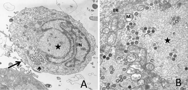
Figure 1.
Ultrastructural analysis of FV3-infected FHM cells. (A) An FV3-infected cell displaying virions budding from the plasma membrane (long arrow) or present within a paracrystalline array (short arrow), a viral assembly site (star), and the nucleus (N) showing chromatin condensation; (B) an enlargement of a viral assembly site (star) showing both full and empty virions and possible assembly intermediates. The assembly site is surrounded by mitochondria (M) and membraneous structures, possibly elements of the endoplasmic reticulum (ER).

