Abstract
We report a case of visceral pentastomiasis caused by Armillifer moniliformis in a 70-year-old aboriginal farmer from rural Malaysian Borneo. The patient complained of upper abdominal pain, jaundice, and loss of weight. Radiological investigations and subsequent histopathological examination revealed an adenocarcinoma of the pancreas with an adjacent liver nodule containing a nymph of A. moniliformis. This report constitutes the first documented human pentastomid infection in the whole of Malaysia after nearly 40 years, and it is the third description from Malaysian Borneo. Cases of human and animal pentastomiasis in Malaysia are discussed.
Introduction
Pentastomiasis is a zoonotic parasitic disease with an increasing number of documented human infections caused by the larval stages (nymphs) of pentastomes (tongue worms).1,2 These vermiform organisms form a unique phylum and are related to branchiurans.1,3 Pentastomes possibly coevolved with their respective vertebrate hosts,1 and adult parasites of most species live in the respiratory tract of snakes and other reptiles.2 Most human infections are caused by tropical and subtropical snake pentastomid parasites of the genus Armillifer, which encompasses species with different geographical distribution. A. armillatus and A. grandis are found in Africa, whereas A. agkistrodontis has been reported in China and A. moniliformis in Southeast Asia.2,4,5 Human infection occurs after accidental ingestion of infective ova, which are shed into the environment by snake secretions and feces. Human pentastomiasis was reported among aborigines in West and East Malaysia in the 1960s (Table 1).6–10 The present report describes the third case of human visceral pentastomiasis in Sabah, Malaysian Borneo, caused by A. moniliformis, and it constitutes the first report of this disease after nearly 40 years in the whole of Malaysia.
Table 1.
Reported cases of human pentastomiasis in Malaysia
| Pentastome species | Organ | Geographic region | Diagnosis | Reference |
|---|---|---|---|---|
| Armillifer sp. (A. moniliformis assumed) | Neck and abdomen | East Malaysia (Sabah, Borneo; two cases, including one European patient) | Radiology (both cases) | Rail8 |
| A. moniliformis | Liver and lung | West Malaysia (Pahang state) | Autopsy | Prathap and others9 |
| Armillifer sp. (A. moniliformis assumed) | Chest and abdomen | West Malaysia (Selangor state) | Radiology | Burns-Cox and others6 |
| A. moniliformis | Liver, lung, mesenteric lymph nodes, and intestinal wall | West Malaysia (10 cases from five different states; most cases from Selangor) | Autopsy | Prathap and others7 |
| Armillifer sp. | Fallopian tube | West Malaysia (state not mentioned) | Histopathology | Ong10 |
Case Report
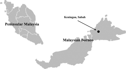
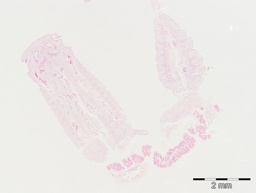
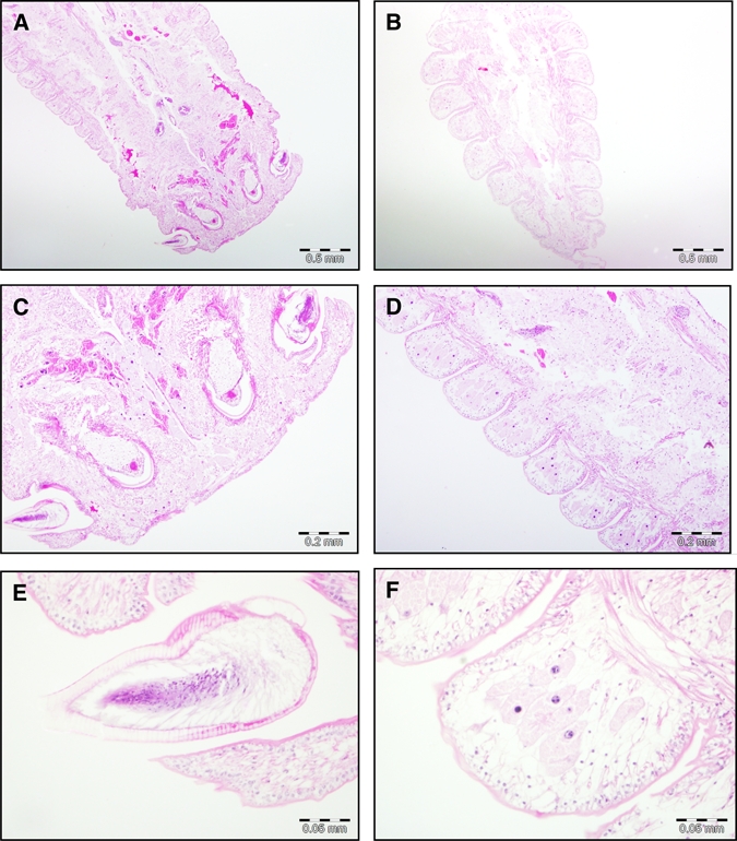
A 70-year-old aboriginal farmer (Orang Asli) from Keningau, Sabah, East Malaysia (Figure 1), was admitted to the Queen Elizabeth Hospital, Kota Kinabalu, in October 2010 with a 1-month history of upper abdominal discomfort, weight loss, anorexia, jaundice, and dark urine. The patient lived in a rural area for his entire life. Imaging techniques showed a tumor of the head of the pancreas and a liver nodule with a diameter of 1–2 cm. No enlarged lymph nodes were seen. The patient underwent Whipple's procedure, and histopathological examination of the pancreas tumor revealed a moderately differentiated adenocarcinoma with lymphatic metastasis. The liver nodule was also excised, and microscopic examination showed a C-shaped vermiform parasite with a body length of approximately 11 mm. The parasite's tegument was pseudosegmented and consisted of 26 annuli. The body tapered into a blunt-pointed cone with four rostral hooks (Figures 2 and 3). In accordance with published data,2,7,11 the parasite was diagnosed as an intact nymph of the Southeast Asian pentastome species A. moniliformis. The liver parenchyma showed obstructive changes within the portal tract, with chronic inflammatory cell infiltration and bile duct proliferation. A thick fibrous capsule associated with chitinous material was present on one free side of the liver tissue. There was intense chronic inflammatory cell infiltration directly adjacent to this fibrous capsule area.
Figure 1.

Map of Malaysia showing the patient's place of residence (5°20′ N, 116°10′ E) in Sabah, Malaysian Borneo.
Figure 2.

C-shaped nymph of A. moniliformis from the patient (hematoxylin and eosin stain) (Original magnification: 1.6×).
Figure 3.

Sections of the nymph of A. moniliformis. (A) Rostral section of the parasite with four oral hooks. (B) Tapering posterior part of the parasite with annulations. (C) Close-up view of A. The mouth is surrounded by four hooks. (D) Annulated rings from the mid-body. (E) Close-up view of the oral hook. (F) Close-up view of a single annulated ring. All sections are stained with haematoxylin and eosin (Original magnification: A and B, 4×; C and D, 10×; E and F, 40×).
The patient was discharged 15 days after surgery but did not return to the follow-up appointments.
Discussion
Most human cases of pentastomiasis are asymptomatic and are incidental findings during surgery or autopsy.2,7,12 If symptoms occur, they depend on the localization of the nymphs and are caused by the death or migration of the larval parasites.2 Most often, the liver, mesenteries, spleen, and lungs are affected,2 and patients report fever, abdominal pain, vomiting, diarrhea, jaundice, and abdominal tenderness.4 Severe and possibly lethal cases, such as massive infection of the liver,13 mechanical ileus,14 and other dissemination,15 are very rare. In the case described here, the symptoms reported were most likely caused by the pancreatic malignancy, and the pentastome infection was an incidental finding. In a series of 30 consecutive autopsies performed on aborigines from five different states in West Malaysia, pentastome larvae were found in 33.3% of the cases, with a prevalence of 45.4% in adults.7 Only two cases of human pentastomiasis caused by A. moniliformis were reported in East Malaysia/Borneo.8 The present report is, thus, the third description of a human pentastome infection in Malaysian Borneo, and it is the first description in the whole of Malaysia after 37 years. Interestingly, all reports about pentastomiasis in all of Malaysia describe the infection in the Orang Asli aborigine population, except for one case; this one case was a European woman who was diagnosed with an A. moniliformis infection in Borneo in 1965.8 In the latter case, consumption of python meat 9 years before the onset of symptoms was revealed. The Indian python (Python molurus) and the Asian reticulated python (P. reticulatus) are known final hosts of A. moniliformis.8,16 In a survey, adult parasites were recovered from two of six P. reticulatus,16 a snake also endemic to Malaysian Borneo. It is well-known that consumption of snake meat is a common practice in some parts of Southeast Asia,16–19 and among the aboriginal tribes in Malaysia, the Temiar, Semai, and Temuan are known habitual python eaters.7 Risk factors for infection include consumption of undercooked contaminated snake meat as well as contact with living snakes and their secretions (i.e., tropical snake farming, pet keeping, harvesting of their skins, or tribal totemism).1,2,20 All of the human A. moniliformis infections in West and East Malaysia were indirectly linked to eating snakes by either the patient's history or affiliation with a certain tribe (ethnicity), and thus, aboriginal people in rural areas are the population with the highest risk. In the case presented, the tribe of the patient was not determined, and no information about possible snake meat consumption was obtained. Another possible source of infection in Malaysian aborigines may be drinking of river water contaminated with snake secretions.7 In Malaysia, visceral pentastomiasis was also reported in wild and domestic animals as intermediate and final hosts (Table 2).6,16,18,19,21–27 In a large survey in 1981, the infection rate of different wild animals with nymphs of A. moniliformis was 1.7% in West Malaysia, with the highest individual number of parasites found in rodents and carnivores. Of the carnivora, 20.7% were infected.16
Table 2.
Pentastomiasis among animals in Malaysia
| Host | Pentastome species | Organ | Reference |
|---|---|---|---|
| Cynomolgus monkey (Macaca irus/fascicularis) | A. moniliformis | n/a | Burns-Cox and others6 |
| Giant swamp rat (Rattus bowersi) | Porocephalus armillatus | Lung, liver, spleen, and mesentery | Liat and Krishnasamy21 |
| House geckoes* | Raillietiella hemidactyli | n/a | Liat and Sen22 |
| Wild animals (rodents, primates, carnivores, and reptiles*) | A. moniliformis | Abdominal cavity and lungs | Krishnasamy and others16 |
| Cat | A. moniliformis | Liver and spleen | Chooi and others23 |
| Wild animals | A. moniliformis | n/a | Krishnasamy and others18 |
| Wild animals (house lizards,* snakes,* and others) | A. moniliformis | Lungs and mesenteries | Krishnasamy and others19 |
| Otter | A. moniliformis | Kidney, liver, spleen, and mesenteries | Cheah and others24 |
| Lizard* | Raillietiella sp. | Lung | Jeffery and others26 |
| Cockroaches | Raillietiella sp. | n/a | Jeffery and others27 |
| Wild rats | A. moniliformis | n/a | Syed-Amez and Mohd Zain25 |
n/a = Information not available.
Final hosts.
Of note, in some human cases, the diagnosis of visceral pentastomiasis was achieved by X-ray and not histology.6,8 Thus, in theory, an Armillifer species different from A. moniliformis could have caused the visceral pentastomiasis in these patients, because the radiological picture cannot discriminate between the parasites involved. However, it is generally accepted that A. moniliformis is the only Armillifer species found in Malaysia, which is underlined by the other reports about human7,9 and animal6,16,18,19,23–25 visceral pentastomiasis with definitive species diagnosis in Malaysia. In one report about rats, the pentastome species responsible was termed Porocephalus armillatus,21 which might have been confused with A. (P.) moniliformis.
In the present report, the parasite was diagnosed as a nymph of A. moniliformis based on morphology. Unfortunately, not enough tissue was available for polymerase chain reaction analysis and phylogenetic positioning of the parasite. Recent analysis revealed that the pentastome snake parasites from Africa (A. armillatus), China (A. agkistrodontis), and the Americas (P. crotali) clustered together in the minimal evolution model, indicating coevolution with their vertebrate final hosts.1 Armillifer nymphs have a body length of 9–23 mm,2 and A. moniliformis has been reported to have a minimal size of 11–12 mm in Malaysia.7,19 The species has about 30 rings,7 but lower counts of 26 rings have also been reported.11 In contrast, the geographically nearest neighbor, A. agkistrodontis, has only 7–9 spiral rings,4 whereas A. armillatus has 18–22 rings.6,7 To assess the full extent of human visceral pentastomiasis in Malaysia, serological prevalence studies in risk populations should be performed; these studies were used in a recent investigation from The Gambia1 and an earlier investigation from the Ivory Coast.28
ACKNOWLEDGMENTS
The authors are grateful to Professor Dato Dr. Khalid Yusoff, Dean of the Faculty of Medicine, Universiti Teknologi MARA for his continuous support and laboratory facilities. Sincere thanks to Mr. Haji Sulaiman Abdullah for his help in tracing the literature about pentastomiasis in Malaysia.
Footnotes
Authors' addresses: Baha Latif, Effat Omar, and Chong Chin Heo, Faculty of Medicine, Universiti Teknologi MARA, Shah Alam, Selangor, Malaysia, E-mails: bahalatif@yahoo.com, effatomar@yahoo.com, and chin@salam.uitm.edu.my. Noriah Othman, Department of Pathology, Hospital Selayang, Batu Caves, Selangor, Malaysia, E-mail: noriaho@selayanghospital.gov.my. Dennis Tappe, Institute of Hygiene and Microbiology, University of Würzburg, Würzburg, Germany, E-mail: dtappe@hygiene.uni-wuerzburg.de.
References
- 1.Tappe D, Meyer M, Oesterlein A, Jaye A, Frosch M, Schoen C, Pantchev N. Transmission of Armillifer armillatus ova at snake farm, The Gambia, West Africa. Emerg Infect Dis. 2011;17:251–254. doi: 10.3201/eid1702.101118. [DOI] [PMC free article] [PubMed] [Google Scholar]
- 2.Tappe D, Büttner DW. Diagnosis of human visceral pentastomiasis. PLoS Negl Trop Dis. 2009;3:e320. doi: 10.1371/journal.pntd.0000320. [DOI] [PMC free article] [PubMed] [Google Scholar]
- 3.Lavrov DV, Brown WM, Boore JL. Phylogenetic position of the Pentastomida and (pan)crustacean relationships. Proc Biol Sci. 2004;271:537–544. doi: 10.1098/rspb.2003.2631. [DOI] [PMC free article] [PubMed] [Google Scholar]
- 4.Chen SH, Liu Q, Zhang YN, Chen JX, Li H, Chen Y, Steinmann P, Zhou XN. Multi-host model-based identification of Armillifer agkistrodontis (Pentastomida), a new zoonotic parasite from China. PLoS Negl Trop Dis. 2010;4:e647. doi: 10.1371/journal.pntd.0000647. [DOI] [PMC free article] [PubMed] [Google Scholar]
- 5.Yao MH, Wu F, Tang LF. Human pentastomiasis in China: case report and literature review. J Parasitol. 2008;94:1295–1298. doi: 10.1645/GE-1597.1. [DOI] [PubMed] [Google Scholar]
- 6.Burns-Cox CJ, Prathap K, Clark E, Gillman R. Porocephaliasis in western Malaysia. Trans R Soc Trop Med Hyg. 1969;3:409–411. doi: 10.1016/0035-9203(69)90021-2. [DOI] [PubMed] [Google Scholar]
- 7.Prathap K, Lau KS, Bolton JM. Pentastomiasis: a common finding at autopsy among Malaysian Aborigines. Am J Trop Med Hyg. 1969;18:20–27. [PubMed] [Google Scholar]
- 8.Rail GA. Porocephaliasis: a description of two cases in Sabah. Trans R Soc Trop Med Hyg. 1967;61:715–717. doi: 10.1016/0035-9203(67)90140-x. [DOI] [PubMed] [Google Scholar]
- 9.Prathap K, Ramachandran CP, Haug N. Hepatic and pulmonary porocephaliasis in a Malaysian Orang Asli (Aborigine) Med J Malaya. 1968;13:92–95. [PubMed] [Google Scholar]
- 10.Ong HC. An unusual case to pentastomiasis of the fallopian tube in an Aborigine woman. J Trop Med Hyg. 1974;77:187–189. [PubMed] [Google Scholar]
- 11.Heymons R, Vitzthum HG. Beiträge zur Systematik der Pentastomiden. Z f Parasitenkunde. 1935;8:1–103. [Google Scholar]
- 12.Yapo Ette H, Fanton L, Adou Bryn KD, Botti K, Koffi K, Malicier D. Human pentastomiasis discovered postmortem. Forensic Sci Int. 2003;137:52–54. doi: 10.1016/s0379-0738(03)00281-0. [DOI] [PubMed] [Google Scholar]
- 13.Adeyekun AA, Ukadike I, Adetiloye VA. Severe pentasomide Armillifer armillatus infestation complicated by hepatic encephalopathy. Ann Afr Med. 2011;10:59–62. doi: 10.4103/1596-3519.76592. [DOI] [PubMed] [Google Scholar]
- 14.Lavarde V, Fornes P. Lethal infection due to Armillifer armillatus (Porocephalida): a snake-related parasitic disease. Clin Infect Dis. 1999;29:1346–1347. doi: 10.1086/313460. [DOI] [PubMed] [Google Scholar]
- 15.Obafunwa JO, Busuttil A, Nwana EJC. Sudden death due to disseminated porocephalosis—a case history. Int J Legal Med. 1992;105:43–46. doi: 10.1007/BF01371237. [DOI] [PubMed] [Google Scholar]
- 16.Krishnasamy M, Singh I, De Witt GF, Ambu S. The natural infection of wild animals in West Malaysia with nymphs of Armillifer moniliformis (Diesing, 1835) Sambon, 1922. Malays J Pathol. 1981;4:29–34. [PubMed] [Google Scholar]
- 17.Lai C, Wang XQ, Lin L, Gao DC, Zhang HX, Zhang YY, Zhou YB. Imaging features of pediatric pentastomiasis infection: a case report. Korean J Radiol. 2010;11:480–484. doi: 10.3348/kjr.2010.11.4.480. [DOI] [PMC free article] [PubMed] [Google Scholar]
- 18.Krishnasamy M, Singh I, Jeffery J. Some new vertebrate host records of Armillifer moniliformis (Diesing, 1835) Sambon, 1922 in Peninsular Malaysia. J Malaysian Soc Hlth. 1984;4:20–21. [Google Scholar]
- 19.Krishnasamy M, Singh I, Jeffery J, Ambu S, Ali JH, Baker ED. Some pentastomes from Malaysia, with special emphasis on those from Peninsular Malaysia. J Malaysian Soc Health. 1985;5:49–56. [Google Scholar]
- 20.Dakubo J, Naaeder S, Kumodji R. Totemism and the transmission of human pentastomiasis. Ghana Med J. 2008;42:165–168. [PMC free article] [PubMed] [Google Scholar]
- 21.Liat LB, Krishnasamy M. A heavy infection of Porocephalus armillatus in Rattus bowersi. Southeast Asian J Trop Med Public Health. 1973;4:282–284. [PubMed] [Google Scholar]
- 22.Liat LB, Sen YH. Pentastomid infection in the house geckoes from Sarawak, Malaysia. Med J Malaysia. 1977;32:59–62. [PubMed] [Google Scholar]
- 23.Chooi KF, Omar AR, Lee JYS. Meloidosis in a domestic cat, with concurrent infestation by nymphs of the cat pentastome, Armillifer moniliformis. Kajian Vet Malaysia. 1982;14:41–44. [Google Scholar]
- 24.Cheah TS, Rajamanickam C, Lazarus K. Natural infection of Armillifer moniliformis in a smooth otter (Lutra perspicillata) Trop Biomed. 1989;6:139–140. [Google Scholar]
- 25.Syed-Amez ASK, Mohd Zain SN. A study on wild rats and their endoparasites fauna from the Endau Rompin National Park Johor. Malaysian J Sci. 2006;25:19–39. [Google Scholar]
- 26.Jeffery J, Mohd Z, Abdullah S, Krishnasamy M. Gekko smithi, a new lizard host of the pentastomid, Raillietiella sp. (Pentastomida: Cephalobaenida) in Peninsular Malaysia. Trop Biomed. 1997;14:145–146. [Google Scholar]
- 27.Jeffery J, Abdullah S, Anuar K, Omar B, Moktar N, Sulaiman S, Hidayatulfathi O, Krishnasamy M. Collection of domicilliary cockroaches and their parasites from the Faculty of Medicine, University of Malaya, Kuala Lumpur. Trop Biomed. 2003;20:165–168. [Google Scholar]
- 28.Nozais JP, Cagnard V, Doucet J. Pentastomosis. A serological study of 193 Ivorians. Med Trop (Mars) 1982;42:497–499. [PubMed] [Google Scholar]


