Abstract
Kinesins are a family of motor proteins conserved throughout eukaryotes. In our present study we characterize a novel kinesin, KinesinCaaX, orthologs of which are only found in the kinetoplastids and not other eukaryotes. KinesinCaaX has the CVIM amino acids at the C-terminus, and CVIM was previously shown to be an ideal signal for protein farnesylation in T. brucei. In this study we show KinesinCaaX is farnesylated using radiolabeling studies and that farnesylation is dependent on the CVIM motif. Using RNA interference, we show KinesinCaaX is essential for T. brucei proliferation. Additionally RNAi KinesinCaaX depleted T. brucei are 4 fold more sensitive to the protein farneysltransferase (PFT) inhibitor LN-59, suggesting that KinesinCaaX is a target of PFT inhibitors' action to block proliferation of T. brucei. Using tetracycline-induced exogenous tagged KinesinCaaX and KinesinCVIMdeletion (non-farnesylated Kinesin) expression lines in T. brucei, we demonstrate KinesinCaaX is farnesylated in T. brucei cells and this farnesylation has functional effects. In cells expressing a CaaX-deleted version of Kinesin, the localization is more diffuse which suggests correct localization depends on farnesylation. Through our investigation of cell cycle, nucleus and kinetoplast quantitation and immunofluorescence assays an important role is suggested for KinesinCaaX in the separation of nuclei and kinetoplasts during and after they have been replicated. Taken together, our work suggests KinesinCaaX is a target of PFT inhibition of T. brucei cell proliferation and KinesinCaaX functions through both the motor and farnesyl groups.
Introduction
Trypanosoma brucei species are the causative agents of Human African Trypanosomiasis (HAT) or African sleeping sickness in humans and the wasting disease, nagana, in cattle. There were 9878 new HAT cases reported to the WHO in 2009 [1], notably the first decline below 10,000 reported cases since 1960 due in part to increased national sleeping sickness control programs [1] and disease mapping [2]. However, as resources are limited in many parts of rural Africa and surveillance in many areas is not yet routine, many cases go unreported. The WHO estimates from 30,000 to 70,000 new cases of HAT occur per year [3]. Trypanosoma species can also infect livestock including goats, sheep, pigs, donkeys and cattle [4]. This has an impact economically as many parts of Africa are unable to raise livestock for consumption and sale due to this parasite [4].
Currently no vaccines are effective at preventing Trypanosoma brucei infections. Existing medical therapies do exist, however many are toxic, require long treatment regimens and are difficult to administer [3]. Drug resistance is also a concern [5]–[8] and new drugs are urgently needed.
In our search for possible drug targets against protozoan parasites we have characterized the enzymes responsible for protein prenylation [9]–[19]. Prenylation is the posttranslational modification of proteins by the covalent addition of the isoprenyl lipid farnesyl or geranylgeranyl [20], [21]. In farnesylation, the fifteen carbon farnesyl group from farnesyl pyrophosphate is added to the C of the CaaX motif, a cysteine-containing four amino acid residue motif at the C-terminus of some proteins. The aa represent two aliphatic residues and the X represents amino acids including serine, methionine, alanine, threonine or glutamine [20]. Geranylgeranylation refers to the addition of a twenty-carbon geranylgeranyl group to the CaaX motif where X is commonly a leucine or phenylalanine. Prenylation modifications create a hydrophobic C-terminus that allows the protein to interact with the cell membrane, membrane-bound organelles, other cellular proteins and hydrophobic surfaces. Addition of the farnesyl or geranylgeranyl groups is mediated in mammalian cells by three heterodimeric enzymes: protein farneysltransferase (PFT), protein geranylgeranyltransferase type I (PGGT-I) and protein geranylgeranyltransferase type II (PGGT-II) [20], [21].
Previously our work has investigated T. brucei PFT (TB-PFT) enzyme as a potential drug target for developing new drugs against T. brucei [9]–[16]. TB-PFT covalently-links a farnesyl group from farnesyl-pyrophosphate to the cysteine on the CaaX motif. In mammals, both PFT and PGGT-I share the α-subunit, but bioinformatic studies suggest that T. brucei lacks a gene encoding the β-subunit of PGGT-I and biochemical studies suggest T. brucei lacks PGGT-I activity [14]. Our group has shown that PFT inhibitors inhibit prenylation of T. brucei proteins and inhibit T. brucei growth [10]–[11]. We have also demonstrated that T. brucei parasites are more sensitive to PFT inhibitors than T. cruzi parasites [18]. We have identified a T. cruzi gene encoding the β-subunit homolog of PGGT-I, Gene DB# Tc00.1047053508817.150 [18]. PGGT-I has been shown to act on many of the same substrates that PFT normally prenylates [20] and T. cruzi PGGT-I may provide a redundant function for PFT and thus explain why T. cruzi is less sensitive to PFT inhibitors than T. brucei. Taken together, these data suggest TB-PFT is a valid target for drug development for HAT therapy.
Conventionally the X amino acids in CaaX motifs modified by mammalian PFT include serine, methionine, alanine and glutamine. In contrast, our work has shown that while TB-PFT readily farnesylates CaaX targets containing a methionine or glutamine in the X position, it does not readily modify CaaX targets with serine, threonine, alanine or cysteine in the X position [12]. We have also identified inhibitors that are ten-fold more potent against TB-PFT than mammalian PFT [11]. This restricted preference to TB-PFT and not the mammalian PFT is desirable for drug design targeted to kill T. brucei in humans but not affect the human form of the enzyme. However, of the PFT inhibitors that had sufficient potency against TB-PFT, none had desirable in vivo pharmacokinetics [15]. Therefore we began the study of the preferred CaaX containing proteins that have the methionine in the X position to investigate proteins downstream of TB-PFT in order to identify new drug targets.
We have shown the CVIM motif is a favored target of TB-PFT [12]. Only two of all known C-terminal CaaX -containing proteins have the CVIM CaaX -motif in T. brucei brucei (Fig. S1). One CVIM-containing gene, Gene DB# Tb10.70.0590, is a predicted ras-like small GTPase and has been previously characterized in T. cruzi [19]. The second, Gene DB# Tb927.10.12440, was annotated as a kinesin-like protein with a predicted molecular weight of 91 kilodaltons. As we had observed a decrease in prenylation of proteins at approximately this molecular weight in protein farnesyl transferase inhibitor (PFTi) -treated T. brucei [11] and because no other proteins with a CaaX motif besides Tb927.10.12440 were between 77 and 98 kilodaltons (Fig. S1), we set out to further investigate this protein. Bioinformatics studies reveal this protein contains motifs common to the kinesin family of motor proteins. Due to the presence of the CaaX motif we have named the protein KinesinCaaX. In our present study we demonstrate this protein is farnesylated in T. brucei cells and this farnesylation has functional effects. With KinesinCaaX protein depletion using RNAi we demonstrate the importance of KinesinCaaX for T. brucei proliferation. We characterize tetracycline-induced exogenous tagged KinesinCaaX expression in T. brucei and localize it in the cell and show this localization to be dependent on prenylation. Through our investigation of cell cycle, nucleus and kinetoplast quantitation and immunofluorescence assays we hypothesize an important role for KinesinCaaX in nuclear and kinetoplast segregation.
Results
KinesinCaaX has motifs consistent with kinesin motor proteins
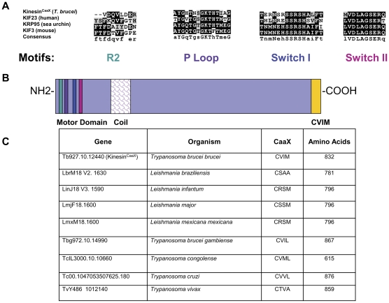
All motifs common to the kinesin family of motor proteins are present in KinesinCaaX. Kinesins are proteins that convert the chemical energy from ATP into a mechanical force to move along microtubules [22]. Kinesins include a diverse range of molecules organized into nearly 20 families based on the location of the motor domain in the protein and regions of homology outside of the motor domain [23]. Kinesins from various families have diverse functions and may function as monomers, dimers or tetramers [23]. Kinesins are implicated in moving a wide range of cellular macromolecules, termed “cargo”, including vesicles, organelles and chromosomes [23]. KinesinCaaX cannot be placed in any known kinesin family due to low homology with previously characterized kinesin families outside the motor domain [24]. KinesinCaaX is similar in domain layout to the canonical kinesin family 1 members that function as homodimers with two large globular motor domains followed by a coiled-coil neck [25]. The globular motor domain interacts with microtubules and the coiled-coil neck is associated with dimerization. Movement along microtubules has been described as stepwise using 8-nm steps in an asymmetric hand-over-hand mechanism [22]. Structural features involved in force generation in the kinesin superfamily are conserved in KinesinCaaX when compared to known kinesins in sea urchins, mice and humans (Fig. 1A) as shown by MUSCLE alignment [26]. The R2 domain, P Loop, Switch I and the relay helix Switch II [27] domains are conserved in KinesinCaaX. KinesinCaaX from T. brucei brucei has 832 residues with the motor domain located at the amino terminus of the molecule, followed by a coil region that is predicted to form a coiled coil upon dimerization and the CaaX motif at the carboxy terminus (Fig. 1B). Based on the presence of a coil domain and the motor domain at the amino-terminus of the protein, we predict KinesinCaaX moves towards the plus-end of microtubules as a dimer.
Figure 1. KinesinCaaX has motifs consistent with plus-end directed kinesin proteins and is conserved in pathogenic kinetoplastids.
(A) Key kinesin domains are conserved in Tb10.389.1270 when sequences are compared to kinesins in sea urchins, mice and humans. The corresponding GenBank accession numbers for each corresponding protein are Homo sapiens NP_612565.1, Mus musculus NP_03246, and Strongylocentrotus purpuratus P46871. (B) Structural features and domain architecture of KinesinCaaX. KinesinCaaX has 832 residues. Represented are the R2 domain (teal), the phosphate binding loop or P Loop (purple), Switch I (blue) and Switch II (magenta) regions, the coil region (fish scales) that facilitates dimerization, and CaaX motif (yellow) that is predicted to act as a farnesylation signal. (C) Comparison of KinesinCaaX orthologs in other kinetoplastids shows CaaX motif and size conservation in other pathogenic kinetoplastid species.
KinesinCaaX is conserved in other pathogenic kinetoplastids but not other eukaryotes
Homology studies utilizing OrthoMCL [28] do not detect orthologs to T. brucei brucei KinesinCaaX in higher eukaryotes, but do predict a single orthologous copy in other pathogenic kinetoplastids including Leishmania braziliensis, Leishmania infantum, Leishmania major, Leishmania mexicana mexicana, Trypanosoma brucei gambiense, Trypanosoma brucei rhodesiense, Trypanosoma congolense, Trypanosoma cruzi and Trypanosoma vivax (Fig. 1C, S2 & S3). The orthologous members vary moderately in molecular weight, yet in each case retain the CaaX motif at the carboxy terminus (Fig. 1C). The evolutionary relationship of KinesinCaaX was analyzed using CLUSTALW Biology Work Bench, Version 3.2 [29]. This analysis demonstrates T. brucei gambiense KinesinCaaX is most closely related to that of T. brucei brucei followed by the other Trypanosoma species and then the Leishmania species (Fig. S3). As KinesinCaaX is conserved throughout kinetoplastids but not other higher eukaryotes, we hypothesize this kinesin performs a conserved role in kinetoplastid cell biology.
KinesinCaaX has an ATP-dependent motor capable of movement with microtubules
Some predicted kinesins in eukaryotic organisms attach to microtubules but do not demonstrate motor activity. Truncated kinesin genes expressing presumably non-motile kinesins have been suggested to be genetic remnants of once functional proteins [30]. Non-motile kinesins may also facilitate other not yet defined microtubule-protein interactions. To test for motor activity in KinesinCaaX, we used an in vitro microtubule motility assay using rhodamine-labeled microtubules and time lapse fluorescence microscopy. Truncated recombinant KinesinCaaX containing the motor domain and the coil dimerization domain (Fig. S4A) led to the processive movement of fluorescent microtubules, in a linear direction, in the presence of ATP (Fig. S4B). Control extracts expressing β-galactosidase, non-motor recombinant protein plus ATP did not lead to the movement of fluorescent microtubules (Fig. S4C). KinesinCaaX without ATP also did not lead to the movement of fluorescent microtubules (data not shown). Thus, both ATP and the KinesinCaaX motor domain were required to move microtubules. These studies demonstrate that KinesinCaaX has motor activity with microtubules in an ATP dependent manner. We hypothesize KinesinCaaX uses this ATP-dependent motor activity to move cargo along the microtubule network within T. brucei cells. As a more sophisticated biophysical study would require a full length protein isolated from T. brucei, we proceeded to studies to elucidate the functional significance of KinesinCaaX in the parasite.
Reduction of KinesinCaaX expression in the bloodstream form inhibits growth and contributes to mitotic delay
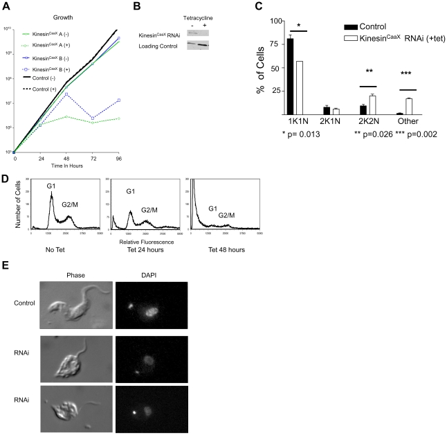
T. brucei and other kinetoplastids have a complex life cycle with various developmental forms. Two forms of T. brucei brucei can be cultured and studied in the lab; the mammalian bloodstream (trypomastigote) form and the insect (procyclic) form. We utilized RNA interference (RNAi) in order to test the effect of reduced KinesinCaaX expression on the phenotype of bloodstream trypomastigotes, as trypomastigotes are the forms most relevant to human and animal disease. We cloned a unique portion of the KinesinCaaX ORF into a tetracycline-inducible construct designed with opposing T7 promoters that induce dsRNA and made stably transfected T. brucei brucei bloodstream parasites. Within 72 hours of tetracycline induction of KinesinCaaX RNAi, a dramatic growth defect occurred in two independently derived clones (Fig. 2A). RT-PCR for KinesinCaaX mRNA reveals levels from induced with tetracycline were reduced to 18% (Clone A) and 32% (Clone B) of uninduced controls by 48 hours. Levels of KinesinCaaX mRNA of no-tetracycline controls were similar to quantities observed in non-transfected controls. Further work was carried out with clone A because of its superior RNA reduction after tetracycline induction. Protein levels of KinesinCaaX clone A were depleted to undetectable levels as shown by Western blot analysis (Fig. 2B). Growth arrest and correlating mRNA and protein depletion of KinesinCaaX support the hypothesis that KinesinCaaX is essential for growth in the bloodstream form.
Figure 2. RNAi of KinesinCaaX in the bloodstream form inhibits growth and contributes to mitotic delay.
(A) Growth of tetracycline-inducible RNAi KinesinCaaX knockdown cells with and without tetracycline induction. RNAi cell lines were constructed with the tetracycline-inducible vector to induce double stranded RNA specific to KinesinCaaX. KinesinCaaX A and B represent two different clones. Untransfected and uninduced (−tet) cells are used as controls. Broken lines indicate tetracycline added to cultures during growth (+). KinesinCaaX RNAi-induction results in a significant growth defect in both A & B clones. (B) Western blot analysis of Clone A whole cell lysates of bloodstream (BSF) probed with rabbit serum from animals immunized with a fragment of recombinant KinesinCaaX. Non-induced (−) and tetracycline induced (+) cells reveal KinesinCaaX protein levels (91 kDa) were diminished by 48 hours of tetracycline-induction. A 21 kDa non-specific band from the Western Blot serves as a loading control. The same loading ratio of − to + tetracycline protein was confirmed in non-transferred Coomassie stained PAGE lanes (data not shown). (C) Fluorescence microscopy of DAPI stained cells demonstrates a shift in kinetoplast and nuclei content of KinesinCaaX-depleted cells. Shown are the mean and SD of 2 experiments at 24 hours after tetracycline induction performed with clone A cells. Control cells are nontransfected controls treated with tetracycline. Similar results were seen with control transfected cells that were not induced with tetracycline (data not shown). Compared with control cells, KinesinCaaX-depleted T. brucei cells have a larger proportion of cells with 2 kinetoplasts and 2 nuclei (2K2N) and an increased number of cells with greater than two nuclei (other). Statistical differences were assessed with a Student T Test. (D) Flow cytometry reveals KinesinCaaX depleted T. brucei cells, compared with non-induced cells, is dominated by the appearance of an increased number of cells with lower than G1 DNA content, particularly evident at 48 hours after induction. The Y axis represents the number of cells and the X axis is DAPI fluorescence, representing DNA content. The lines represent histograms of cells. Peaks representing G1 and G2/M are indicated. (E) Photomicrographs of phase and DAPI-stained T. brucei blood-stage cells. Depletion of KinesinCaaX (RNAi) leads to cell morphology changes with cells demonstrating a rounded cell body and detached flagella compared to the non-induced cells (Control). The RNAi examples represent two typical cells, with altered morphology, observed at 48 hrs after tetracycline induction.
T. brucei cells cannot be easily synchronized to study the cell cycle, but kinetoplast and nuclear DNA quantitative analysis by fluorescence microscopy of DAPI stained fixed cells can be used to detect alterations in cell cycle progression. G1 cells are diploid and have 1 kinetoplast and 1 nucleus (1K1N). Kinetoplast replication and division occurs before nuclear DNA replication is completed, such that cells undergoing nuclear DNA synthesis (S phase) can have an elongated kinetoplast (still 1K1N) or 2 kinetoplasts and 1 nucleus (2K1N) [30]. Cells with 2 kinetoplasts and 2 nuclei (2K2N) have completed mitotic division and are in G2 or mitosis (G2/M) phase. Cells with greater than 2K2N have not completed cytokinesis. Quantitative analysis reveals cells undergoing KinesinCaaX RNAi for 24 hours, compared to uninduced cells, demonstrate a decrease of 1K1N cells from 81% to 57% (p = 0.013 Student T test), an increase in 2K2N cells from 9.5% to 20% (p = 0.026 Student T test) and an increase in of cells with greater than 2K2N (other) 1.5% to 17% (p = 0.002 Student T test)(Fig. 2C). No 1K0N cells were observed. Flow cytometry reveals RNAi depletion of KinesinCaaX results in a slight proportional increase in G2/M cells and a decrease in G1 cells, at 24 hours (Fig. 2D). However, the bulk of the events at 48 hours have low DAPI fluorescence (Fig. 2D, right panel), suggesting that there is an abundance of cell fragments with reduced DNA, likely representing degenerating and dying cells. 1K1N cells undergoing RNAi at 48 hours have a rounded cell body with detached flagella and the DAPI staining appears weaker than non-induced cells (Fig. 2E). Taken together, these data suggest KinesinCaaX is essential for growth in the bloodstream form.
KinesinCaaX is farnesylated in T. brucei
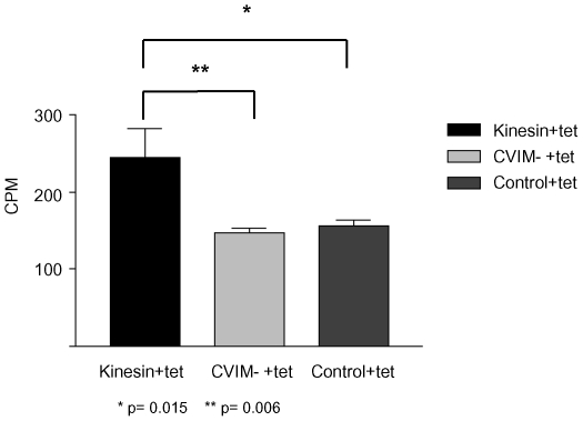
Farnesyl pyrophosphate is used by T. brucei protein farnesyl transferase to add farnesyl groups to CaaX-motifs and is generated in a series of enzymatic steps from mevalonate [20]. In the presence of simvastatin, mevalonate synthesis is inhibited, and T. brucei cells incorporate scavenged mevalonalactone. Scavenged mevalonalactone is incorporated into farnesyl-pyrophosphate and used to farnesylate proteins [11]. We engineered a tetracycline-inducible plasmid to ectopically express an amino-terminal hemagglutinin-epitope-tagged full length copy of KinesinCaaX (HA-KinesinCaaX). We made a second construct using site directed mutagenesis, we deleted the twelve nucleotides corresponding to the CVIM (CaaX) from the tetracycline-inducible HA-KinesinCaaX construct and transfected the plasmid to make a tetracycline inducible HA-KinesinCVIM deletion expressing T. brucei line. Tet-inducible HA-KinesinCaaX, HA-KinesinCVIM deletion and control (untransfected) T. brucei lines were grown in the presence of 3H-mevalonalactone, simvastatin and tetracycline. Immunoprecipitation with anti-HA antibodies shows tetracycline-induced HA-KinesinCaaX immunoprecipitates had a statistically significant increase in radioactive counts per minute (cpm) than control immunoprecipitates (Fig. 3) (p = 0.015, Student T test) and HA-KinesinCVIM deletion (p = 0.006, Student T test) while KinesinCVIM deletion did not have a statistically significant difference to the control (p = 0.10, Student T test). These data support our previous finding that TB-PFT adds farnesyl to CVIM peptides and is highly suggestive that CVIM of HA-KinesinCaaX is farnesylated by TB-PFT in T. brucei. This supports the hypothesis that native KinesinCaaX is farnesylated by TB-PFT in T. brucei cells.
Figure 3. KinesinCaaX is farnesylated in T. brucei.
Radioactivity (CPM: counts per minute) found in HA-antibody immunoprecipitates from induced HA-KinesinCaaX (Kinesin+tet), induced HA-KinesinCVIM deletion (CVIM- +tet), and control non-transfected cells (Control+tet) extracts from bloodstream form cells. Cells were treated with tetracycline, simvastatin and incubated with 3H mevalonolactone for 48 hours. Radioactivity above background is found only in the induced HA-KinesinCaaX –immunoprecipitates. Whole cell lysates pre-IP were compared using a scintillaton counter to ensure equal lysate concentrations per reaction. Statistical differences were assessed with a Student T Test.
KinesinCaaX localizes near dividing kinetoplasts and nuclei
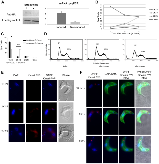
To shed light on the localization of KinesinCaaX, we made an antibody specific to a unique sequence in KinesinCaaX in T. brucei cells and performed immunofluorescence. Despite trying multiple fluorescent signal amplification protocols, expression of native levels of KinesinCaaX appeared to be too weak to reliably and reproducibly detect by immunofluorescence. We used the HA-KinesinCaaX exogenous expression lines to study growth and morphology. qPCR shows a 4.3 fold (SD = 0.56) induction of mRNA (Fig. 4A). Western blot using an anti-HA antibody reveals induction of HA-KinesinCaaX following tetracycline (tet) addition (Fig. 4A). Fluorescence microscopy of DAPI stained cells reveals that induction of HA-KinesinCaaX results in an increase of cells with the 2K1N configuration from 8% to 44% (p = 0.0019, Student T test) compared to non-induced cells, with a relative decrease in 1K1N cells from 81% to 45.5% by 48 hours (Fig. 4B & 4C) (p = 0.006, Student T test) (Fig. 4C). Flow cytometry analysis on this tetracycline-inducible HA-KinesinCaaX-line reveals a slight decrease in the G1 peak and a slight increase of events between the G1 and G2/M peaks by 48 hours after tetracycline induction (Fig. 4D) consistent with the increase in 2K1N cells. Induced cells have a similar growth rate to control cells and uninduced cells (Fig. S5). Additionally, very few cells with low amounts of DNA (presumably dead cells, Fig. 4D) were seen after KinesinCaaX induction compared with RNAi induction (Fig. 2D).
Figure 4. KinesinCaaX localizes near the nucleus and kinetoplast and is enriched in post-mitotic cells.
(A) Left Western blot of whole cell lysates of bloodstream form (BSF) T. brucei tagged HA- KinesinCaaX cell line either induced (+) or non-induced (−) for 48 hours. While more material is loaded in the (−) non-induced lane, much more HA-Kinesin is observed in the (+) induced lane. Right Quantitative PCR (qPCR) showed induced cells had 4.2 times more mRNA for KinesinCaaX than non-induced controls (error bars are SD). (B) DAPI staining and fluorescence microscopy reveals a larger proportion of cells with 2 kinetoplasts and one nucleus (2K1N) and a reduction in 1K1N cells in tetracycline-induced KinesinCaaX T. brucei cells compared with non-induced cells, with the effect becoming prominent at 48 hours (C) The mean and SD of 2 experiments at 48 hours. Statistical analysis was performed using a Student T test. (D) Ectopic expression of KinesinCaaX results in a slight decrease in G1 cells and a slight increase in cells between the G1 and G2/M peaks at 48 hours after tetracycline induction. (E) Cells were stained with DAPI to stain nuclear and kinetoplast DNA (blue), anti-HA rat mAb primary and polyclonal anti-Rat IgG Cy5.5 (red) secondary 24 hours post-tetracycline induction. Cells with 2K1N and 2K2N DNA content have an increased amount of KinesinCaaX relative to 1K1N cells. (F) Cells were stained with DAPI to stain nuclear and kinetoplast DNA (blue), FITC-anti-mouse IgG to localize the KMX mouse monoclonal antibody (mAb) that binds β- tubulin (green) and anti-HA rat mAb primary and polyclonal anti-rat IgG Cy5.5 (red) secondary that localize HA-KinesinCaaX 24 hours post tetracycline induction. β-tubulin localization demonstrates these cells have normal morphology. The cell marked 1Kdiv1N has an elongated kinetoplast consistent with a kinetoplast that is undergoing division.
As we could not consistently detect native KinesinCaaX using immunofluorescence, we used the HA-tagged-expression line for localization studies. Exogenously expressed HA-KinesinCaaX localizes between the nucleus and kinetoplast in a small percentage of 1K1N containing cells but is not present in all 1K1N cells (Fig. 4E & F). In general, 1K1N cells that have detectable HA-KinesinCaaX have elongated and V-shaped kinetoplasts (Fig. 4F 1Kdiv1N), indicative of initiation of kinetoplast replication [31]. Cells with 2K1N and 2K2N DNA content have more easily detectable HA-KinesinCaaX relative to 1K1N cells (Fig. 4E & 4F). In 2K1N containing cells, HA-KinesinCaaX localizes around the nucleus and is enriched near the anterior kinetoplast (Fig. 4E) and in post-mitotic 2K2N cells, KinesinCaaX is found near the anterior kinetoplast, between the nuclei, and surrounding the nuclei (Fig. 4E). Using the anti-β-tubulin antibody KMX [32] we monitored KMX epitope localization before and after HA-KinesinCaaX induction (Fig. 4F). KMX localizes to β-tubulin in the microtubule corset of T. brucei cells (Fig. 4F). Though the KMX β-tubulin localization looked similar in uninduced cells and induced cells, HA-KinesinCaaX appeared enriched in the β-tubulin between the nuclei of 2K2N cells (Fig. 4F). These localization data support a role for KinesinCaaX in separating nuclei and kinetoplasts.
KinesinCaaX localization is disrupted with CaaX deletion
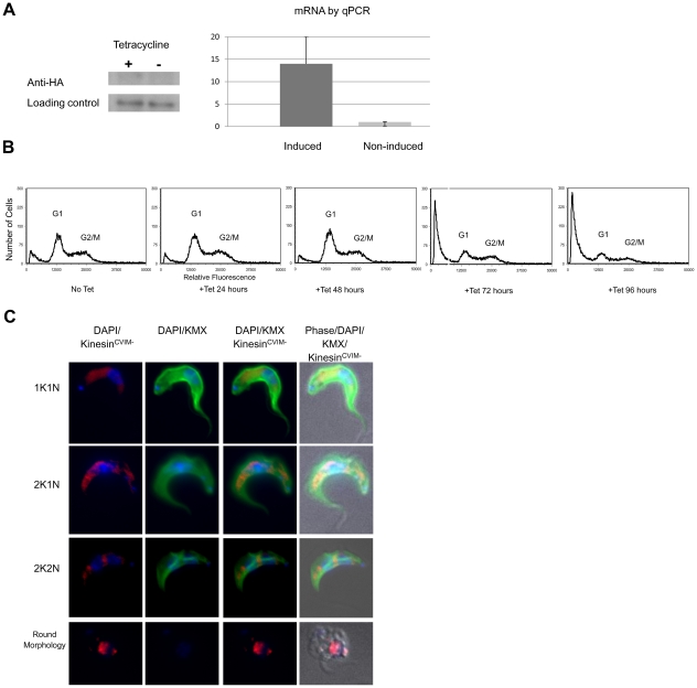
Upon tetracycline induction KinesinCVIM deletion mRNA is induced 13.9 fold (SD = 6.5) as shown by RT-PCR and protein is detected and Western blot analysis (Fig. 5A). HA-KinesinCVIM deletion expressing-cells demonstrate a similar growth rate as control cells for the first 72 hours and then demonstrate a slowed growth rate by 96 hours post-induction (Fig. S5). Upon tetracycline induction, HA-KinesinCVIM deletion cells have a very similar nuclear and kinetoplast quantitative profile to control unstimulated cells at 24 and 48 hours (data not shown) consistent with flow cytometry profiles of G1 and G2/M (Fig. 5B). While profiles of the 24 and 48 hour time points after induction are very similar to the non-induced control by 72 hours and at 96 hours after induction, a large population of cells containing less than a G1 complement of DNA becomes apparent, suggestive of dying and degenerating cells. This is consistent with the observed decrease in growth by 96 hours (Fig. S5). Immunofluorescence microscopy shows the HA-KinesinCVIM deletion has a more diffuse localization in 1K1N, 2K1N and 2K2N cells than the HA-tagged KinesinCaaX cells at 24 hours post-induction (Fig. 5C). Additionally we observed cells with a rounded cell body some of which disrupted the KMX antibody β-tubulin binding (Fig. 5C). This cell morphology is similar to what is observed in cells undergoing KinesinCaaX RNAi (Fig. 2E). Thus the exogenous expression of KinesinCVIMdeletion led to a death phenotype similar to that seen in RNAi depleted cells, but at later time points than that seen with RNAi. Furthermore, KinesinCVIM deletion was found much more diffusely localized throughout the cells than KinesinCaaX, suggesting the farnesyl group contributes to proper localization.
Figure 5. Deletion of CVIM amino acids from KinesinCaaX results in a more diffuse localization of KinesinCVIM deletion.
(A) Left Western blot of whole cell lysates of bloodstream form (BSF) T. brucei tagged HA- KinesinCVIM deletion cell line either induced (+) or non-induced (−) for 48 hours. Right Quantitative PCR (qPCR) showed induced cells had 13.9 fold increase in mRNA for KinesinCVIM deletion than non-induced controls (error bars are SD). (B) Flow cytometry reveals little change at up to 48 hours post tetracycline induction of exogenous HA-KinesinCaaX expression. Fragmented cells are seen at 72–96, similar to the phenotype observed in RNAi cells at 48 hours. (C) Cells tetracycline-induced for 24 hours, fixed and stained with DAPI to stain nuclear and kinetoplast DNA (blue), FITC-anti-mouse IgG to stain mouse anti-β tubulin KMX antibody(green) and anti-HA rat mAb primary and polyclonal anti-IgG Cy5.5 (red) secondary.
Depletion of KinesinCaaX leads to enhanced sensitivity to protein farnesyl transferase inhibitors but not other classes of drugs
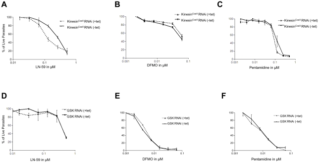
Our group has characterized and optimized protein farneysltransferase inhibitor molecules based on the tetrahydroquinoline (THQ) scaffold [33]. LN-59 is a THQ compound (Structure 4 g in reference 32) that has potent activity against mammalian PFT (IC50 of 3.2 nM) and Plasmodium falciparum PFT (IC50 of 1.1 nM) [33]. Additionally LN-59 has activity against TB-PFT (IC50 of 35 nM) and against T. brucei 427 bloodstream-form parasites in whole cell screens (EC50 of 63 nM) suggesting TB-PFT is the target of growth inhibition. In proliferation assays, T. brucei depleted of KinesinCaaX by RNAi had enhanced sensitivity to LN-59 (Fig. 6A). In five independent experiments, effective concentrations of LN-59 to reduce proliferation by 50% (EC50s) were 4.2 fold less in the KinesinCaaX -depleted cells compared with non-induced controls (SD of 1.0, Student T Test p<0.001). T. brucei depleted of KinesinCaaX by RNAi were not hypersensitive to DFMO (Fig. 6B) or pentamidine (Fig. 6C), compounds that do not act through inhibition of TB-PFT. DFMO and pentamidine were tested three times each, and the same EC50s were observed whether tet-induced to reduce KinesinCaaX or not. Additionally we tested another RNAi line, which gave similar growth reduction to that of KinesinCaaX upon tetracycline induction, a glycogen synthase kinase (GSK) RNAi line [34]. T. brucei depleted of GSK by RNAi were not hypersensitive to LN-59 (Fig. 6D), DFMO (Fig. 6E), or pentamidine (Fig. 6F). These assays demonstrate that cells with reduced KinesinCaaX are more sensitive to a PFT inhibitor but not two drugs from other classes. These data demonstrate that depletion of a CaaX-containing molecular target sensitizes T. brucei to protein farnesyltransferase inhibition of cell growth.
Figure 6. RNAi of KinesinCaaX show enhanced sensitivity to protein farnesyl transferase inhibitors but not other drugs.
(A) LN-59 (B) DFMO and (C) Pentamidine mean % growth (normalized to no drug) in induced and noninduced KinesinCaaX RNAi cells. (D) LN-59 (E) DFMO and (F) Pentamidine mean % growth in induced and noninduced GSK RNAi line. Values normalized to no drug with error bars representing SD.
Discussion
Our analysis reveals an important role for KinesinCaaX in the cell growth of T. brucei. In this study we have demonstrated KinesinCaaX has motor activity along microtubules and is essential for growth in the bloodstream form. KinesinCaaX depleted cells (RNAi-induced cells) demonstrate an increase of cells with less than a G1 complement of DNA, which are presumably dying. KinesinCaaX-depleted cells have more rounded cell bodies compared to control uninduced T. brucei cells. This change in morphology could be a direct effect of reduced KinesinCaaX or an indirect effect of cell death. Supporting that rounded cells were dying, the rounded cells that predominated during RNAi appeared to have weaker DAPI staining, implying their nuclei were undergoing degradation. Exogenous expression cell lines show HA-KinesinCaaX is highly enriched near the anterior kinetoplast and near the nucleus in 2K1N containing cells and between post-mitotic nuclei in 2K2N containing cells prior to cytokinesis. In our exogenous expression lines, cells with 2K1N and 2K2N content had more easily detectable KinesinCaaX relative to 1K1N cells perhaps suggesting a role in separation of nuclei and kinetoplasts. HA-KinesinCaaX was found to overlay β-tubulin in this area between 2 nuclei in 2K2N cells and this localization may also support a role for KinesinCaaX in the separation of nuclei. It is possible that post-transcriptional regulation of KinesinCaaX levels, such as protein instability, could be responsible for the reduced detection of KinesinCaaX in 1K1N cells. Kinesin Family 5 and Family 7 members responsible for chromosome separation in higher organisms are lacking in T. brucei [24]. Due to localization and increased abundance of HA-KinesinCaaX in cells near kinetoplast and nuclear DNA, we hypothesize that KinesinCaaX may facilitate nuclear and kinetoplast segregation.
Our radiolabeling studies show that HA-KinesinCaaX but not HA-KinesinCVIM deletion is a target for prenylation in T. brucei cells. To uncover the role of farnesylation in KinesinCaaX, we localized ectopically-expressed HA-KinesinCVIM deletion. HA-KinesinCVIM deletion expression results in a more diffuse localization pattern compared with HA-KinesinCaaX. This supports the hypothesis that farnesylation helps KinesinCaaX appropriately localize in the cell. Prolonged ectopic expression of HA-KinesinCVIM deletion leads to cells with rounded cell bodies and reduced DNA content, a phenotype we also observed in the RNAi experiments. We speculate this apparent increase in cell death occurs due to an accumulation of KinesinCVIM deletion molecules without prenylation, outcompeting the endogenous KinesinCaaX for functional interactions. We speculate the delayed 96 hour KinesinCVIM deletion effect, compared to the early phenotype that is evident at 48 hours in the RNAi experiments, may be due to heterodimers of native KinesinCaaX and HA-KinesinCVIM deletion being functional at 48 hours after tet-induction and homodimers of HA-KinesinCVIM deletion that accumulate 96 hours after tet-induction may not localize or function properly. We hypothesize KinesinCaaX interacts with cellular cargo via the farnesyl group at the C-terminus, and the motor domain at the N-terminus allows the KinesinCaaX to move along microtubules of the cell. Future experiments are needed define the cargo of KinesinCaaX.
We also show KinesinCaaX RNAi cells have an increased sensitivity to a protein farnesyl transferase inhibitor (PFTi) but not other classes of drugs when compared to cells with wildtype levels of KinesinCaaX. Collectively these data suggest that farnesylation of KinesinCaaX is integral to the function of KinesinCaaX. This is the first study of a molecular target downstream of T. brucei protein farnesyl transferase (TB-PFT). While there are many proteins with CaaX motifs in T. brucei that may be modified by TB-PFT (Fig. S1), our data, of increased sensitivity to PFTi when KinesinCaaX is reduced, suggest that inhibition of farnesylation of KinesinCaaX contributes substantially to the growth inhibition of PFTi-treated T. brucei.
An analysis comparing the kinesins of various organisms, including T. brucei, was unable to group KinesinCaaX with other known kinesin family members [35]. In our work, we show the essential motor motifs are intact in KinesinCaaX (Fig. 1) and demonstrate ATP-dependent motor activity characteristic of kinesins (Fig. S4B). Based on the localization pattern during various stages of the cell cycle, we speculate KinesinCaaX may be facilitating factors key to kinetoplast and nuclear DNA segregation. Centromeric protein E (CENP-E) is a kinesin in higher eukaryotic organisms that contains a CaaX box, is farnesylated and has been shown to be an N-terminal processive motor protein [36]–[38], all properties similar to KinesinCaaX. CENP-E is a Kinesin-7 family member and is involved in chromosome movement during mitosis and links centromeres to spindle microtubules. Similar to the observed levels in expression of HA-KinesinCaaX, CENP-E has the maximum level during late G2 and minimal levels in G1 [37]. CENP-E localization to kinetochores occurs from early premetaphase through anaphase. CENP-E is important during all phases of mitotic chromosome movement and affects kinetochore-microtubule capture. CENP-E loss of function by interfering RNA results in cell cycle arrest [37]. Additionally the use of protein farnesyl transferase inhibitors has been shown to block the association of CENP-E with microtubules [38]. Currently no CENP-E homolog or any Kinesin-7 family member has been uncovered in the Tritryp genomes by bioinformatic studies [24]. Several other kinesins have recently been characterized in T. brucei including the C-terminal Kinesin-13 proteins [39], [40] TbKif13-1 is associated with the nucleus and nuclei in a non cell-cycle dependent manner [40]. Depletion of TbKif13-1 in T. brucei results in an increase in G2/M cells by 48 hours and causes altered minichromosome segregation [39].
We speculate KinesinCaaX may play a role in nuclear and kinetoplast segregation. Kinetochores, the multimeric protein structures that attach at the centromere spindle microtubules to chromosomes during cell division, have not been observed in T. brucei. Peripheral microtubules structures that are suggestive of pole-kinetochore microtubules have been observed in T. brucei. These peripheral microtubules terminate in electron dense structures and these structures may be serving as conventional kinetochores. However, there is a discrepancy between the number of large chromosomes, 22 in the diploid set of T. brucei and the 10 electron dense structures seen in T. brucei cells [41]. Minichromosomes are hypothesized to segregate from the microtubule spindle through tracking along microtubules that extend to the poles rather than via a conventional kinetochore attachment [41]. In mammalian cells, the inner kinetochore attaches to the DNA at the centromeric region while the outer kinetochore proteins interact with the spindle microtubules. Bioinformatic analysis of the Tritryp (T. brucei, L. major, T. cruzi) genomes does not reveal homologs for most of the proteins of the outer kinetochore including CENP-E, CENP-F, HEC1/Ndc80, Nuf2 or for inner kinetochore components including CENP-C, CENP-G, Cep3p, Mis12, Nde10p and Ctf13p [24]. One outer kinetochore protein, TOG/MOR1 and two inner kinetochore proteins, MCAK and Skp1p have been annotated in the TriTryps [24] yet their roles in T. brucei have not been characterized. As Tritryps do not encode homologs to the majority of conventional kinetochore components other proteins must facilitate successful segregation of their DNA. Our work demonstrates the same pattern of accumulation of KinesinCaaX throughout the cell cycle as seen with other reported studies on CENP-E. KinesinCaaX and the associated farnesyl group may function in a similar manner as CENP-E creating additional interactions with microtubules at the C-terminus of KinesinCaaX via the farnesyl group, and thus move microtubules associated with chromosomes, nuclei or kinetoplasts along other microtubules. Alternatively, the farnesyl group may provide a hydrophobic attachment to a membrane target, such as nuclear membranes, allowing nuclear movement during cytokinesis. Thus, CENP-E shares many similarities with KinesinCaaX and we hypothesize that this kinesin performs functions in trypanosomatids that CENP-E performs in higher eukaryotes.
In preparation of this manuscript, KinesinCaaX was identified among a list of proteins in a palmitoylation screen [42]. We speculate based on the cysteine conserved three amino acids before the CaaX motif that this protein is both palmitoylated and farnesylated. We note that the cysteine residue three residues upstream of the CaaX motif is also conserved in all pathogenic kinetoplastids (Fig. S2). While we did not remove the cysteine where palmitoylation may occur, palmitoylation in this context usually requires farnesylation which does not occur if the CaaX motif is removed. Thus we hypothesize that our HA-KinesinCVIMdeleted construct lacks both post-translational modifications and both modifications contribute to proper localization.
In conclusion our work characterizes a kinetoplastid-specific kinesin that has a site for farnesylation, undergoes farneyslation in T. brucei and requires farnesylation for proper function. We hypothesize this farnesylation could facilitate interactions with microtubules, as is the case with CENP-E and H-ras and N-ras in mammalian cells, or with other cargo such as the nuclear membrane. Our work suggests inhibition of farnesylation of KinesinCaaX contributes to the molecular mechanism of growth arrest in PFTi-treated T. brucei. This is the first study to demonstrate motor activity of a T. brucei kinesin and the first study to characterize farnesylation of a kinesin in T. brucei. Further understanding of KinesinCaaX and of interacting partners of KinesinCaaX in T. brucei and other pathogenic kinetoplasts may be helpful to finding new therapeutic interventions for these parasitic pathogens.
Materials and Methods
Cell maintenance
The single marker bloodstream form (BSF) strain T. brucei brucei 427 cell line that expresses T7 RNA polymerase and Tet-repressor [43] was maintained in HMI-9 medium containing 10% tetracycline-free fetal bovine serum (Atlanta Biologicals). Cultures were maintained at 37°C with 5% CO2 as previously described [39]. Cells were diluted into fresh medium once cell density achieved 2×10⁶ cells/mL.
Generation of KinesinCaaX RNAi cell lines
Using the primer prediction algorithm, RNAit [44], primers were selected to minimize off-target effects. The 600 nucleotide sequence was amplified from the full length KinesinCaaX ORF of the expression plasmid and confirmed to contain KinesinCaaX by dye deoxy sequencing. The 600 base pair PCR product was analyzed by gel electrophoresis and compared with the predicted product size. The RNAi vector p2T7TAblue [45] (gift from David Horn) was cut to remove the stuffer region and gel purified. The PCR product was then ligated into p2T7TAblue and transformed into E. coli DH5alpha. Plasmids were screened by restriction enzyme digestion and verified by dye deoxy sequencing prior to trypanosome transformation. Midlog T. brucei (2.5×10⁷) were resuspended in cytomix (120 mM KCl, 0.15 mM CaCl2, 10 mM K2HPO4/KH2PO4 pH 7.6, 25 mM HEPES, 2 mM Na2EDTA, 5 mM MgCl2) containing 12 µg of NotI linearized p2T7TAblue DNA. The mixture was electroporated in a 4 mm gap cuvette with 1.6 kV and 24Ω resistance. Cells were resuspended in HMI-9 media. After 6 hours of recovery, selective drug (2.5 µg/mL hygromycin and 2.5 µg/mL G418) was applied and serial dilutions were made into 24-well plates. Transfected cells grew out from limit diluted cells in 6 to 8 days after electroporation.
Construction of KinesinCaaX expression strains
The complete open reading frame of the T. brucei KinesinCaaX was amplified using T. brucei brucei genomic DNA, strain 427, using primers with an added HindIII and BamHI sequences for subcloning and a N terminal double HA tag using primers 5′CCAAAAAGTAAAATTCACTATCCATATGACGTCCCAGACTCTGCCTATCCATATGACGTCCCAGACTCTGCCAAGCTTATGTCGGGTATATATGCG3′ and antisense 5′CCGGCTACATTATTATTACACAGTAATGGCAACCC3′. The HA tag was engineered with the AatI restriction site (GACGTC) to test for insertion. The PCR product was analyzed by gel electrophoresis and gel purified. The expression vector pHD538 [46] (gift from Christine Clayton) was cut to remove the stuffer region and gel purified. The PCR product was then ligated into the vector and transformed into E. coli DH5alpha. Plasmids were isolated using QIAGEN kits, screened by restriction enzyme digestion, verified by dye deoxy sequencing, analyzed using Vector NTI Suite 9.0 (Invitrogen) and linearized with Not I prior to trypanosome transformation. Trypanosome transformation and drug selection were performed as described above.
Removal of CVIM from KinesinCaaX
Using the pHD538 KinesinCaaX as template we performed site directed mutagenesis using the primers 5′TAAACAGGGTTGCCATTACTAGGGATCCAATTTTCCCC′3
5′ GGGGAAAATTGGATCCCTAGTAATGGCAACCCTGTTTA′3 and the Pfu Ultra polymerase to remove the entire CaaX motif, in order to explore the function of KinesinCaaX when it was not farnesylated as previously described [47]. As the protease that acts after farnesylation removes the VIM residues, we felt that removing the entire CVIM terminus would lead to a better physiologic demonstration of what functionally happens to KinesinCaaX without prenylation. Amplification conditions were 5 minutes at 95°C, followed by 12 cycles of 1 minute at 95°C, 1 minute at 55°C and 18 minutes at 68°C. The PCR reaction was then Dpn I treated overnight to digest the parental plasmid DNA and transformed into E. coli XL10-Gold cells (Invitrogen). Plasmids were screened by restriction enzyme digestion, verified by dye deoxy sequencing to have the desired deletion, analyzed using Vector NTI Suite 9.0 (Invitrogen) and linearized with Not I prior to trypanosome transformation as described above for KinesinCaaX.
RT-PCR
Total RNA was extracted from mid-logarithmic phase cultures of T. brucei using the Qiagen RNeasy Mini kit. mRNA was isolated using the Qiagen Oligotex system from 10 µg of total RNA. Reverse transcription using 25 ng of mRNA per reaction using random hexamers with TaqMan Reverse Transcription Reagents (Applied Biosystems) yielded cDNA. Samples without reverse transcriptase were included to confirm the absence of contaminating genomic DNA. cDNA reactions were diluted two-fold and amplified in 25 µL reactions containing 2.5 µL of each cDNA, 5 µL each of 1.5 µM sense and antisense primers, and 12.5 µL SYBR Green PCR Master Mix (Applied Biosystems) in 96-well plates using the Opticon 2 Real-Time PCR Detection System (BioRad Life Sciences). Amplification conditions were 2 minutes at 50°C and 10 minutes at 95°C, followed by 45 cycles of 15 seconds at 95°C, 1 minute at 55°C and 1 minute 30 seconds at 60°C, 1 second at 80°C. Using the primers corresponding to the 254–353 basepair region, sense 5′-GCTCCAACCGAACGTAACTC-3′ and antisense 5′-CACTTCCCGCTAAGTCAACC-3′. Additionally tubulin primers used as a control were sense 5′-TTCCGCACCCTGAAACTGA-3′ and antisense 5′-TGACGCCGGACACAACAG-3′. Relative changes were determined after normalization to β-tubulin mRNA and expressed as relative mRNA abundance from the respective control cells.
Production of Polyclonal antibody against KinesinCaaX protein
The T. brucei KinesinCaaX amino acids 409–525 corresponding to the least similar region to other T. brucei proteins, as analyzed by BLAST [48], was amplified using the full length gene from the expression plasmids as template using the LIC primers sense 5′CTCACCACCACCACCACCAT3′ and antisense 5′ATCCTATCTTACTCACTTA3′ coding for an N-terminally 6Xhistidine-tagged KinesinCaaX fragment. Nine amino acid segments were checked against the T. brucei genome using BLAST to ensure the specificity to KinesinCaaX. The PCR product was then put into the LIC expression vector Bg1861 and transformed into E. coli NovaBlue cells (Novagen). Plasmids were screened by restriction enzyme digestion, verified by dye deoxy sequencing and analyzed using Vector NTI Suite 9.0 (Invitrogen). The recombinant plasmid was then transformed into E. coli BL21cells (Promega) and grown and induced using IPTG for protein production. Bacteria were harvested by centrifugation at 2,500 g for 10 minutes at 4°C. Bacterial cells were lysed in lysis buffer (50 mM Tris, 100 mM NaCl, 1 mM MgCl2, and Protease Inhibitor Cocktail (Sigma-P8430), pH 7.0) and purified using Nickel-NTA resin (QIAGEN). The purified recombinant protein was dialyzed against PBS. The purified recombinant protein was sequenced using trypsin digestion and mass spectrometry to verify peptide purity. The purified recombinant protein was used to raise rabbit polyclonal antibodies (R&R Rabbitry). Rabbit serum was preblocked with E. coli lysate to reduce nonspecific binding prior to Western blot.
Western Blot
Western blots were performed using 12 µg of total protein induced or noninduced T. brucei cultures run on 12% SDS PAGE gels and transferred to PVDF membrane.
Membranes were blocked with 5% nonfat dry milk in Tris buffered saline with 0.1% Tween 20 (TBS-T) on an orbital shaker overnight at 4°C. Rabbit serum was preabsorbed with E. coli lysates overnight. Preabsorbed rabbit serum was diluted 1∶500 in blocking buffer and incubated with the membranes on a shaker for 1 hour at RT. Membranes were washed three times with TBS-T for 10 minutes. Next anti-rabbit secondary antibody conjugated to HRP (Pierce) was added and incubated on a shaker for 1 hour at RT. The membrane was rinsed with TBS-T three times with TBS-T for 10 minutes and developed using ECL Plus (Amersham). Anti-HA Westerns were performed with 3F10 (Roche) 1∶1000 as a primary antibody and secondary anti-Rat antibody, ab6517 (abcam), 1∶15∶000.
Immunofluorescense microscopy
Cells were washed with PBS, applied to poly-lysine-coated Teflon slides and allowed to adhere. Cells were fixed with 4% paraformaldehyde (PFA) for 5 minutes, washed three times with PBS and permeabilized with 0.05% Triton X at room temperature. Samples were then blocked overnight at 4°C with 5% BSA supplemented with 5% goat serum. The following antibodies were used: The KMX-1 β-tubulin monoclonal antibody [32] (gift from Keith Gull); the High Affinity anti-HA rat mAb 3F10 (Roche); goat polyclonal to rat IgG-H&L Cy5.5 (abcam), FITC-conjugated anti-rabbit IgG (Sigma-Aldrich), anti-mouse IgG 568 (abcam).
Fixed cells were incubated with primary antibodies at room temperature for 60 minutes, washed three times and incubated with FITC-conjugated and Cy5.5 conjugated antibodies for another 60 minutes. After washing three more times, the cells were stained with 1.0 µg/uL of 4, 6-diamino-2-phenlyindole (DAPI) and the slides were mounted with Fluoromount G (Sigma-Aldrich). Cells were examined using a Nikon TE2000 inverted microscope equipped with a Photometrics CoolSNAP HQ2 camera for fluorescence and phase contrast images. Images were analyzed using SoftwoRX software and processed for publication using Adobe Photoshop (Adobe Systems Inc., San Jose, CA) and ImageJ. Secondary antibody only controls were performed and gave only background, low-level fluorescence which was used to set the software for signal detection.
Flow cytometry
Bloodstream form cells were inoculated at 1×10⁵/mL and allowed to grow. The cultures were harvested at various time points, fixed with 4% paraformaldehyde for five minutes, washed once with PBS and permeabilized with 0.05% Triton X. Lastly DAPI was added and cells were analyzed with an Influx analytical flow cytometer (BD Biosciences). Histograms were created using the FCS Express Version 3 software.
Drug Assays
T. brucei KinesinCaaX RNAi line cells were inoculated into 96-well plates at 2.5×10⁴/well. To induce dsRNA, 1 µg/mL tetracycline was added and cells were allowed to grow with various concentrations of the LN-59 protein farnesyl transferase inhibitor drug for 48 hours followed by Alamar Blue analysis to follow cell growth as previously described [43]. Pentamidine and difluoromethyl ornithine or DFMO (Sigma-Aldrich) were used as control drugs and compared to cells in media alone. Experiments were done in triplicate. Best fit curves and EC50 values were fitted using Prism 3.0 (Graphpad) and Kaleidagraph 4.0 (Synergy Software). Drug concentrations are expressed in µM.
Prenylation Determination by Radio-labeling
Double HA-tagged KinesinCaaX expressing, double HA-tagged KinesinCVIM deletion and control T. brucei cell lines were plated into 6-well plates at 1×10⁶/well and treated with 1 µg/mL tetracycline, 1.25 µM simvastatin and 6.7 µM (100 µCi) [3H]-mevalonolactone. Cells were incubated for 48 hours and then processed for immunoprecipitations. [3H]-counts were detected using the MicroBeta Scintillation detector (MicroBeta TriLux, Perkin Elmer).
Immunoprecipitation
T. brucei cells were incubated in lysis buffer (25 mM Tris-Cl, pH 7.6, 100 mM NaCl, 1% Nonidet P-40, 1 mM dithiothreitol and protease inhibitor cocktail) for 30 minutes on ice and cleared by centrifugation. The cleared lysate was pre-incubated with protein G Sepharose beads at 4°C for 1 hour. Beads were removed by centrifugation and the bead-cleared supernatant was incubated with anti-HA polyclonal antibody and protein G Sepharose beads at 4°C for two hours. The immune-precipitates collected were then analyzed in the MicroBeta Scintillation detector (MicroBeta TriLux, Perkin Elmer).
Supplemental Materials and Methods
Expression and purification of active KinesinCaaX in E. coli
The T. brucei KinesinCaaX motor domain was amplified using the full length gene from the expression plasmids as template and the ligation independent cloning (LIC) primers 5′CTCACCACCACCACCACCATATGTCAGAGGTACTCGATGCGC 3′ and antisense 5′ATCCTATCTTACTCACTTACTCACTTGCAAGAGTACCACTAAG 3′ coding for a N-terminally 6Xhistidine-tagged KinesinCaaX motor domain protein. The PCR product was placed into the LIC expression vector Bg1861 [49] and transformed into E. coli NovaBlue cells (Novagen). Plasmids were screened by restriction enzyme digestion, checked by dye deoxy sequencing, and the sequence verified using Vector NTI Suite 9.0 (Invitrogen). The recombinant plasmid was then transformed into E. coli BL21cells (Promega) and grown and induced using IPTG for protein production. Bacteria were harvested by centrifugation at 2,500 g for 10 minutes at 4°C. Bacterial cells were lysed in lysis buffer (100 mM NaCl, 1 mM MgCl2, 1 mM ATP, lysozyme and Protease Inhibitor Cocktail (Sigma-P8430), pH 7.0) and soluble lysates prepared by centrifugation at 14,000× g for 10 minutes. Soluble lysates were used in the motility assay as described below.
Gliding Motility Assays
Fluorescent microtubules (MT) were prepared using an 80∶1 ratio of unlabeled bovine-to Alexa 568-labeled bovine tubulin, BRB80 buffer (80 mM Pipes/KOH, pH 6.9, 1 mM EGTA, 1 mM MgCl2), 4 mM MgCl2, 1 mM GTP, and 5% DMSO at 37°C. After 30 minutes the MT polymers, 5–20 µm in length were stabilized by diluting 200-fold into room-temperature BRB80 containing 10 µM taxol. Microscope slide flow cells were made using a microscope slide fitted with two pieces of double sided tape with 1 cm gap and a coverslip. Motility buffer (BRB80 supplemented with 0.2 mg/mL casein, 20 µM D-glucose, 0.02 mg/mL glucose oxidase, 0.008 mg/mL catalase, 0.5% 2-mercaptoethanol and 32 nM of the final polymerized microtubule reaction) with and without+ 1 mM MgATP was added to the microscope slide coated with E. coli soluble lysates containing the recombinant KinesinCaaX motor protein or the β-galactosidase non-motor control. Time lapse photography was taken every 4 seconds, with a one second exposure, using a Nikon TE2000 inverted microscope, 100× objective, with a Photometrics CoolSNAP HQ2 camera for fluorescence images. Images were analyzed using SoftwoRX software and processed for publication using Adobe Photoshop (Adobe Systems Inc.).
Supporting Information
List of CaaX motif containing proteins in T. brucei . Gene ID, putative name and CaaX motif are listed.
(PDF)
KinesinCaaX is conserved in other pathogenic kinetoplastids. Shown are multiple sequence alignments using MUSCLE version 3.7 [23]. LmxM18.1600 from Leishmania mexicana mexicana, LbrM18_V2.1630 from Leishmania braziliensis, LinJ18_V3.1590 from Leishmania infantum, LmjF18.1600 from Leishmania major, Tc00.1047053507625.180 from Trypanosoma cruzi, TvY486_1012140 from Trypanosoma vivax, TcIL3000.10.10660 from Trypanosoma congolense, Tbg972.10.14990 from Trypanosoma brucei gambiense, and Tb927.10.12440, herein referred to as KinesinCaaX, from Trypanosoma brucei brucei.
(PDF)
Dendrogram of KinesinCaaX and orthologs from other pathogenic kinetoplastids. The evolutionary relationship of KinesinCaaX was analyzed using Biology Work Bench, Version 3.2, DRAWGRAM and PHYLIP (Phylogeny Inference Package) version 3.5c [26]. This analysis demonstrates the T. brucei gambiense homolog to be most closely related to T. brucei brucei KinesinCaaX followed by the other Trypanosoma species and then the Leishmania species. For gene ID and species key, see Legend of Figure S2. Horizontal distances represent relative genetic distance.
(PDF)
KinesinCaaX has motor activity in the presence of ATP in vitro . (A) Recombinant His-tagged truncated N-terminal 340 AA-construct of KinesinCaaX restricted to the predicted motor domain and the coil region predicted to facilitate dimerization. (B) Coomassie stained PAGE showing E.coli soluble lysate used for the motor assays in (C) with the increased band at the expected molecular weight (44 kDa) for KinesinCaaX motor-domain upon IPTG jnduction (+). (C) An in vitro gliding motility assay using truncated KinesinCaaX and fluorescent microtubules. Lysates containing recombinant truncated KinesinCaaX was allowed to adhere to glass microscope slides. Next rhodamine-labeled microtubules were added with or without ATP to the slide and microtubule motility was monitored by time-lapse fluorescence microscopy. Selected frames from time lapse microscopy are represented. The positions of rhodamine-labeled microtubules over time are marked with arrows. (D) Control cell lysates containing recombinant β-galactosidase non-motor protein control showed no microtubule movement in the presence of ATP. The positions of two stationary microtubules are marked with arrows.
(TIF)
Growth rate of T. brucei exogenously expressing KinesinCaaX is not affected after induction but KinesinCVIMdeletion induced cells begin to slow their growth rate at 96 hrs. Shown are growth curves of blood-stream T. brucei parasites grown either with (+) or without (−) tetracycline induction. Control parasites are “Single Marker” parasites without an exogenous expression plasmid. KinesinCaaX and KinesinCVIMdeletion are parasites transfected with the exogenous expression vector expressing these proteins under control of tetracycline induction. Parasite numbers are shown on the Y axis and time after tetracycline induction in hours is shown on the X axis. The experiment shown represents a single clone from each transfection but the same results were also observed with a second clone from each transfection (data not shown).
(TIF)
Acknowledgments
We thank Richard Eastman for initiating these studies and performing the initial bioinformatic analysis of the T. brucei CaaX containing proteins, Linda Wordeman for providing motility assay reagents, Donna Prunkhard, Christal Worthen and Molly Reid for assistance with flow cytometry, Greg Martin in the University of Washington Keck Imaging Center for help with immunofluorescence assays and ImageJ analysis, Michael Gelb and Laxman Nallen for the LN-59 compound and TB-PFT enzyme, David Horn for the p2T7TAblue vector, Christine Clayton for the pHD538 vector, Keith Gull for the KMX antibody, and Lynn Barrett, Marilyn Parsons, Charles Asbury, and Barry Stoddard for discussions, advice and technical assistance.
Footnotes
Competing Interests: The authors have declared that no competing interests exist.
Funding: Sources of funding include the Molecular and Cellular Biology Program and the Royalty Research Fund at the University of Washington, R01CA52874, 1R01AI089441 and 1R01AI080625. No additional external funding received for this study. The funders had no role in study design, data collection and analysis, decision to publish, or preparation of the manuscript.
References
- 1.World Health Organization Report. Human African trypanosomiasis: number of new cases drops to historically low level in 50 years (2010). Available: http://www.who.int/neglected_diseases/integrated_media/integrated_media_hat_june_2010/en/index.html Accessed 2010 Dec 15.
- 2.Simarro PP, Cecchi G, Paone M, Franco JR, Diarra A, et al. The Atlas of human African trypanosomiasis: a contribution to global mapping of neglected tropical diseases. International J of Health Geographics. 2010;9:57. doi: 10.1186/1476-072X-9-57. Available: http://www.ij-healthgeographics.com/content/9/1/57. [DOI] [PMC free article] [PubMed] [Google Scholar]
- 3.World Health Organization African trypanosomiasis (sleeping sickness) Fact sheet N°259. 2010. Available: http://www.who.int/mediacentre/factsheets/fs259/en/. Accessed 2010 Dec 15.
- 4.Conner RJ. Rome: Food and Agricultural Organization of the United Nations Publications Division; 1992. Programme for the control of African animal trypanosomiasis and related development.199 [Google Scholar]
- 5.Maser P, Sutterlin C, Kralli A, Kaminsky R. A nucleoside transporter from Trypanosoma brucei involved in drug resistance. Science. 1999;5425:242–244. doi: 10.1126/science.285.5425.242. [DOI] [PubMed] [Google Scholar]
- 6.Matovu E, Seebeck T, Enyaru JC, Kaminsky R. Drug resistance in Trypanosoma brucei spp., the causative agents of sleeping sickness in man and nagana in cattle. Microbes Infect. 2001;9:763–770. doi: 10.1016/s1286-4579(01)01432-0. [DOI] [PubMed] [Google Scholar]
- 7.Maser P, Luscher A, Kaminsky R. Drug transport and drug resistance in African trypanosomes. Drug Resist Update. 2003;5:281–290. doi: 10.1016/j.drup.2003.09.001. [DOI] [PubMed] [Google Scholar]
- 8.Matovu E, Stewart ML, Geiser F, Brun R, Maser P, et al. Mechanisms of arsenical and diamidine uptake and resistance in Trypanosoma brucei. Eukaryot Cell. 2003;5:1003–1008. doi: 10.1128/EC.2.5.1003-1008.2003. [DOI] [PMC free article] [PubMed] [Google Scholar]
- 9.Yokoyama K, Lin Y, Stuart KD, Gelb MH. Prenylation of proteins in Trypanosoma brucei. Mol Biochem Parasitol. 1997;1:61–69. doi: 10.1016/s0166-6851(97)00043-1. [DOI] [PubMed] [Google Scholar]
- 10.Yokoyama K, Trobridge P, Buckner FS, Scholten J, Stuart KD, et al. The effects of protein farnesyltransferase inhibitors on trypanosomatids: Inhibition of protein farnesylation and cell growth. Mol Biochem Parasitol. 1998;1:87–97. doi: 10.1016/s0166-6851(98)00053-x. [DOI] [PubMed] [Google Scholar]
- 11.Yokoyama K, Trobridge P, Buckner FS, Van Voorhis WC, Stuart KD, et al. Protein farnesyltransferase from Trypanosoma brucei. A heterodimer of 61- and 65-kda subunits as a new target for antiparasite therapeutics. J Biol Chem. 1998;41:26497–26505. doi: 10.1074/jbc.273.41.26497. [DOI] [PubMed] [Google Scholar]
- 12.Buckner FS, Yokoyama K, Nguyen L, Grewal A, Erdjument-Bromage H, et al. Cloning, heterologous expression, and distinct substrate specificity of protein farnesyltransferase from Trypanosoma brucei. J Biol Chem. 2000;29:21870–21876. doi: 10.1074/jbc.M000975200. [DOI] [PMC free article] [PubMed] [Google Scholar]
- 13.Buckner FS, Kateete DP, Lubega GW, Van Voorhis WC, Yokoyama K. Trypanosoma brucei prenylated-protein carboxyl methyltransferase prefers farnesylated substrates. Biochem J. 2002;367:809–816. doi: 10.1042/BJ20020277. [DOI] [PMC free article] [PubMed] [Google Scholar]
- 14.Gelb MH, Van Voorhis WC, Buckner FS, Yokoyama K, Eastman R, et al. Protein farnesyl and N-myristoyl transferases: piggy-back medicinal chemistry targets for the development of antitrypanosomatid and antimalarial therapeutics. Mol Biochem Parasitol. 2003;2:155–163. doi: 10.1016/s0166-6851(02)00282-7. [DOI] [PubMed] [Google Scholar]
- 15.Buckner FS, Eastman RT, Yokoyama K, Gelb MH, Van Voorhis WC. Protein farnesyl transferase inhibitors for the treatment of malaria and African trypanosomiasis. Curr Opin Investig Drugs. 2005;8:791–797. [PubMed] [Google Scholar]
- 16.Gelb MH, Brunsveld L, Hrycyna CA, Michaelis S, Tamanoi F, et al. Therapeutic intervention based on protein prenylation and associated modifications. Nat Chem Biol. 2006;10:518–528. doi: 10.1038/nchembio818. [DOI] [PMC free article] [PubMed] [Google Scholar]
- 17.Fletcher S, Cummings CG, Rivas K, Katt WP, Horney C, et al. Potent, plasmodium-selective farnesyltransferase inhibitors that arrest the growth of malaria parasites: Structure-activity relationships of ethylenediamine-analogue scaffolds and homology model validation. J Med Chem. 2008;51:5176–5197. doi: 10.1021/jm800113p. [DOI] [PMC free article] [PubMed] [Google Scholar]
- 18.Yokoyama K, Gillespie JR, Van Voorhis WC, Buckner FS, Gelb MH. Protein geranylgeranyltransferase-I of Trypanosoma cruzi. Mol Biochem Parasitol. 2008;157:32–43. doi: 10.1016/j.molbiopara.2007.09.006. [DOI] [PMC free article] [PubMed] [Google Scholar]
- 19.Nepomuceno-Silva JL, Yokoyama K, de Mello LD, Mendonca SM, Paixão JC, et al. TcRho1, a farnesylated Rho family homologue from Trypanosoma cruzi: cloning, trans-splicing, and prenylation studies. J Biol. 2001;Chem276:29711–29718. doi: 10.1074/jbc.M102920200. [DOI] [PubMed] [Google Scholar]
- 20.McTaggart SJ. Isoprenylated proteins. Cell Mol Life Sci. 2006;3:255–267. doi: 10.1007/s00018-005-5298-6. [DOI] [PMC free article] [PubMed] [Google Scholar]
- 21.Sinensky M. Recent advances in the study of prenylated proteins. Biochim Biophys Acta. 2000;1484:93–106. doi: 10.1016/s1388-1981(00)00009-3. [DOI] [PubMed] [Google Scholar]
- 22.Asbury CL, Fehr AN, Block SM. Kinesin moves by an asymmetric hand-over-hand mechanism. Science. 2003;302:2130–2133. doi: 10.1126/science.1092985. [DOI] [PMC free article] [PubMed] [Google Scholar]
- 23.Wickstead B, Gull K. A “holistic” kinesin phylogeny reveals new kinesin families and predicts protein functions. Mol Biol Cell. 2006;17:1734–1743. doi: 10.1091/mbc.E05-11-1090. [DOI] [PMC free article] [PubMed] [Google Scholar]
- 24.Berriman M, Ghedin E, Hertz-Fowler C, El-Sayed NM. The genome of the African Trypanosome Trypanosoma brucei. Science. 2005;5733:416–422. doi: 10.1126/science.1112642. [DOI] [PubMed] [Google Scholar]
- 25.Lawrence CJ, Malmberg RL, Muszynski MG, Dawe RK. Maximum likelihood methods reveal conservation of function among closely related kinesin families. J Mol Evol. 2002;54:42–53. doi: 10.1007/s00239-001-0016-y. [DOI] [PubMed] [Google Scholar]
- 26.Edgar RC. MUSCLE: multiple sequence alignment with high accuracy and high throughput. Nucl Acids Res. 2004;5:1792–1797. doi: 10.1093/nar/gkh340. [DOI] [PMC free article] [PubMed] [Google Scholar]
- 27.Gull FJ, Endow S. Kinesin; switch I & II and the motor mechanism. J Cell Sci. 2002;115:15–23. doi: 10.1242/jcs.115.1.15. [DOI] [PubMed] [Google Scholar]
- 28.Li L, Stoeckert CJ, Roos DS. OrthoMCL identification of ortholog groups for eukaryotic genomes. Genome Res. 2003;13:2178–2189. doi: 10.1101/gr.1224503. [DOI] [PMC free article] [PubMed] [Google Scholar]
- 29.Thompson JD, Higgins DG, Gibson TJ. CLUSTAL W: improving the sensitivity of progressive multiple sequence alignment through sequence weighting, position-specific gap penalties and weight matrix choice. Nucl Acids Res. 1994;22:4673–4680. doi: 10.1093/nar/22.22.4673. [DOI] [PMC free article] [PubMed] [Google Scholar]
- 30.Dagenbach EM, Endow SA. A new kinesin tree. J Cell Sci. 2004;117:3–7. doi: 10.1242/jcs.00875. [DOI] [PubMed] [Google Scholar]
- 31.Hammarton TC, Monnerat S, Mottram JC. Cytokinesis in trypanosomatids. Curr Opin Micro. 2007;10:520–527. doi: 10.1016/j.mib.2007.10.005. [DOI] [PubMed] [Google Scholar]
- 32.Birkett CR, Foster KE, Johnson L, Gull K. Use of monoclonal antibodies to analyse the expression of a multi-tubulin family. FEBS Letters. 1985;187:211–218. doi: 10.1016/0014-5793(85)81244-8. [DOI] [PubMed] [Google Scholar]
- 33.Nallan L, Bauer KD, Bendale P, Rivas K, Yokoyama K, et al. Protein farnesyltransferase inhibitors exhibit potent antimalarial activity. J Med Chem. 2005;48:3704–3713. doi: 10.1021/jm0491039. [DOI] [PubMed] [Google Scholar]
- 34.Ojo KK, Gillespie JR, Riechers AJ, Napuli AJ, Verlinde CL, et al. Glycogen synthase kinase 3 is a potential drug target for African trypanosomiasis therapy. Antimicrob Agents Chemother. 2008;10:3710–3717. doi: 10.1128/AAC.00364-08. [DOI] [PMC free article] [PubMed] [Google Scholar]
- 35.Wickstead B, Gull K. A “holistic” kinesin phylogeny reveals new kinesin families and predicts protein functions. Mol Biol Cell. 2006;4:1734–43. doi: 10.1091/mbc.E05-11-1090. [DOI] [PMC free article] [PubMed] [Google Scholar]
- 36.Wood KW, Sakowicz R, Goldstein LSB, Cleveland DW. CENP-E is a plus end-directed kinetochore motor required for metaphase chromosome alignment. Cell. 1997;91:357–366. doi: 10.1016/s0092-8674(00)80419-5. [DOI] [PubMed] [Google Scholar]
- 37.Wood KW, Chua P, Sutton D, Jackson JR. Centromere-Associated Protein E: A motor that puts the brakes on the mitotic checkpoint. Clin Cancer Res. 2008;23:7588–7592. doi: 10.1158/1078-0432.CCR-07-4443. [DOI] [PubMed] [Google Scholar]
- 38.Ashar HR, James L, Gray K, Carr D, Black S, et al. Farnesyl transferase inhibitors block the farnesylation of CENP-E and CENP-F and alter the association of CENP-E with microtubules. J Biol Chem. 2000;39:30451–30457. doi: 10.1074/jbc.M003469200. [DOI] [PubMed] [Google Scholar]
- 39.Chan KY, Matthews KR, Ersfeld K. Functional characterisation and drug target validation of a mitotic kinesin-13 in Trypanosoma brucei. PLoS Pathog. 2010;6(8) doi: 10.1371/journal.ppat.1001050. pii: e1001050. [DOI] [PMC free article] [PubMed] [Google Scholar]
- 40.Wickstead B, Carrington JT, Gluenz E, Gull K. The expanded Kinesin-13 repertoire of trypanosomes contains only one mitotic Kinesin indicating multiple extra-nuclear roles. PLoS. 2010;11:e15020. doi: 10.1371/journal.pone.0015020. [DOI] [PMC free article] [PubMed] [Google Scholar]
- 41.Ogbadoyi E, Ersfeld K, Robinson D, Sherwin T, Gull K. Architecture of the Trypanosoma brucei nucleus during interphase and mitosis. Chromosoma. 1999;108:501–513. doi: 10.1007/s004120050402. [DOI] [PubMed] [Google Scholar]
- 42.Emmer BT, Nakayasu ES, Souther C, Choi H, Sobreira TJP, et al. Global Analysis of Protein Palmitoylation in African Trypanosomes. Euk Cell. 2011;10:455–463. doi: 10.1128/EC.00248-10. [DOI] [PMC free article] [PubMed] [Google Scholar]
- 43.Arakaki TL, Buckner FS, Gillespie JR, Malmquist NA, Phillips MA, et al. Characterization of Trypanosoma brucei dihydroorotate dehydrogenase as a possible drug target; structural, kinetic and RNAi studies. Mol Micro. 2008;68:37–50. doi: 10.1111/j.1365-2958.2008.06131.x. [DOI] [PMC free article] [PubMed] [Google Scholar]
- 44.Redmond S, Vadivelu J, Field MC. RNAit: an automated web-based tool for the selection of RNAi targets in Trypanosoma brucei. Mol Biochem Parasitol. 2003;128:115–118. doi: 10.1016/s0166-6851(03)00045-8. [DOI] [PubMed] [Google Scholar]
- 45.Alibu VP, Storm L, Haile S, Clayton C, Horn D. A doubly inducible system for RNA interference and rapid RNAi plasmid construction in Trypanosoma brucei. Mol Biochem Parasitol. 2005;139:75–82. doi: 10.1016/j.molbiopara.2004.10.002. [DOI] [PubMed] [Google Scholar]
- 46.Wirtz E, Leal S, Ochatt C, Cross GAM. A tightly regulated inducible expression system for conditional gene knock-outs and dominant-negative genetics in Trypanosoma brucei. Mol Biochem Parasitol. 1999;99:89–101. doi: 10.1016/s0166-6851(99)00002-x. [DOI] [PubMed] [Google Scholar]
- 47.Ayonga L, DaSilvaa T, Mausera J, Allen CM, Chakrabartia D. Evidence for prenylation-dependent targeting of a Ykt6 SNARE in Plasmodium Falciparum. Mol Biochem Parasitol. 2011;175:162–168. doi: 10.1016/j.molbiopara.2010.11.007. [DOI] [PMC free article] [PubMed] [Google Scholar]
- 48.Altschul SF, Gish W, Miller W, Myers EW, Lipman DJ. Basic local alignment search tool. J Mol Biol. 1990;215:403–410. doi: 10.1016/S0022-2836(05)80360-2. [DOI] [PubMed] [Google Scholar]
- 49.Mehlin C, Boni E, Buckner FS, Engel L, Feist T, et al. Heterologous Expression of Proteins from Plasmodium falciparum: Results from 1000 Genes. Mol Biochem Parasitol. 2006;148:144–160. doi: 10.1016/j.molbiopara.2006.03.011. [DOI] [PubMed] [Google Scholar]
Associated Data
This section collects any data citations, data availability statements, or supplementary materials included in this article.
Supplementary Materials
List of CaaX motif containing proteins in T. brucei . Gene ID, putative name and CaaX motif are listed.
(PDF)
KinesinCaaX is conserved in other pathogenic kinetoplastids. Shown are multiple sequence alignments using MUSCLE version 3.7 [23]. LmxM18.1600 from Leishmania mexicana mexicana, LbrM18_V2.1630 from Leishmania braziliensis, LinJ18_V3.1590 from Leishmania infantum, LmjF18.1600 from Leishmania major, Tc00.1047053507625.180 from Trypanosoma cruzi, TvY486_1012140 from Trypanosoma vivax, TcIL3000.10.10660 from Trypanosoma congolense, Tbg972.10.14990 from Trypanosoma brucei gambiense, and Tb927.10.12440, herein referred to as KinesinCaaX, from Trypanosoma brucei brucei.
(PDF)
Dendrogram of KinesinCaaX and orthologs from other pathogenic kinetoplastids. The evolutionary relationship of KinesinCaaX was analyzed using Biology Work Bench, Version 3.2, DRAWGRAM and PHYLIP (Phylogeny Inference Package) version 3.5c [26]. This analysis demonstrates the T. brucei gambiense homolog to be most closely related to T. brucei brucei KinesinCaaX followed by the other Trypanosoma species and then the Leishmania species. For gene ID and species key, see Legend of Figure S2. Horizontal distances represent relative genetic distance.
(PDF)
KinesinCaaX has motor activity in the presence of ATP in vitro . (A) Recombinant His-tagged truncated N-terminal 340 AA-construct of KinesinCaaX restricted to the predicted motor domain and the coil region predicted to facilitate dimerization. (B) Coomassie stained PAGE showing E.coli soluble lysate used for the motor assays in (C) with the increased band at the expected molecular weight (44 kDa) for KinesinCaaX motor-domain upon IPTG jnduction (+). (C) An in vitro gliding motility assay using truncated KinesinCaaX and fluorescent microtubules. Lysates containing recombinant truncated KinesinCaaX was allowed to adhere to glass microscope slides. Next rhodamine-labeled microtubules were added with or without ATP to the slide and microtubule motility was monitored by time-lapse fluorescence microscopy. Selected frames from time lapse microscopy are represented. The positions of rhodamine-labeled microtubules over time are marked with arrows. (D) Control cell lysates containing recombinant β-galactosidase non-motor protein control showed no microtubule movement in the presence of ATP. The positions of two stationary microtubules are marked with arrows.
(TIF)
Growth rate of T. brucei exogenously expressing KinesinCaaX is not affected after induction but KinesinCVIMdeletion induced cells begin to slow their growth rate at 96 hrs. Shown are growth curves of blood-stream T. brucei parasites grown either with (+) or without (−) tetracycline induction. Control parasites are “Single Marker” parasites without an exogenous expression plasmid. KinesinCaaX and KinesinCVIMdeletion are parasites transfected with the exogenous expression vector expressing these proteins under control of tetracycline induction. Parasite numbers are shown on the Y axis and time after tetracycline induction in hours is shown on the X axis. The experiment shown represents a single clone from each transfection but the same results were also observed with a second clone from each transfection (data not shown).
(TIF)


