Abstract
Background
Coronaviruses (CoVs) can be classified into alphacoronavirus (group 1), betacoronavirus (group 2), and gammacoronavirus (group 3) based on diversity of the protein sequences. Their 3C-like protease (3CLpro), which catalyzes the proteolytic processing of the polyproteins for viral replication, is a potential target for anti-coronaviral infection.
Methodology/Principal Findings
Here, we profiled the substrate specificities of 3CLpro from human CoV NL63 (group 1), human CoV OC43 (group 2a), severe acute respiratory syndrome coronavirus (SARS-CoV) (group 2b) and infectious bronchitis virus (IBV) (group 3), by measuring their activity against a substrate library of 19×8 of variants with single substitutions at P5 to P3' positions. The results were correlated with structural properties like side chain volume, hydrophobicity, and secondary structure propensities of substituting residues. All 3CLpro prefer Gln at P1 position, Leu at P2 position, basic residues at P3 position, small hydrophobic residues at P4 position, and small residues at P1' and P2' positions. Despite 3CLpro from different groups of CoVs share many similarities in substrate specificities, differences in substrate specificities were observed at P4 positions, with IBV 3CLpro prefers P4-Pro and SARS-CoV 3CLpro prefers P4-Val. By combining the most favorable residues at P3 to P5 positions, we identified super-active substrate sequences ‘VARLQ↓SGF’ that can be cleaved efficiently by all 3CLpro with relative activity of 1.7 to 3.2, and ‘VPRLQ↓SGF’ that can be cleaved specifically by IBV 3CLpro with relative activity of 4.3.
Conclusions/Significance
The comprehensive substrate specificities of 3CLpro from each of the group 1, 2a, 2b, and 3 CoVs have been profiled in this study, which may provide insights into a rational design of broad-spectrum peptidomimetic inhibitors targeting the proteases.
Introduction
A number of coronaviruses (CoVs) have been identified as causative agents of respiratory tract and gastroenteritis diseases in mammals and birds [1], [2], [3], [4], [5], [6], [7], [8], [9], [10], [11]. Sequence analysis suggests that these coronaviral strains can be classified into three main groups – alphacoronavirus (group 1), betacoronavirus (group 2), and gamacoronavirus (group 3) [12]. The sequence of severe acute respiratory syndrome coronavirus (SARS-CoV), discovered in 2003, was found to be diverse from any existing groups of CoVs. The group 2 CoVs are then further divided into 2a and 2b sub-groups, with the original group 2 CoVs assigned to group 2a and SARS-CoV to group 2b [13], [14]. Most of coronaviral strains are group 1 and 2a members. They include the four human coronaviruses (HCoVs) strains, NL63, 229E, OC43 and HKU1, that associate with up to 5% of total respiratory tract disease cases [15], [16]. The most infamous strain in group 3 is infectious bronchitis virus (IBV), which can cause lethal infections in birds [17], [18].
3C-like protease (3CLpro), which is also named main protease, is responsible for the processing of the viral polyproteins into at least 15 non-structural proteins, most of which are constituents of the viral replication and transcription complex. The cleavage process can be acted in cis and in trans [19]. This enzyme is a good drug target for anti-coronaviral infection, as inhibiting the autocleavage process can inhibit viral replication and reduce virus-induced cytopathic effects on host cells [20], [21], [22], [23]. A detailed knowledge of substrate specificity of 3CLpro is helpful in the rational design of inhibitors. Substrate specificity of SARS-CoV 3CLpro was extensively investigated after the outbreak of SARS in 2003. Fan et al. measured the protease activity against 34 single-substituted variants at P5 to P1' positions, while Goetz et al. profiled the specificity at P4 to P1 positions by using a fully degenerated library of tetrapeptide mixtures [24], [25]. Chuck et al. profiled the substrate preference of SARS-CoV 3CLpro by measuring the activity of 3CLpro against substrate variants with single substitutions at P5 to P3' positions [26].
On the other hand, reports describing the substrate specificities of 3CLpro in group 1, 2a, and 3 are scarce. Only the activity of 3CLpro from HCoV-229E (group 1), transmissible gastroenteritis coronavirus (group 1) and mouse hepatitis virus (group 2a) against three to four of their own autocleavage sequences have been measured by Hegyi et al. [27]. Comprehensive study on substrate specificities of group 1, 2a and 3 3CLpro is lacking. Here, we profiled the substrate specificities of selected 3CLpro from group 1, 2a, 2b and 3 CoVs. Activities of 3CLpro from HCoV-NL63 (group 1), HCoV-OC43 (group 2a), SARS-CoV (group 2b) and IBV (group 3) against a substrate library of 19×8 variants were measured by fluorescence resonance energy transfer (FRET) assay [26]. Similarities and differences in substrate specificities among different 3CLpro are discussed.
Results
Profiling substrate specificities of 3CLpro from group 1, 2a, 2b, and 3 CoVs
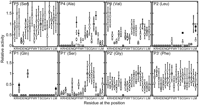
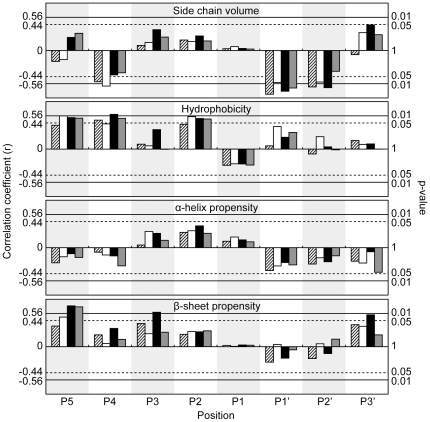
We have previously created a 19×8 substrate library by performing saturation mutagenesis at P5 to P3' positions on the wild type (WT) sequence (SAVLQ↓SGF), which corresponds to the autocleavage sequence at the N-terminus of SARS-CoV 3CLpro [26]. The values of kobs/[3CLpro] of the proteases against this WT sequence were 443±11, 124±13, 180±5 and 174±19 mM-1 min-1 for HCoV-NL63 (group 1), HCoV-OC43 (group 2a), SARS-CoV (group 2b), and IBV (group 3), respectively. That all proteases can cleave the WT sequence efficiently justifies that we can use our substrate library to profile the substrate specificities of 3CLpro from other groups of CoVs. Based on the FRET assay we developed, we measured the activities of 3CLpro from HCoV-NL63, HCoV-OC43, SARS-CoV and IBV against the 19×8 substrate variants (Figure 1, Table S1) [26]. To identify the structural basis of substrate preferences for different CoVs, the protease activities were correlated with side chain volume [28], hydrophobicity [29], and α-helix and β-sheet propensities [30] as described [26]. The correlations were quantified in terms of correlation coefficients and p-values (Figure 2, Table S2).
Figure 1. Substrate specificity of 3CLpro at P5 to P3' positions.
Relative protease activity of 3CLpro from HCoV-NL63 (circles, group 1), HCoV-OC43 (squares, group 2a), SARS-CoV (diamond, group 2b) and IBV (triples, group 3) against 19×8 of substrate variants were measured by FRET assay. Relative activities that are significantly (p-value<0.001) higher than the rest are represented as filled symbols.
Figure 2. Correlation between 3CLpro activities and structural properties of substituting residues.
The relative protease activities of 3CLpro from HCoV-NL63 (shaded, group 1), HCoV-OC43 (white, group 2a), SARS-CoV (black, group 2b) and IBV (grey, group 3), were correlated with structural properties of substituting residue properties, including side chain volume [28], hydrophobicity [29] and α-helix and β-sheet propensities [30]. Correlation coefficients of +/−0.56 and +/−0.44 correspond to p-values of 0.01 and 0.05 respectively.
Differences in substrate specificities among 3CLpro
We then tested if the relative activities of 3CLpro from any CoV strains were significantly different from the other by analysis of variance. Substitutions that resulted in significantly higher relative activities (p<0.001) were indicated as filled symbol in Figure 1. IBV 3CLpro (Figure 1, triangles) was the most efficient in cleaving A4P and A4F with relative activities of 1.09±0.24 and 0.58±0.14, respectively, while SARS 3CLpro (Figure 1, diamonds) preferred A4V with relative activity of 1.39±0.19. HCoV-OC43 3CLpro (Figure 1, squares) appeared to be the most versatile in accepting substitutions at P1 and P2 positions, and could cleave Q1H, Q1M, L2M and L2C, significantly better than 3CLpro from other strains. No significant differences were observed for other substitutions, suggesting that 3CLpro from different CoVs shares many similarities in substrate preferences.
Substrate preferences that are common to all 3CLpro
The most preferred P1 residue is Gln (Figure 1), which forms hydrogen-bonds with the side-chain of an invariant His residue and the backbone carbonyl group of an invariant Phe residue (His-163 and Phe-140 in SARS-CoV 3CLpro) in the P1 binding pocket. Interestingly, our results showed that 3CLpro from all groups of CoVs can cleave His at P1 position reasonably well. The relative activities for 3CLpro from HCoV-NL63, HCoV-OC43, SARS-CoV, and IBV were 0.26±0.08, 0.47±0.08, 0.19±0.03 and 0.25±0.12, respectively (Table S1). Consistent with this observation, His is found natively at P1 positions in the polyproteins from group 1 and 2a CoVs (Table S3). Taken together, the ability to cleave His at P1 position is a conserved property for all 3CLpro. Moreover, we showed that all 3CLpro can cleave Q1M, albeit at an even lower rate, and all other substitutions resulted in undetected activity.
The protease activities correlate positively with the hydrophobicity of substituting residues at P2 position (Figure 2). In fact, among the P2 variants, only L2M, L2C, L2F, L2I and L2V were cleavable, suggesting that P2 position favors hydrophobic residues. However, substitution with β-branched residues, Val or Ile, led to >10-folds decreases in the activity (Figure 1, Table S1). Considering that Leu, Val and Ile share similar hydrophobicity and side chain volume, the large differences in activities suggest that β-branched residues are not preferred in all 3CLpro, probably due to steric clashes with the P2 binding pocket. Taken together, P2 position prefers hydrophobic residues without β-branch, and the most preferred residue is Leu.
At P3 position, the protease activities on Arg/Lys-substituting variants were 5 to 14 fold higher than that on Asp/Glu-substituting variants (Figure 1, Table S1). This observation suggests that P3 position prefers positively charged residues over negatively charged one. In the active site of 3CLpro, there is no substrate-binding pocket for P3 residue. Molecular modeling showed that there is an invariant Glu residue (Glu-166 in SARS-CoV 3CLpro) in the active site of 3CLpro that may form favorable charge-charge interactions with a positively charged residue at the P3 position, which may explain why Arg/Lys are favored over Asp/Glu at this position (Figure S1). Moreover, no cleavage was observed for substrate containing Pro-substitution at P3 position.
The protease activities correlate negatively with side chain volume, and positively with the hydrophobicity of substituting residues at P4 position (Figure 2). The correlations with hydrophobicity were more evident (with correlation coefficients >0.89) when only small residues (Ala, Asn, Asp, Cys, Gly, Ser, and Thr) with side chain volumes <70 Å3 (Figure 3) were included in the analysis. This result suggests that as long as the side chain can fit into the P4 binding pocket, the protease activity is directly proportional to the hydrophobicity of the substituting residues. On the other hand, charged residues like Lys, Arg, His, Asp and Glu were not cleavable, presumably due to the unfavorable burial of charges in the hydrophobic P4 pocket.
Figure 3. All 3CLpro prefer small hydrophobic residues at P4 position.
All 3CLpro activities are highly correlated to hydrophobicity of residues with side chain volumes of <70 Å3 (filled symbols). The correlation coefficients and the corresponding p-values are indicated.
In general, the activities of 3CLpro correlate positively with the hydrophobicity and β-sheet propensity of substituting residues at P5 position (Figure 2). The correlations are significant (p<0.05) for group 2a, 2b, and 3 CoVs, but are weaker for group 1 CoV. Like the P3 position, there is no substrate-binding pocket for P5 residue. In the crystal structure of SARS-CoV 3CLpro in complex with a peptide substrate, the P5 residue adopts an extended β-strand conformation to avoid clashing of P5-P6 residues with the protease [31]. Residues with high β-sheet propensity may stabilize the extended conformation at P5 and improve enzyme-substrate interaction. As shown in Figure 1, a number of substitutions at P5 position resulted in a substrate better than the WT sequence (i.e. with relative activity >1). Consistent with the suggestion that P5 position favors residues with high hydrophobicity and β-sheet propensity, Val-substitution consistently yielded substrates with higher than WT activities for all 3CLpro. On the other hand, negatively charged residues (Asp/Glu) were not favored at P5 position, with significantly lower activities (0.16 to 0.50).
At P1' position, the protease activities correlate negatively with side chain volume of substituting residues (Figure 2). In fact, the relative activities for substrates with the smallest residues (Gly, Ala, Ser, and Cys) at P1' position were in the range of 0.64 to 1.40, which were consistently higher than those for other larger residues (Figure 1). At P2' position, all variants, except G2'P, could be cleaved with relative activities of 0.17 to 1.04 (Figure 1). The protease activities also correlate negatively with the side chain volume (Figure 2), but the difference in the protease activities was relatively small (Figure 1). At P3' position, no obvious substrate preference was observed.
The effect of combining multiple favorable substitutions
Our profiling analysis showed that all CoV 3CLpro prefer P5-Val and P3-Arg (Figure 1). To test if we can combine two favorable substitutions to create a more active substrate, we have created a doubly-substituted substrate variant ‘VARLQ↓SGF’. The protease activities of HCoV-NL63, HCoV-OC43, SARS-CoV and IBV against the doubly-substituted sequence were 1.70±0.07, 1.87±0.17, 1.70±0.12 and 3.24±0.37, respectively (Table 1). The results suggest that the increase in activity is additive, and the sequence ‘VARLQ↓SGF’ can represent a good broad-spectrum substrate for all 3CLpro.
Table 1. 3CLpro activities against doubly- and triply-substituted substrate variants. WT substrate was substituted at P3 to P5 positions to generate doubly- and triply-substituted variants. The relative activities of 3CLpro on these substrate variants are reported.
| Variant sequence | HCoV-NL63 | HCoV-OC43 | SARS-CoV | IBV |
| VAVLQ↓SGF | 1.23±0.40 | 1.55±0.30 | 1.80±0.31 | 1.58±0.27 |
| SARLQ↓SGF | 1.14±0.24 | 1.36±0.17 | 0.97±0.12 | 1.72±0.22 |
| VARLQ↓SGF | 1.70±0.07 | 1.87±0.17 | 1.70±0.17 | 3.24±0.37 |
| SPVLQ↓SGF | 0.06±0.01 | 0.29±0.07 | 0.61±0.10 | 1.09±0.24 |
| VPRLQ↓SGF | 0.15±0.04 | 0.91±0.12 | 0.99±0.12 | 4.33±0.98 |
| SVVLQ↓SGF | 0.76±0.10 | 0.59±0.07 | 1.39±0.19 | 0.59±0.09 |
| VVVLQ↓SGF | 1.23±0.06 | 0.60±0.05 | 1.97±0.19 | 0.86±0.05 |
| VVRLQ↓SGF | 1.63±0.07 | 0.55±0.04 | 2.50±0.51 | 2.19±0.13 |
On the other hand, our profiling analysis suggests that 3CLpro from SARS-CoV and IBV have different substrate preferences at P4 position – SARS-CoV prefers P4-Val (relative activity = 1.09±0.24) while IBV prefers P4-Pro (relative activity = 1.39±0.10) (Figure 1, Table S1). To see if we can exploit this distinct substrate preference at P4 position to create a substrate more specific for IBV 3CLpro, we have created the triply-substituted variant ‘VPRLQ↓SGF’. The protease activity of IBV 3CLpro against this sequence was boosted to 4.33±0.98, while that of the other strains were significantly reduced, demonstrating that this substrate sequence can represent a specific substrate-sequence for IBV 3CLpro (Table 1). Similarly, the protease activity of SARS-CoV 3CLpro against the triply-substituted sequence ‘VVRLQ↓SGF’ was boosted to 2.50±0.51, while that of the other strains were reduced (Table 1). Taken together, these results suggest that one can combine the substrate preference profiled in this study to create a better substrate sequences.
Discussion
This study provides the first comprehensive profiling of substrate specificities of 3CLpro from group 1, 2a, and 3 CoVs. We showed that the substrate specificities of these 3CLpro share many similarities to those of 3CLpro from SARS-CoV (group 2b) reported previously by us [26]. Table 2 summarizes the substrate specificities that are common to all 3CLpro. Although the substrate specificities for 3CLpro from different groups of CoVs share a number of similarities, unique substrate preferences were identified in this study. In particular, we showed that only IBV 3CLpro, but not other proteases, prefers P4-Pro (Figure 3).
Table 2. Summary of substrate specificities that are common among all 3CLpro.
| Position | Substrate preferences |
| P5 | No strong preference |
| P4 | Small hydrophobic residues |
| P3 | Positively charged residues |
| P2 | High hydrophobicity and absence of β-branch |
| P1 | Gln |
| P1' | Small residues |
| P2' | Small residues |
| P3' | No strong preference |
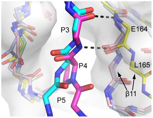
To understand the structural basis of this unique substrate preference, we compared the structures of IBV 3CLpro with other coronaviral 3CLpro. We noticed that strand-11 of IBV 3CLpro is positioned further away from the P4 and P5 substrate-binding site compared to other 3CLpro (Figure 4) [31], [32], [33]. This results in a wider substrate-binding pocket in IBV 3CLpro. We further docked the substrate variant A4P into the substrate-binding pocket of IBV 3CLpro. Due to the cyclic structure of Pro residue, the backbone Ø dihedral angle of the P4 residue is restrained to ca. −60°, which causes the substrate peptide to bend towards the strand-11 of 3CLpro. Such conformation of substrate is much better accommodated by IBV 3CLpro, which has a wider substrate-binding pocket near the P4 and P5 positions. This observation justifies why only IBV 3CLpro cleaves P4-Pro efficiently.
Figure 4. IBV 3CLpro has a wider substrate-binding pocket to accommodate substrate containing P4-Pro.
The structure of IBV 3CLpro (PDB: 2Q6D, yellow cartoon and white surface) is superimposed with 3CLpro from HCoV-229E (PDB: 1P9S, light blue), HCoV-HKU1 (PDB: 3D23, light green), and SARS-CoV (PDB: 2Q6G, pink) [31], [32], [33]. The structure of WT substrate (magenta) is derived from crystal structure of SARS-CoV 3CLpro in complex with the autocleavage sequence (TSAVLQ↓SGFRKM) (PDB: 2Q6G) [31]. The structure of the A4P substrate variant (cyan) was modeled based on the crystal structure of IBV 3CLpro in complex with its own autocleavage sequence (PDB: 2Q6D) [31]. Note that strand-11 of IBV 3CLpro is positioned further away from P4 to P5 positions, resulting in a wider substrate-binding pocket.
Similarities in substrate specificity suggest that it is feasible to create a broad-spectrum inhibitor that targets all 3CLpro. A broad-spectrum inhibitor is desirable for a first line defense against coronaviral infection because CoVs are capable of generating novel strains with high virulence through high frequency of mutations and recombination [34], [35], [36], [37].. Based on the autocleavage sequence of SARS-CoV 3CLpro (i.e. AVLQ↓), Rao and co-workers designed broad-spectrum peptidomimetic inhibitors that can inhibit 3CLpro from different groups of CoVs [20]. Their results are consistent with our observation that the autocleavage sequence of SARS-CoV 3CLpro can be well cleaved by all 3CLpro. The substrate preferences profiled in this study will provide a rational basis to improve the broad-spectrum 3CLpro inhibitors. For example, by combining favorable substitutions at P3 to P5 positions, we identified a substrate sequence ‘VARLQ↓SGF’ that can be cleaved with high relative activities by 3CLpro from all groups of CoVs (Table 1). This substrate sequence may serve as a good starting point of the design of broad-spectrum peptidomimetic inhibitors for 3CLpro.
Although it is generally accepted that substrate specificity provides insights into the design of peptidomimetic protease inhibitors, there are exceptions to the dogma that good peptidomimetic inhibitors should be derived from good substrate sequences. For example, Hilgenfeld and co-workers showed that the P2 position of peptide aldehyde inhibitors can accommodate aspartate or serine, which are poor substrates for SARS-CoV 3CLpro [38].
In the FRET assay developed by us, all 3CLpro can efficiently cleave the WT sequence of ‘SAVLQ↓SGF’ with activity of 120–440 mM−1 min−1, and the activity can be further improved by 1.7 to 3.2 fold using the substrate sequence of ‘VARLQ↓SGF’. Because the substrate sequences can be cleaved by all 3CLpro with high efficiency, one could use the FRET assay to screen for broad-spectrum inhibitors targeting 3CLpro from all groups of CoVs.
Materials and Methods
Cloning, Expression and Purification of 3CLpro and the Substrate Library
Cloning, expression and purification of SARS-CoV 3CLpro were described previously [26]. Codon-optimized DNA sequences encoding HCoV-NL63 (GenBank AY567487) and HCoV-OC43 (GenBank AAX85666), and IBV (GenBank M95169) 3CLpro were purchased from Mr. Gene (http://mrgene.com). The coding sequences of 3CLpro from HCoV-NL63, HCoV-OC43 and IBV were sub-cloned and expressed in E. coli strain BL21 (DE3) pLysS as fusion proteins with N-terminal tags of poly-histidine-small ubiquitin-related modifier (His6-SUMO) or poly-histidine-maltose binding protein (His6-MBP). Protein expression was induced by addition of 0.1 mM of isopropyl β-D-1-thiogalactopyranoside. After overnight incubation at 25°C, cells were harvested by centrifugation and resuspended in buffer A (20 mM Tris, pH 7.8, 150 mM NaCl and 1 mM tris(2-carboxyethyl)phosphine) with 30 mM imidazole and disrupted by sonication. Soluble fraction was subject to immobilized metal ion affinity chromatography for purification as described for SARS-CoV 3CLpro [26]. The His6-SUMO or His6-MBP tags were removed by protease digestion using sentrin-specific protease 1 or factor Xa, respectively, followed by immobilized metal ion affinity chromatography. Native 3CLpro were finally purified by G75 size exclusion column and stored in buffer A. Elution profiles of size exclusion chromatography indicated that all 3CLpro purified were dimeric.
The construction, expression and purification of the substrate library were described previously [26]. In brief, the WT substrate sequence ‘TSAVLQ↓SGFRKM’ was inserted between the cyan fluorescent protein and the yellow fluorescent protein to create the substrate protein. Saturation mutagenesis was performed at each of the P5 to P3' positions to generate a substrate library of 19×8 variants.
FRET assay for 3CLpro activity measurement
The protease activity of 3CLpro was measured by the FRET assay we developed previously [26]. Purified 3CLpro at 0.2 to 2 µM were mixed with 35 µM of the substrate protein in buffer A. Cleavage of the substrate protein leads to a decrease in fluorescence at 530 nm when the reaction mixture was excited at 430 nm. The fluorescence intensity, monitored by EnVision 2101 Multilabel Plate Reader, was fitted to single exponential decay to obtain the observed rate constant (kobs). The protease activity against variant substrates was normalized against the WT activity to yield the relative activity. The assay was repeated in triplicate.
Correlation analysis
Structural properties of substituting residues, including side chain volume [28], hydrophobicity [29], and α-helix and β-sheet propensities [30], were correlated with relative activity to determine correlation coefficients (r) and p-values.
Supporting Information
Molecular modeling showing P3-Arg may interact with Glu-166 of 3CLpro. The model was based on the crystal structure of 3CLpro (grey) in complex with a peptide substrate ‘TSAVLQ↓SGFRK’ (yellow). P3-Val was replaced by P3-Arg using the program PyMOL. As shown, the invariant Glu-166 is in close proximity to P3-Arg, and may form favorable charge-charge interaction to P3-Arg.
(TIF)
Relative activities of HCoV-NL63, HCoV-OC43, SARS-CoV and IBV 3CLpro. ND stands for non-detectable cleavage. The average and the standard deviation of three measurements are shown.
(DOC)
Correlation between activity of 3CLpro and structural properties of substituting residues. The correlation coefficients and p-values (bracketed) are reported.
(DOC)
Autocleavage sequences of 3CLpro. PEDV, TGEV, MHV, PHEV stand for porcine epidemic diarrhoea coronavirus, transmissible gastroenteritis coronavirus, mouse hepatitis coronavirus and porcine hemagglutinating encephalomyelitis coronavirus respectively.
(DOC)
Footnotes
Competing Interests: The authors have declared that no competing interests exist.
Funding: This work was supported by the Research Fund for the Control of Infectious Diseases, Hong Kong SAR Government (project number: 09080282) (http://www.fhb.gov.hk/grants/english/funds/funds_rfcid/funds_rfcid_abt/funds_rfcid_abt.html). The funders had no role in study design, data collection and analysis, decision to publish, or preparation of the manuscript.
References
- 1.van der Hoek L, Pyrc K, Jebbink MF, Vermeulen-Oost W, Berkhout RJ, et al. Identification of a new human coronavirus. Nat Med. 2004;10:368–373. doi: 10.1038/nm1024. [DOI] [PMC free article] [PubMed] [Google Scholar]
- 2.Hamre D, Procknow JJ. A new virus isolated from the human respiratory tract. Proc Soc Exp Biol Med. 1966;121:190–193. doi: 10.3181/00379727-121-30734. [DOI] [PubMed] [Google Scholar]
- 3.Woo PC, Lau SK, Chu CM, Chan KH, Tsoi HW, et al. Characterization and complete genome sequence of a novel coronavirus, coronavirus HKU1, from patients with pneumonia. J Virol. 2005;79:884–895. doi: 10.1128/JVI.79.2.884-895.2005. [DOI] [PMC free article] [PubMed] [Google Scholar]
- 4.Kuiken T, Fouchier RA, Schutten M, Rimmelzwaan GF, van Amerongen G, et al. Newly discovered coronavirus as the primary cause of severe acute respiratory syndrome. Lancet. 2003;362:263–270. doi: 10.1016/S0140-6736(03)13967-0. [DOI] [PMC free article] [PubMed] [Google Scholar]
- 5.Peiris JS, Lai ST, Poon LL, Guan Y, Yam LY, et al. Coronavirus as a possible cause of severe acute respiratory syndrome. Lancet. 2003;361:1319–1325. doi: 10.1016/S0140-6736(03)13077-2. [DOI] [PMC free article] [PubMed] [Google Scholar]
- 6.Cheever FS, Daniels JB, Pappenheimer AM, Bailey OT. A murine virus (JHM) causing disseminated encephalomyelitis with extensive destruction of myelin. J Exp Med. 1949;90:181–210. doi: 10.1084/jem.90.3.181. [DOI] [PMC free article] [PubMed] [Google Scholar]
- 7.Binn LN, Lazar EC, Keenan KP, Huxsoll DL, Marchwicki RH, et al. Proc Annu Meet U S Anim Health Assoc; 1974. Recovery and characterization of a coronavirus from military dogs with diarrhea. pp. 359–366. [PubMed] [Google Scholar]
- 8.Poon LL, Chu DK, Chan KH, Wong OK, Ellis TM, et al. Identification of a novel coronavirus in bats. J Virol. 2005;79:2001–2009. doi: 10.1128/JVI.79.4.2001-2009.2005. [DOI] [PMC free article] [PubMed] [Google Scholar]
- 9.Beaudette FR, Hudson CB. Cultivation of the virus of infectious bronchitis. J Am Vet Med Assoc. 1937;90:51–58. [Google Scholar]
- 10.Doyle LP, Hutchings LM. A transmissible gastroenteritis virus in pigs. J Am Vet Assoc. 1946;108:257–259. [PubMed] [Google Scholar]
- 11.Tyrrell DA, Bynoe ML. Cultivation of viruses from a high proportion of patients with colds. Lancet. 1966;1:76–77. doi: 10.1016/s0140-6736(66)92364-6. [DOI] [PubMed] [Google Scholar]
- 12.Woo PC, Lau SK, Huang Y, Yuen KY. Coronavirus diversity, phylogeny and interspecies jumping. Exp Biol Med (Maywood) 2009;234:1117–1127. doi: 10.3181/0903-MR-94. [DOI] [PubMed] [Google Scholar]
- 13.Snijder EJ, Bredenbeek PJ, Dobbe JC, Thiel V, Ziebuhr J, et al. Unique and conserved features of genome and proteome of SARS-coronavirus, an early split-off from the coronavirus group 2 lineage. J Mol Biol. 2003;331:991–1004. doi: 10.1016/S0022-2836(03)00865-9. [DOI] [PMC free article] [PubMed] [Google Scholar]
- 14.Gorbalenya AE, Snijder EJ, Spaan WJ. Severe acute respiratory syndrome coronavirus phylogeny: toward consensus. J Virol. 2004;78:7863–7866. doi: 10.1128/JVI.78.15.7863-7866.2004. [DOI] [PMC free article] [PubMed] [Google Scholar]
- 15.Gaunt ER, Hardie A, Claas EC, Simmonds P, Templeton KE. Epidemiology and clinical presentations of the four human coronaviruses 229E, HKU1, NL63, and OC43 detected over 3 years using a novel multiplex real-time PCR method. J Clin Microbiol. 2010;48:2940–2947. doi: 10.1128/JCM.00636-10. [DOI] [PMC free article] [PubMed] [Google Scholar]
- 16.Dominguez SR, Robinson CC, Holmes KV. Detection of four human coronaviruses in respiratory infections in children: a one-year study in Colorado. J Med Virol. 2009;81:1597–1604. doi: 10.1002/jmv.21541. [DOI] [PMC free article] [PubMed] [Google Scholar]
- 17.Cavanagh D. Coronavirus avian infectious bronchitis virus. Vet Res. 2007;38:281–297. doi: 10.1051/vetres:2006055. [DOI] [PubMed] [Google Scholar]
- 18.Ignjatovic J, Sapats S. Avian infectious bronchitis virus. Rev Sci Tech. 2000;19:493–508. doi: 10.20506/rst.19.2.1228. [DOI] [PubMed] [Google Scholar]
- 19.Lin CW, Tsai CH, Tsai FJ, Chen PJ, Lai CC, et al. Characterization of trans- and cis-cleavage activity of the SARS coronavirus 3CLpro protease: basis for the in vitro screening of anti-SARS drugs. FEBS Lett. 2004;574:131–137. doi: 10.1016/j.febslet.2004.08.017. [DOI] [PMC free article] [PubMed] [Google Scholar]
- 20.Yang H, Xie W, Xue X, Yang K, Ma J, et al. Design of wide-spectrum inhibitors targeting coronavirus main proteases. PLoS Biol. 2005;3:e324. doi: 10.1371/journal.pbio.0030324. [DOI] [PMC free article] [PubMed] [Google Scholar]
- 21.Li SY, Chen C, Zhang HQ, Guo HY, Wang H, et al. Identification of natural compounds with antiviral activities against SARS-associated coronavirus. Antiviral Res. 2005;67:18–23. doi: 10.1016/j.antiviral.2005.02.007. [DOI] [PMC free article] [PubMed] [Google Scholar]
- 22.Wu CY, Jan JT, Ma SH, Kuo CJ, Juan HF, et al. Small molecules targeting severe acute respiratory syndrome human coronavirus. Proc Natl Acad Sci U S A. 2004;101:10012–10017. doi: 10.1073/pnas.0403596101. [DOI] [PMC free article] [PubMed] [Google Scholar]
- 23.Chen L, Gui C, Luo X, Yang Q, Gunther S, et al. Cinanserin is an inhibitor of the 3C-like proteinase of severe acute respiratory syndrome coronavirus and strongly reduces virus replication in vitro. J Virol. 2005;79:7095–7103. doi: 10.1128/JVI.79.11.7095-7103.2005. [DOI] [PMC free article] [PubMed] [Google Scholar]
- 24.Fan K, Ma L, Han X, Liang H, Wei P, et al. The substrate specificity of SARS coronavirus 3C-like proteinase. Biochem Biophys Res Commun. 2005;329:934–940. doi: 10.1016/j.bbrc.2005.02.061. [DOI] [PMC free article] [PubMed] [Google Scholar]
- 25.Goetz DH, Choe Y, Hansell E, Chen YT, McDowell M, et al. Substrate specificity profiling and identification of a new class of inhibitor for the major protease of the SARS coronavirus. Biochemistry. 2007;46:8744–8752. doi: 10.1021/bi0621415. [DOI] [PubMed] [Google Scholar]
- 26.Chuck CP, Chong LT, Chen C, Chow HF, Wan DC, et al. Profiling of substrate specificity of SARS-CoV 3CLpro. PLoS ONE. 2010;5:e13197. doi: 10.1371/journal.pone.0013197. [DOI] [PMC free article] [PubMed] [Google Scholar]
- 27.Hegyi A, Ziebuhr J. Conservation of substrate specificities among coronavirus main proteases. J Gen Virol. 2002;83:595–599. doi: 10.1099/0022-1317-83-3-595. [DOI] [PubMed] [Google Scholar]
- 28.Lee S, Tikhomirova A, Shalvardjian N, Chalikian TV. Partial molar volumes and adiabatic compressibilities of unfolded protein states. Biophys Chem. 2008;134:185–199. doi: 10.1016/j.bpc.2008.02.009. [DOI] [PubMed] [Google Scholar]
- 29.Kyte J, Doolittle RF. A simple method for displaying the hydropathic character of a protein. J Mol Biol. 1982;157:105–132. doi: 10.1016/0022-2836(82)90515-0. [DOI] [PubMed] [Google Scholar]
- 30.Chou PY, Fasman GD. Prediction of the secondary structure of proteins from their amino acid sequence. Adv Enzymol Relat Areas Mol Biol. 1978;47:45–148. doi: 10.1002/9780470122921.ch2. [DOI] [PubMed] [Google Scholar]
- 31.Xue X, Yu H, Yang H, Xue F, Wu Z, et al. Structures of two coronavirus main proteases: implications for substrate binding and antiviral drug design. J Virol. 2008;82:2515–2527. doi: 10.1128/JVI.02114-07. [DOI] [PMC free article] [PubMed] [Google Scholar]
- 32.Anand K, Ziebuhr J, Wadhwani P, Mesters JR, Hilgenfeld R. Coronavirus main proteinase (3CLpro) structure: basis for design of anti-SARS drugs. Science. 2003;300:1763–1767. doi: 10.1126/science.1085658. [DOI] [PubMed] [Google Scholar]
- 33.Zhao Q, Li S, Xue F, Zou Y, Chen C, et al. Structure of the main protease from a global infectious human coronavirus, HCoV-HKU1. J Virol. 2008;82:8647–8655. doi: 10.1128/JVI.00298-08. [DOI] [PMC free article] [PubMed] [Google Scholar]
- 34.Jenkins GM, Rambaut A, Pybus OG, Holmes EC. Rates of molecular evolution in RNA viruses: a quantitative phylogenetic analysis. J Mol Evol. 2002;54:156–165. doi: 10.1007/s00239-001-0064-3. [DOI] [PubMed] [Google Scholar]
- 35.Lai MM. RNA recombination in animal and plant viruses. Microbiol Rev. 1992;56:61–79. doi: 10.1128/mr.56.1.61-79.1992. [DOI] [PMC free article] [PubMed] [Google Scholar]
- 36.Pasternak AO, Spaan WJ, Snijder EJ. Nidovirus transcription: how to make sense...? J Gen Virol. 2006;87:1403–1421. doi: 10.1099/vir.0.81611-0. [DOI] [PubMed] [Google Scholar]
- 37.Duffy S, Shackelton LA, Holmes EC. Rates of evolutionary change in viruses: patterns and determinants. Nat Rev Genet. 2008;9:267–276. doi: 10.1038/nrg2323. [DOI] [PubMed] [Google Scholar]
- 38.Zhu L, George S, Schmidt MF, Al-Gharabli SI, Rademann J, et al. Antiviral Res; 2011. Peptide aldehyde inhibitors challenge the substrate specificity of the SARS-coronavirus main protease. doi: 10.1016/j.antiviral.2011.08.001. [DOI] [PMC free article] [PubMed] [Google Scholar]
Associated Data
This section collects any data citations, data availability statements, or supplementary materials included in this article.
Supplementary Materials
Molecular modeling showing P3-Arg may interact with Glu-166 of 3CLpro. The model was based on the crystal structure of 3CLpro (grey) in complex with a peptide substrate ‘TSAVLQ↓SGFRK’ (yellow). P3-Val was replaced by P3-Arg using the program PyMOL. As shown, the invariant Glu-166 is in close proximity to P3-Arg, and may form favorable charge-charge interaction to P3-Arg.
(TIF)
Relative activities of HCoV-NL63, HCoV-OC43, SARS-CoV and IBV 3CLpro. ND stands for non-detectable cleavage. The average and the standard deviation of three measurements are shown.
(DOC)
Correlation between activity of 3CLpro and structural properties of substituting residues. The correlation coefficients and p-values (bracketed) are reported.
(DOC)
Autocleavage sequences of 3CLpro. PEDV, TGEV, MHV, PHEV stand for porcine epidemic diarrhoea coronavirus, transmissible gastroenteritis coronavirus, mouse hepatitis coronavirus and porcine hemagglutinating encephalomyelitis coronavirus respectively.
(DOC)


