Gymnastics requires conversion of the upper limb into a load-bearing extremity, leading to upper extremity, especially wrist, injuries. The “gymnast's wrist” or the Madelung-like deformity involves repetitive excessive loading of immature bones leading to premature closure of the distal radial physis with associated ulna-plus variance and wrist pain.1–3 The significant physical demands of gymnastics and the frequency of injuries especially in elite gymnasts has led some to state that chronic gymnastic injuries should more properly be referred to as consequences of participation in the sport, implying the inevitability of chronic injuries.4
Up to 88% of gymnasts experience wrist pain.1 This is associated with older age, increased training hours and higher skill level.5 Elite gymnasts have a significantly greater rate of injuries than lower level gymnasts.6 Therefore they need to be considered as a separate group from casual participants in terms of sport-specific stresses and resulting injuries.
The scapholunate interosseus ligament (SLIL) is involved in maintaining the stability of the complex structure of the wrist. Wrist injuries commonly involve the SLIL.7 However, there is no reported literature on SLIL injuries in gymnasts. It may be that these injuries have been overlooked in the past. The wrist pain associated with these injuries is usually chronic and is less severe than a fracture, so athletes may interpret it as a nonsignificant sprain and may not seek medical attention. Physicians may overlook these injuries for similar reasons.
The mechanism of injury is generally believed to be impact loading with the wrist in extension and ulnar deviation,8,9 resulting in the capitate being driven between the scaphoid and lunate bones. This can occur with either a fall on an outstretched hand or repeated loading with weight bearing through the upper extremities. Clinical presentation is usually pain over the dorsal and radial aspects of the wrist with associated loss of motion or grip strength, or both.
Radiologically, scapholunate diastasis is defined as greater than 3 mm with a normal scapholunate distance of 2 mm or less on the other wrist. Diastasis is present in static scapholunate dissociation but is not visible in dynamic instability unless elicited on stress views, which can be performed with fist clenching or ulnar deviation of the wrist. Additional radiographic signs include the cortical “ring” sign due to the volar flexion of the scaphoid, a scapholunate angle greater than 70° (normal 30°–60°), and a radiolunate angle greater than 5°, which indicates a dorsal intercalated segmental instability (DISI) pattern with a dorsiflexed lunate. Radiographs in most scapholunate injuries appear normal, the radiographic findings already described being seen mostly in complete ligament tears or chronic partial tears.
Further evaluation can include MRI, which can better visualize scapholunate diastasis, ligament tears, occult fractures, and bone bruising. Arthrography has been used but often now is bypassed in favour of arthroscopy, which continues to be the standard method for evaluating SLIL injuries.
We describe 3 patients who had tears of the scapholunate ligament.
Case reports
Case 1
A 21-year-old right-hand dominant man from the Canadian national gymnastics team presented with a 3-month history of pain over the dorsal aspect of his right wrist that made training impossible. The pain was worsened by activities that axially loaded his wrist, such as the pommel horse. Physical examination revealed ulnocarpal joint tenderness. No effusion was palpable. CT showed a small minimally displaced fracture on the dorsal aspect of the triquetrum. Wrist radiographs appeared normal.
Bone scanning showed mild uptake of radoactive material at the radiocarpal junction and mild–moderate uptake in the triquetrum/pisiform region and the radial intercarpal region of the right wrist. Subsequently, magnetic resonance arthrography showed a scapholunate ligament tear, with contrast material extending into the midcarpal joint and associated DISI. Contrast material also extended along the medial aspect of the distal ulna consistent with an ulnar collateral ligament tear. A small avulsed bone fragment was confirmed dorsal to the triquetrum.
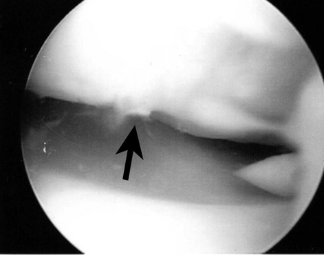
Arthroscopy of the wrist (Fig. 1) revealed that the scapholunate ligament was approximately 50% torn, with a scapholunate distance of approximately 4 mm. Osteoarthritis of the scapholunate joint and synovitis were debrided. Joint debris was removed. The triangular fibrocartilage complex (TFCC) was not torn.

FIG. 1. Case 1. Wrist arthroscopy demonstrating disruption of the scapholunate interosseus ligament.
Wrist exercises were prescribed and the man subsequently obtained a full range of motion of his wrist. Recommendations were made to wait 3 months before resuming training.
Case 2
A 19-year-old man from the Canadian national gymnastics team presented with pain over the dorsal aspect of his left wrist. There was dorsal tenderness on the ulnar side but not over the TFCC. Radiographs appeared normal.
Magnetic resonance arthrography indicated a possible focal tear within the mid-or volar portion of the scapholunate ligament. A minute amount of contrast material entered the mid-carpal joint. Ultrasonography confirmed that the dorsal portion of the scapholunate ligament was intact.
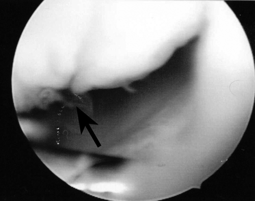
He continued to have symptoms and was unable to continue his training. Arthroscopy was performed for diagnostic reasons. A partial scapholunate ligament tear was discovered along with chondromalacia of the scaphoid, lunate and distal radius (Fig. 2). Therefore, arthroscopic debridement was performed.

FIG. 2. Case 2. Wrist arthroscopy demonstrates chondromalacia of the scaphoid and lunate as well as disruption of the scapholunate interosseus ligament.
His symptoms improved and he was able to resume training.
Case 3
A 15-year-old boy from the Canadian national gymnastics team presented with ulnar-sided left wrist pain. His pain was worsened with activities such as the pommel horse that axially loaded his wrist. There was tenderness and a full range of motion of the wrist. Radiographs appeared normal.
Magnetic resonance arthrography showed an area of altered signal within the lunate that could not exclude a small bone bruise or avascular necrosis. A subsequent bone scan appeared normal.
He continued to train and compete but his significant wrist pain persisted. Arthroscopy detected a partial full-thickness tear of the scapholunate ligament, chondromalacia of the lunate and fraying of the ulnar collateral ligament.
After arthroscopic debridement his wrist symptoms improved and he was able to continue training.
Discussion
The high physical demands of elite gymnastics on the upper extremity produce the potential for significant injuries. In addition to acute wrist injuries, skeletally immature athletes can suffer chronic wrist injuries, ranging from stress fractures through the physis of the distal radius to ulnar-plus deformities related to premature growth arrest due to excessive loading through the physis. Ligamentous injuries can occur either acutely or chronically and affect either the extrinsic or intrinsic ligaments of the wrist. These injuries are considered as minor injuries or overuse syndromes.
We hypothesize that the ligament injuries and degenerative changes we have reported in our 3 cases are related to twisting, dismount-type activities in gymnastics that place maximal stress on the radial column of the wrist.
All 3 athletes were managed with arthroscopy and debridement after appropriate imaging had been conducted. Wrist supports limiting excessive dorsiflexion of the wrist were recommended and worn by each athlete for training and competitions. All athletes were able to return to full training. Given the likely mechanism of injury of dorsiflexion and ulnar deviation of the wrist and the frequent upper extremity loading occurring in this posture in gymnastics, it may be advisable in the future to consider the use of dorsiflexion-limiting wrist supports to prevent scapholunate ligament tears.
This study was conducted at McMaster University Medical Centre, Hamilton, Ont.
Competing interests: None declared.
Correspondence to: Dr. Jung Mah, 674 Upper James St., Hamilton ON L9C 2Z6; fax 905 575-1799
References
- 1.Dobyns JH, Gabel GT. Gymnast's wrist. Hand Clin 1990;6:493-505. [PubMed]
- 2.De Smet L, Claissens A, Fabry G. Gymnast wrist. Acta Orthop Belg 1993;59:377-80. [PubMed]
- 3.Tolat AR, Sanderson PL, De Smet L, et al. The gymnast's wrist: acquired positive ulnar variance following chronic epiphyseal injury. J Hand Surg [Br] 1992;17:678-81. [DOI] [PubMed]
- 4.Gabel GT. Gymnastic wrist injuries. Clin Sports Med 1998;17:611-21. [DOI] [PubMed]
- 5.DiFiori JP, Puffer JC, Mandelbaum BR, et al. Factors associated with wrist pain in the young gymnast. Am J Sports Med 1996;24:9-14. [DOI] [PubMed]
- 6.McAuley E, Hudash G, Shields K, et al. Injuries in women's gymnastics. The state of the art. Am J Sports Med 1987;15:558-65. [DOI] [PubMed]
- 7.Jones WA. Beware the sprained wrist: the incidence and diagnosis of scapholunate instability. J Bone Joint Surg Br 1988;70:293-7. [DOI] [PubMed]
- 8.Mayfield JK, Johnson RP, Kilcoyne RK. Carpal dislocations: pathomechanics and progressive perilunar instability. J Hand Surg [Am] 1980;5:226-41. [DOI] [PubMed]
- 9.Johnson RP. The acutely injured wrist and its residuals. Clin Orthop Relat Res 1980;(149):33-44. [PubMed]


