

Abstract
A 67-year-old morbidly obese female with a background of stage 4 chronic renal failure, ischaemic heart disease, congestive cardiac failure, atrial fibrillation and type 2 diabetes mellitus presented with sepsis and necrotic lesions of the proximal lower limbs. Initial histological findings were consistent with the clinical diagnosis of calciphylaxis and supportive treatment was commenced with addition of a phosphate binder and dietary restriction. Due to high anaesthetic risk, her wounds were managed with larva therapy in the first instance, however, ultimately surgical debridement was the required. Repeat histology from a further biopsy revealed necrosis secondary to numerous thrombi in the cutaneous vessels and a new diagnosis of purpura fulminans was made, likely secondary to her sepsis. Unfortunately, despite aggressive medical and surgical treatment measures, this patient died of multiple organ dysfunction following a prolonged admission.
Background
Purpura fulminans (PF) is a rare acute-onset disorder of cutaneous haemorrhage and necrosis usually precipitated by an episode of severe sepsis, which is associated with high morbidity and mortality 1. This case is atypical in that it presented in the proximal lower limbs of an adult rather than the more typical presentation in the distal lower limbs of a child.
In addition, this case is made more unusual as it is likely that the patient had a co-existing condition of calciphylaxis as supported by initial histological findings. Calciphylaxis is also a rare and often fatal syndrome of skin necrosis secondary to vascular calcification and thrombosis, often associated with diabetes mellitus and end-stage renal failure 2.
Case presentation
A 67-year-old morbidly obese patient with a background of stage 4 chronic renal failure without a renal replacement requirement, ischaemic heart disease, congestive cardiac failure, atrial fibrillation and type 2 diabetes mellitus presented with symptomatic anaemia, sepsis of unknown origin and urinary retention with acute on chronic renal failure. She was retired, independent of activities of daily living and lived with her husband.
On admission it was noted that she had painful, well-demarcated necrotic lesions on her left outer hip and both inner thighs surrounded by a border of erythema (figure 1).
Figure 1.
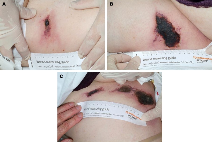
(A) Necrotic skin lesion (left outer thigh). (B) Necrotic skin lesion (left inner thigh). (C) Necrotic skin lesion (right inner thigh).
Following a dermatology referral, an initial clinical diagnosis of calciphylaxis with secondary infection was made given the history of renal failure, diabetes, long-term warfarin usage and the typical appearance and distribution of the wounds on the thighs.
Investigations
The clinical diagnosis of calciphylaxis was supported following an initial tissue biopsy of the presenting lesions, which on histological examination, revealed infarcted changes with focal dystrophic calcification and a mild diffused mixed inflammatory infiltrate.
A repeat biopsy was performed and histopathology revealed numerous thrombi in cutaneous vessels. Consequently, a diagnosis of purpura fulminans was made, likely to be secondary to her initial sepsis. At this stage, it was deemed that the acute phase of the dermatological insult was complete leaving the large and deep necrotic areas to the lower limbs.
Differential diagnosis
-
▶
PF
-
▶
Calciphylaxis (calcific uraemic arteriolopathy)
-
▶
Warfarin-induced skin necrosis (WISN)
-
▶
Henoch-Schönlein purpura (HSP)
-
▶
Thrombocytopenic purpura (TP)
As the patient was on long-term warfarin anticoagulant therapy for atrial fibrillation, the diagnosis of WISN was considered. Typically, WISN presents within 3–6 days of commencing therapy and is associated with administration of large doses of the drug, however it can present many years after commencing warfarin 3. In this case, warfarin was stopped in the first week of admission in view of worsening liver failure. Given the lack of improvement in the skin lesions on stopping warfarin, which would typically be expected in WISN, and on the basis of histological findings in addition to the delayed onset, this differential diagnosis was excluded.
The diagnosis of HSP is clinical and there is no specific biochemical test that can be used to confirm it. It commonly presents in children following an upper-respiratory tract infection with a symmetrical macular on the buttocks, posterior aspect of the legs and ulnar aspect of the arms. The rash evolves into purpura within 24 h. It is typically, although not always, associated with arthritis commonly involving the knee and ankle joints, gastrointestinal disturbance, low-grade fever and nephritis. TP presents with spontaneous non-palpable purpura and petechiae on the extremities and possibly bleeding diatheses. Unlike the high- and low-grade fever typically found in PF and HSP, respectively, in TP the patient is typically afebrile.
Differential diagnoses of HSP and TP were excluded on clinical grounds. First, the appearance of the skin necrosis is much less severe in HSP and TP than was observed in this case. Second, both the distribution of the lesions particularly to inner thighs and the age of the patient would be atypical for HSP. Furthermore, the absence of a reduced platelet count confirmed the exclusion of TP, either of autoimmune aetiology or secondary to an underlying disease process.
Treatment
This patient was actively managed under the medical team with a 1-week course of broad-spectrum intravenous antibiotics in addition to supportive treatment with diuretic therapy and transfusions of packed red cells.
With regard to her cutaneous lesions, before a histological diagnosis was available, the patient was clinically diagnosed with calciphylaxis and as such treated supportively with a phosphate-restricted diet and a phosphate binder was commenced. The surrounding cellulitic skin was treated with intravenous flucloxacillin and she required intravenous opioid analgesia. Due to her multiple comorbidities, she was of high anaesthetic risk. Consequently, following tissue viability advice it was decided to treat the deep slough of the wounds with larva therapy in order to clean the ulcers and prevent systemic infection, a treatment often found to be beneficial in the early stages of calciphylaxis 4 5. Despite best efforts to debride the affected areas with larva therapy, success was only achieved with the left leg, while the more extensive necrosis of the right leg did not respond well.
Following the secondary diagnosis of PF after the repeat histopathology report, treatment was unchanged and remained supportive. However, in the face of intractable sepsis unresponsive to several weeks of intravenous flucloxacillin, after review by the plastic surgeons, a plan was made for surgical debridement of the remaining necrotic tissue to remove the focus of sepsis. The considerable risks of surgery made this a difficult decision, given the patient’s numerous comorbidities, sepsis and consequent high anaesthetic risk. The patient survived the general anaesthetic and surgical procedure, however required transfusion of several units of blood perioperatively.
The aim of surgery in sepsis-associated PF is to remove areas of extensive skin necrosis while preserving as much tissue and limb function as possible. As there is a high risk of postoperative complications, optimal surgical treatment is best achieved in a regional burns unit and surgical treatment protocols in PF continue to evolve 6. Surgical options include either debridement of full-thickness skin loss and muscle damage or limb amputation. Early-stage fasciotomies may also be used to limit muscle loss in some cases. Conservative management of purpuric lesions should be initially used in PF and debridement is best performed once there is demarcation of necrotic tissue margins 7 or earlier if there is associated wound purulence. For both debridement and amputation, temporary allograft reconstruction during the initial procedure is often required, followed by definitive autograft reconstruction and potential revision surgery.
Outcome and follow-up
In this case, despite the multidisciplinary management including early medical management of her renal and cardiac failure, administration of intravenous antibiotics and other supportive treatment measures, multiple organ dysfunction ensued and the patient died 3 months following the initial presentation of sepsis.
Discussion
PF is a rare dermatological condition with an associated mortality of 50% 1. It is a disorder characterised by haemorrhagic infarction of the skin as a result of disseminated intravascular coagulation and thrombosis of the small cutanenous vessels. In particular, there is only a mild inflammatory component differentiating the condition from purpuric conditions such as HSP. Causes of PF can be classified into acute infectious, either inherited or acquired coagulopathy, for example, protein C or protein S deficiency and more rarely, idiopathic 8.
This particular presentation of PF is somewhat unusual in that the disease typically manifests in the distal limbs of babies or young children secondary to meningococcal and other gram-negative septicaemias 9. Less frequently, it presents in adults following a pneumococcal sepsis.
There are several published case reports discussing presentations of PF in both adults and children. Briefly, these include PF secondary to acute infection and idiopathic presentation in paediatric patients 9 and in the adult population following haemophilus influenzae sepsis 10 and Streptococcus pneumoniae sepsis in a bariatric patient 11. This case report discusses a patient who presented with histological features of both calciphylaxis and PF on tissue biopsy.
Learning points.
-
▶
Both PF and calciphylaxis are rare systemic diseases of an acute onset, which carry high mortality clinically manifesting as necrotic skin lesions. Both require a multidisciplinary approach to treatment involving supportive medical management and may require surgical intervention following initial medical treatment.
-
▶
Purpura fulminans: is a result of haemorrhagic necrosis of the cutaneous tissues resulting in necrotic lesions of the skin typically affecting distal limbs of children although not exclusively.
-
▶
Usually associated with history of severe sepsis often meningitis.
-
▶
Full cutaneous examination of patients presenting with sepsis is essential to early diagnosis, which is confirmed by biopsy and histology.
-
▶
Treatment involves initial medial management with aggressive control of sepsis, replacement of deficient clotting factors and potentially subsequent surgical debridement and limb amputation1.
-
▶
Larval debridement is an alternative in patients unfit for surgery.
-
▶
Calciphylaxis: is a result of systemic calcification of tunica media of the arteries and arterioles resulting in often extensive necrotic lesions of the skin.
-
▶
Commonly associated with diabetes mellitus and end-stage renal failure in patients undergoing haemodialysis.
-
▶
Is a clinical diagnosis supported by histology of skin biopsy.
-
▶
Treatment involves rigorous calcium and phosphate balance12, increased frequency of haemodialysis, followed by definitive surgical2 or larval debridement4 or and in some cases hyperbaric oxygen therapy to minimise tissue or limb loss13.
Footnotes
Competing interests None.
Patient consent Obtained.
References
- 1.Betrosian AP, Berlet T, Agarwal B. Purpura fulminans in sepsis. Am J Med Sci 2006;332:339–45 [DOI] [PubMed] [Google Scholar]
- 2.Weenig RH, Sewell LD, Davis MD, et al. Calciphylaxis: natural history, risk factor analysis, and outcome. J Am Acad Dermatol 2007;56:569–79 [DOI] [PubMed] [Google Scholar]
- 3.Essex DW, Wynn SS, Jin DK. Late-onset warfarin-induced skin necrosis: case report and review of the literature. Am J Hematol 1998;57:233–7 [DOI] [PubMed] [Google Scholar]
- 4.Tittelbach J, Graefe T, Wollina U. Painful ulcers in calciphylaxis - combined treatment with maggot therapy and oral pentoxyfillin. J Dermatolog Treat 2001;12:211–4 [DOI] [PubMed] [Google Scholar]
- 5.Pliquett RU, Schwock J, Paschke R, et al. Calciphylaxis in chronic, non-dialysis-dependent renal disease. BMC Nephrol 2003;4:8. [DOI] [PMC free article] [PubMed] [Google Scholar]
- 6.Lowery K, Shirley R, Shelley OP, et al. Purpura fulminans skin loss: surgical management protocols at a regional burns centre. J Plast Reconstr Aesthet Surg 2008;61:1520–3 [DOI] [PubMed] [Google Scholar]
- 7.Dinh TA, Friedman J, Higuera S. Plastic surgery management in pediatric meningococcal-induced purpura fulminans. Clin Plast Surg 2005;32:117–21 [DOI] [PubMed] [Google Scholar]
- 8.Adcock DM, Brozna J, Marlar RA. Proposed classification and pathologic mechanisms of purpura fulminans and skin necrosis. Semin Thromb Hemost 1990;16:333–40 [DOI] [PubMed] [Google Scholar]
- 9.Nolan J, Sinclair R. Review of management of purpura fulminans and two case reports. Br J Anaesth 2001;86:581–6 [DOI] [PubMed] [Google Scholar]
- 10.Gast T, Kowal-Vern A, An G, et al. Purpura fulminans in an adult patient with Haemophilus influenzae sepsis: case report and review of the literature. J Burn Care Res 2006;27:102–7 [DOI] [PubMed] [Google Scholar]
- 11.Cone LA, B Waterbor R, Sofonio MV. Purpura fulminans due to Streptococcus pneumoniae sepsis following gastric bypass. Obes Surg 2004;14:690–4 [DOI] [PubMed] [Google Scholar]
- 12.Goel SK, Bellovich K, McCullough PA. Treatment of severe metastatic calcification and calciphylaxis in dialysis patients. Int J Nephrol 2011;2011:701603. [DOI] [PMC free article] [PubMed] [Google Scholar]
- 13.Basile C, Montanaro A, Masi M, et al. Hyperbaric oxygen therapy for calcific uremic arteriolopathy: a case series. J Nephrol 2002;15:676–80 [PubMed] [Google Scholar]