Abstract
Happy Puppet syndrome is characterised by a partial deficit of paired autosomal chromosome 15. It is a neuro-genetic disorder characterised by intellectual and developmental delay, sleep disturbance, seizures, jerky movements (especially hand-flapping), frequent laughter or smiling and usually a happy demeanour. It is also called as Angelman syndrome (AS). People with AS are sometimes known as ‘angels’, both because of the syndrome’s name and because of their youthful, happy appearance. A 6.5-year-old girl is presented and clinical suspicion of AS was raised at the age of 4 years when she presented with mental retardation and epilepsy, absence of speech, inability to gait and unprovoked episodes of laughter. Fluorescent in situ hybridisation chromosomal analysis revealed micro deletion on the maternally derived allele of 15q chromosome.
Background
This syndrome is characterised by bouts of laughter, intellectual retardation, cerebella ataxia and severe developmental delay, malformations of the skull and facial bones, seizures. Such characteristics are due to a partial deficit on a region of paired chromosome 15. The chromosome defect is responsible for the dysfunction of the GABA (γ-amino butyric acid) receptor, as well as the damage to the synthesis and secretion of GABA. The GABA receptor is a common channel for the action of many drugs used for general anaesthesia. Patients with Angelman syndrome (AS) have an impairment of this receptor.1
A report of patient with AS and its different features helps our dentist to have clinical presentation, early diagnosis with preventive approach and management of such child in dental clinic. We describe the 6.5-year-old female patient with AS who underwent treatment for dental caries. The basic factors that needed to be considered when treating this patient were epilepsy, significant dominance of the vagal tone, craniofacial abnormalities and peripheral muscular atrophy. The syndrome has oral manifestations such as diastemas, tongue thrusting, sucking/swallowing disorder, mandibular prognathism, a wide mouth, frequent drooling and excessive chewing behaviour. The dental literature on the syndrome is scarce. The purpose of paper is to describe the interesting aspects of the dental treatment of a child with AS.2 3
Case presentation
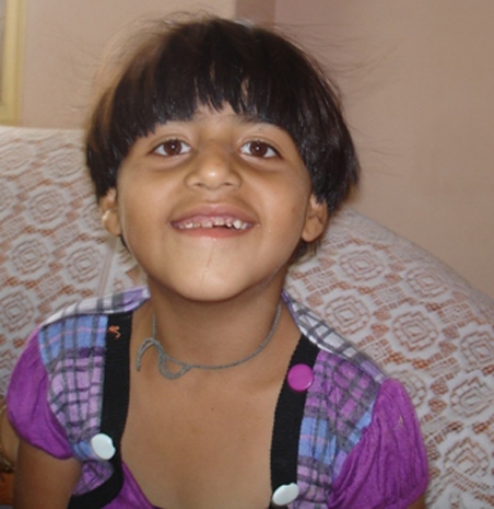
A 6-year and 5 month old girl reported with her parents to the Department of Pedodontics and Preventive Dentistry of Modern Dental College and Hospital, Indore, India. The patient’s history gathered from parents revealed that the patient is unable to understand, has severe learning disability and speak with the mental retardation since birth, on further interaction with parents, the chief complaint was decayed teeth in upper front region of mouth (figures 1 and 2). She was frequently laughing or smiling, and presented with a happy demeanour (figure 3). Psychological status of child represented a diagnosis based on gene mapping as a case of Angleman syndrome (figure 1). Further studying the literature and case reports, comparison of feature is made. A clinical suspicion of AS was raised at the age of 5 years when she presented with mental retardation and epilepsy; first attack at 1 year of age, second at 3 years of age, last at 6 years of age, absence of speech, extreme irritable nature, fascination with water, history revealed delayed milestone, walking at 3 years of age, normal birth history, the patient was alright till the age of 8 months until she had fever of unknown origin, which lasted for 4 months and had first episode of epileptic attack and unprovoked episodes of laughter, the patient is on antiepileptic drugs continuously under paediatrician supervision. At age of 5 years, the patient was referred to Hinduja Hospital, Mumbai for fluorescent in situ hybridisation (FISH) chromosomal analysis which revealed micro deletion on the maternally derived allele of 15q chromosome and confirmed the case of AS.
Figure 1.

Girl with Happy Puppet syndrome.
Figure 2.

Carious upper primary anterior teeth.
Figure 3.

Happy demeanour of patient.
Dental examination was done with physical restraining, includes smooth surface caries with upper incisors, occlusal caries with 75, 85, proximal caries with 64, heavy plaque and stain deposits indicate poor oral hygiene; all the primary teeth are present. As the patient was unable to understand and express herself, it was decided to treat the child under sedation or general anaesthesia. The patient was referred to her anaesthesiologist for an opinion. Consent was not given on the basis that any kind of pharmacological procedure is relatively contraindicated because of recent episode of epileptic attack. Then it was decided to perform the restorative procedure under physical restraining.
Investigations
-
▶
Electro encephalograms – (1) Persistent rhythmic 4–6/s activity reaching more than 200 mV, not associated with drowsiness, (2) Prolonged runs of rhythmic (triphasic) 2–3/s activity with an amplitude of 200–500 mV, maximal over the frontal regions and normally mixed with spikes or sharp waves, and (3) Spikes mixed with 3–4/s components, usually of more than 200 mV mainly posteriorly and facilitated by or only seen on eye closure.
-
▶
FISH analysis and by methylation analysis of the small nuclear ribonucleoprotien in polypeptide N (SNRPN) promoter which lies within a CpG island at 15q11-13.
Differential diagnosis
Prader Willi syndrome–In a normal individual, the maternal allele is expressed and the paternal allele is silenced. If the maternal contribution is lost or mutated, the result is AS. And when the paternal contribution is lost, by similar mechanisms, the result is Prader Willi syndrome.4
Treatment
In the present case of AS, glass ionomer cement restorations (figure 4) were done followed by prescription of fluoridated tooth paste, electronic and figure brush, anticipatory guidance to parents and preventive programs planned with regular follow-ups.
Figure 4.

Postoperative view.
Outcome and follow-up
Patient was kept under follow-up.
Discussion
The original patients reported by Angelman had severe learning disability, epileptic seizures, ataxia, absent speech and dysmorphic facial features with a prominent chin, deep set eyes, wide mouth with protruding tongue and microcephaly with a flat occiput. They were also hypo pigmented with fair hair and blue eyes. Some patients may not have the characteristic dysmorphic facies and have minimal ataxia. Several patients with AS are able to speak, although speech is always limited. The behavioural features seen in AS are perhaps the most consistent clinical feature.5
Oral features include flat occiput, occipital groove, protruding tongue, tongue thrusting, suck/swallowing disorders, feeding problems during infancy, prognathia, wide mouth, widely spaced teeth, frequent drooling excessive chewing/mouthing behaviours, strabismus hypo pigmented skin, light hair and eye colour (compared to family), seen only in deletion cases and fascination with water.3 4
Neurological features include slow gait, stiff legged and ataxic and the arms are raised and held flexed at the wrists and elbows. Hand-flapping is common when walking and if excited. Muscle tone is abnormal with truncal hypotonia and hypertonicity of the limbs and reflexes are brisk. Thoracic scoliosis occurs in approximately 10% of children but is a major problem in the majority of adult patients. In childhood, a variety of seizures can be observed, ranging from tonic-clonic seizures, atypical absences, complex partial, myoclonic, atonic and tonic seizures to status epilepticus. The epilepsy of AS is difficult to control with antiepileptic drugs, especially in childhood.6
The commonest genetic mechanism giving rise to AS, occurring in approximately 70–75% of patients, is an interstitial deletion of chromosome 15q11-13.7
A recent report by Cox et al has suggested that intracytoplasmic sperm injection may be a mechanism which interferes with establishment of the maternal imprint in an oocyte and might therefore predispose to AS. For parents of children with imprinting defects who wish to pursue prenatal testing during future pregnancies, Glenn et al showed that methylation analysis at the SNRPN locus of DNA extracted from chorionic villus or amniocytes will give a reliable result.8
No specific treatment is available for AS. Symptomatic management is based on physiotherapy, education, early stimulation, enrichment program, speech and communication therapy. Sleep disorders have been treated with sedatives or neuroleptics; melatonine.
In epilepsy, benzylpiperazine are drugs of choice, either in monotherapy or in association with valproic acid. Scoliosis can be treated with braces and surgical correction.4
Parents should emphasise the importance of good dental hygiene and supervise and assist with any tooth brushing carried out by patient. A curved (Collis curve) tooth brush may be effective. Regular visit to local dentist for preventive care is very important. Meticulous dental hygiene is imperative when any treatment which reduces saliva production is used.9
Learning points.
-
▶
Normal prenatal and birth history, normal head circumference at birth, no major birth defects.
-
▶
Delayed attainment of developmental milestones without loss of skills.
-
▶
Speech impairment, with minimal to no use of words; receptive language skills and non-verbal communication skills higher than expressive language skills.
-
▶
Movement or balance disorder, usually ataxia of gait and/or tremulous movement of the limbs.
-
▶
Behavioural uniqueness, including any combination of frequent laughter/smiling; apparent happy demeanour; excitability, often with hand-flapping movements; hypermotoric behaviour; short attention span.
Footnotes
Competing interests None.
Patient consent Obtained.
References
- 1.Kim BS, Yeo JS, Kim SO. Anesthesia of a dental patient with Angelman syndrome-a case report. Korean J Anesthesiol 2010;58:207–10 [DOI] [PMC free article] [PubMed] [Google Scholar]
- 2.Guerrini R, Carrozzo R, Rinaldi R, et al. Angelman syndrome: etiology, clinical features, diagnosis, and management of symptoms. Paediatr Drugs 2003;5:647–61 [DOI] [PubMed] [Google Scholar]
- 3.Williams CA, Angelman H, Clayton-Smith J, et al. Angelman syndrome: consensus for diagnostic criteria. Angelman Syndrome Foundation. Am J Med Genet 1995;56:237–8 [DOI] [PubMed] [Google Scholar]
- 4.Campos-Castello J. Angelman syndrome. Orphanet Encyclopedia 2004 [Google Scholar]
- 5.Fiumara A, Pittalà A, Cocuzza M, et al. Epilepsy in patients with Angelman syndrome. Ital J Pediatr 2010;36:31. [DOI] [PMC free article] [PubMed] [Google Scholar]
- 6.Clayton-Smith J, Laan L. Angelman syndrome: a review of the clinical and genetic aspects. J Med Genet 2003;40:87–95 [DOI] [PMC free article] [PubMed] [Google Scholar]
- 7.Williams CA, Driscoll DJ, Dagli AI. Clinical and genetic aspects of Angelman syndrome. Genet Med 2010;12:385–95 [DOI] [PubMed] [Google Scholar]
- 8.Laura AEM, Haeringen A, Oebele F, et al. Angelman syndrome, (AS, MIM 105830). Eur J Hum Genet 2009;17:1367–73 [DOI] [PMC free article] [PubMed] [Google Scholar]
- 9.Murakami C, Nahás Pires Corrêa MS, Nahás Pires Corrêa F, et al. Dental treatment of children with Angelman syndrome: a case report. Spec Care Dentist 2008;28:8–11 [DOI] [PubMed] [Google Scholar]


