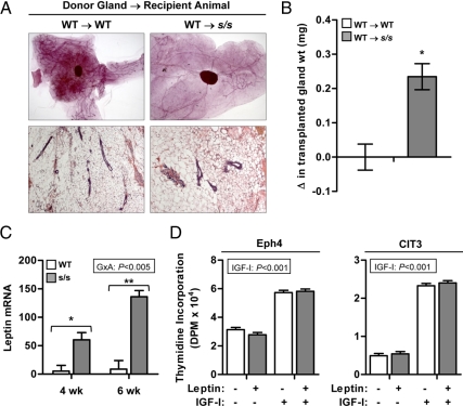
Fig. 5.
Effect of lack of leptin-dependent STAT3 signaling on the intramammary leptin system. A, WT or heterozygous (referred to as WT) mammary glands were transplanted into the subscapular region of female mice lacking leptin-dependent STAT3 signaling (s/s) or their WT counterparts (n = 5–7 transplantations per recipient category). Donor and recipient mice were 25 d of age at the time of transplantation. After 10 wk, the transplanted mammary glands were dissected and analyzed by whole-mount preparations and H&E staining. Representative glands are shown. B, The change (Δ) in the weight of the transplanted gland was calculated as the difference between final and initial weight. Each bar represents the mean ± se of five to seven transplantations. *, P < 0.05. C, MFPs were collected from female s/s mice or their WT counterparts at 4 and 6 wk of age. Total RNA was analyzed by real-time PCR for the mRNA abundance of leptin. The significant interaction between genotype × age (G×A) is shown. Pairwise comparisons performed within each age are significant at P < 0.05 (*) or P < 0.001 (**). Each bar represents the mean ± se of six to eight mice. D, Proliferation of Scp2 and Eph4 mouse mammary epithelial cells was measured using thymidine incorporation. Cells were incubated under basal conditions in the absence or presence of leptin (100 ng/ml) and IGF-I (100 ng/ml). The significant effect of IGF-I is indicated. Each bar represents the mean ± se of three wells.

