

Abstract
Bochdalek hernia, a type of congenital diaphragmatic hernia usually presents in the immediate neonatal period with respiratory distress. Presentation in an adult is rare and asymptomatic. We report one such case of Bochdalek hernia, in a 35 year old male, who presented only with mild chest discomfort on left side. Radiological investigations were suggestive of a huge left side Bochdalek hernia with stomach and intestines in the left thorax. This case emphasizes the rarity of presentation of Bochdalek hernia in adults and the importance of high clinical suspicion and the role of imaging and surgery in the accurate diagnosis of this abnormality.
Keywords: Congenital diaphragmatic hernia, Bochdalek hernia
Bochdalek hernia is the commonest congenital diaphragmatic hernia, which manifests primarily in neonates. It is a developmental defective disorder due to failure of fusion of pleuroperitoneal canal that closes usually by the 8th week of gestation. Its occurrence is rare in adults, quite often being asymptomatic, usually diagnosed as an incidental finding in abdominal CT scan [1]. Commonly identified on plain chest radiograph as an area of eventration, this defect on a CT scan is characteristically associated with protrusion of the omental or the retroperitoneal fat [2]. The ipsilateral lung is invariable hypoplastic with deviation of the mediastinum [3]. Until now, only around 100 cases of occult asymptomatic Bochdalek hernia in adults have been reported in the world literature [4].
A 35-year-old male presented with a mild discomfort on the left side of the chest for 1 week duration. He had no respiratory problems or gastrointestinal symptoms. Examination of the chest revealed mediastinal shift to the right and bowel sounds in the left hemithorax with reduced air entry. Chest radiograph revealed a raised left hemidiaphragm, with its outlines not well discernible, mediastinal shift to the right and presence of air filled bowel loops in the left thoracic cavity (Fig. 1). Contrast CT showed left-side diaphragmatic hernia with stomach and other viscera in the left thorax, and left lung was compressed and displaced (Fig. 2).
Fig. 1.
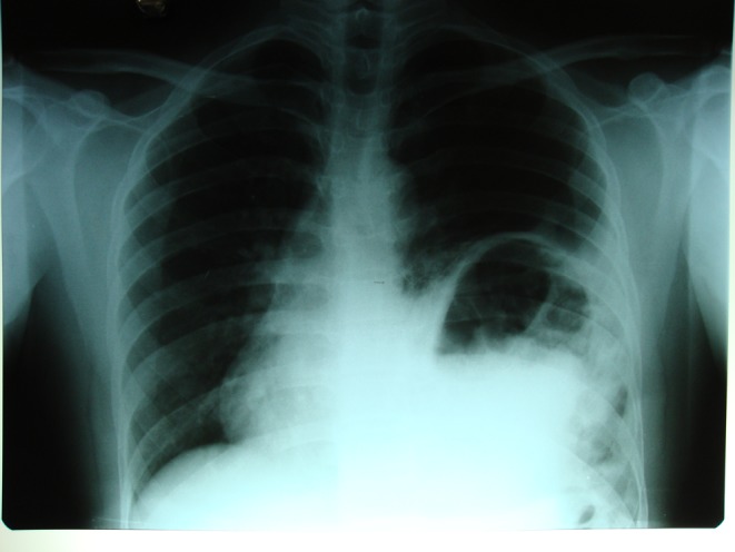
Chest X ray
Fig. 2.

CT scan
The patient was taken up for surgery, abdominal approach through a left subcostal incision showed a defect of about 12 × 8 cm in the left posterolateral aspect of the diaphragm with part of the stomach, small intestine, and colon herniating into the thorax suggestive of a Bochdalek hernia. The contents were reduced and there was no hernial sac. The defect was closed with interrupted nonabsorbable sutures, reinforced with a polypropylene mesh, and the thoracic cavity was drained by a single chest tube. The patient had an uneventful postoperative recovery. A repeat chest radiograph after 5 days of surgery revealed fully expanded left lung with no evidence of any herniation of bowel loops in the thoracic cavity.
References
- 1.Alam WCA, Chander BN. Adult Bochdalek hernia. MJAFI. 2005;61:284–286. doi: 10.1016/S0377-1237(05)80177-7. [DOI] [PMC free article] [PubMed] [Google Scholar]
- 2.Naidich DP, Webb WR, Muller NL, Vlahos I, Krinsky GA. Computed tomography and magnetic resonance of the thorax. 4. Philadelphia: Lippincott Williams & Wilkins; 2007. [Google Scholar]
- 3.Murfitt J. The normal chest: methods of investigation and differential diagnosis. In: Sutton D, editor. Textbook of radiology and imaging. 7. Edinburgh: Churchill Livingstone; 2003. p. 53. [Google Scholar]
- 4.Kumar A, Maheshwari V, Ramakrishnan TS, Sahu S. Caecal perforation with faecal peritonitis – unusual presentation of Bochdalek hernia in an adult: a case report and review of literature. World J Emerg Surg. 2009;4:16. doi: 10.1186/1749-7922-4-16. [DOI] [PMC free article] [PubMed] [Google Scholar]