Figure 6.
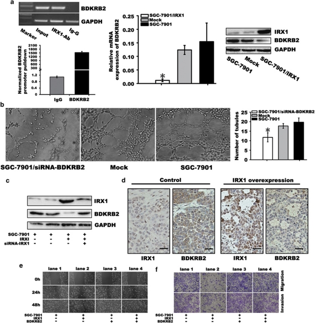
Analyzing the association between BDKRB2 with IRX1 and biological functions. (a) By chromatin immunoprecipitation, BDKRB2, a downstream target gene of IRX1 is identified (left). The mRNA (middle, *P<0.001) and protein levels (right) of BDKRB2 were significantly decreased by IRX1 overexpression. (b) After 48 h treatment by siRNA-BDKRB2, a significant reduction of endothelial tube formation was identified, compared with mock or controls by counting the tubular number (11.75±2.36 vs 17.75±1.25 or 19.75±2.21, *P<0.001). The data represent the mean±s.d. of three independent experiments. (c) Overexpression of IRX1 downregulated BDKRB2 expression. Whereas knocking down IRX1 by siRNA on IRX1-overexpressed cells can reverse the expressing level of BDKRB2. (d) Immunohistochemical staining for BDKRB2 and IRX1 on peritoneal tumor tissues of nude mice. A significant reduction of BDKRB2 expression in tumor tissue was observed at IRX1 overexpressing group, compared with control (the bar represents 20 μm). (e) In wound healing assay, overexpression of IRX1 alone suppressed the wound healing significantly (lane 2) and overexpression of BDKRB2 alone enhanced wound healing significantly (lane 3). Co-expression of BDKRB2 and IRX1 revealed the effect observed as control (lane 4). The mean wound distances of 7901/IRX1, 7901/BDKRB2 and 7901/IRX1/BDKRB2 were 322.88±22.45, 53.67±4.16 and 80.33±3.06, respectively, at 48 h (P<0.001). (f) The similar effects on cell migration and invasion were also observed by transwell assay. The amount of migrated cells in 7901/IRX1, 7901/siRNA-BDKRB2 and 7901/IRX1/BDKRB2 was 31±4.00 vs 165±8.54 and 123±3.05, respectively (P<0.001). The amount of invasive cells in 7901/IRX1, 7901/BDKRB2 and 7901/IRX1/BDKRB2 was 28.67±4.04 vs 168.33±3.06 and 134.33±2.52, respectively (P<0.001). The data represent the mean±s.d. of three independent experiments.

