Abstract
In order to augment responses to respiratory vaccines in swine, various adjuvants were intranasally coadministered with a foot-and-mouth disease virus (FMDV) antigen to pigs. Detoxified Escherichia coli enterotoxins LTK63 and LTR72 enhanced antigen-specific mucosal and systemic immunity, demonstrating their efficacy as adjuvants for nonreplicating antigens upon intranasal immunization in swine.
TEXT
Most pathogens initiate infections through contact with the mucosal surfaces of their hosts, thereby leading to colonization of the epithelium and/or invasion of tissues. Vaccination strategies that induce the production of mucosal immunity are desirable, as this approach can reduce the contact of pathogens with epithelial cells and possibly prevent dissemination to peripheral sites of the body. This is particularly important in the respiratory tracts of animals, as many of the potent innate barrier defenses present in the digestive system (e.g., low stomach pH, digestive enzymes, etc.) are not found in the airways. Given that the mucosae are inherently toleragenic (1), the use of adjuvants is essential to induce robust responses for nonreplicating mucosal vaccines (4). Unfortunately, the efficacy of many adjuvants in domestic animals (including pigs) has not been thoroughly tested, particularly for vaccines administered via the intranasal (i.n.) route.
The best-characterized mucosal adjuvants are the heat-labile enterotoxin (LT) produced by Escherichia coli and the closely related cholera toxin (CT) elaborated by Vibrio cholerae. Regrettably, these toxins are unsafe for use in humans (6), which dramatically limits their use in human and veterinary vaccines. To address this issue, detoxified mutants of LT, known as LTR72 and LTK63, were previously generated and have minimal residual toxicity (5, 8) but retain mucosal adjuvant activity in some animal species (10). Recently, several other adjuvant candidates have been developed in an attempt to elicit more potent mucosal immune responses. The interaction between bacterial CpG DNA and Toll-like receptor 9 (TLR 9) has made this motif a promising candidate for boosting both systemic and mucosal immunity. Another adjuvant that has been shown to augment mucosal and systemic immune responses is chitosan, a polysaccharide derived from the exoskeleton of crustaceans. More recently, chitosan nanoparticles harboring CpG motifs induced mucosal and systemic immune responses in mice (3, 11).
In this work, four mucosal adjuvants (wild-type CT [20 μg], LTK63 [100 μg], LTR72 [100 μg], and CpG/chitosan [200 μg] [manufacturer's recommended dosages shown in brackets]) were tested to assess their efficacy upon coadministration with a model foot-and-mouth disease virus (FMDV) antigenic peptide via the intranasal route. Groups of five female Yorkshire pigs (6 weeks old; John Correy, Scotland, CT) received the mucosal adjuvants admixed with a peptide derived from FMDV serotype O1-BFS VP1 G-H loop (which contains at least one important neutralizing epitope of the virus [9]). This peptide is referred to as the “TCA” peptide, representing a well-conserved VP4 T helper cell epitope, VP1 site C, and VP1 site A epitopes. The animals were intranasally inoculated with 100 μg of TCA peptide (reconstituted in 400 μl of water, the volume given to each animal) (Genemed Synthesis Inc., South San Francisco, CA) at weeks 1, 2, 3, and 5, with a parenteral boost given at week 4 with MPL+TDM+CWS RIBI adjuvant system (100 μg; administered intramuscularly [i.m.]; Sigma-Aldrich, St. Louis, MO). The parenteral boost was included to augment serum antibody and virus neutralization titers, since mucosal immunity alone may not adequately control viral infection/dissemination (7). Additionally, control groups included pigs sham immunized i.n. with chicken ovalbumin (OVA) plus RIBI adjuvant (Sigma-Aldrich, St. Louis, MO) at week 0, with a week 4 i.m. boost of OVA and RIBI or with TCA peptide plus RIBI adjuvant administered i.m. at weeks 0 and 4. Even though CT retains toxicity, it has been shown to be efficacious when administered i.n. to pigs, and we thus utilized this adjuvant as a “gold standard” for comparative purposes.
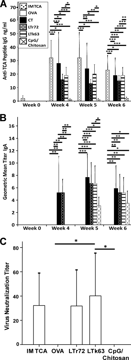
Serum samples were collected for assessment of anti-TCA peptide IgG responses as measured by an enzyme-linked immunosorbent assay (ELISA) (previously described [2]), and the antibody concentration was calculated by the method of Barrette et al. (2) (statistical significance for all analyses was determined using a one-way analysis of variance [ANOVA] with post hoc multiple pairwise comparisons by Fisher's least significant difference [LSD] test). A negligible IgG response was observed in all i.n. immunized groups prior to week 4; however, the i.m. TCA peptide-immunized group did produce a notable response at week 3 (data not shown). Animals i.n. inoculated with CT, LTR72, or LTK63 or i.m. inoculated with the TCA peptide produced statistically significant IgG antipeptide responses by week 4 relative to those inoculated with OVA or CpG/chitosan (Fig. 1A), and this tendency was observed throughout the remainder of the experiment (the responses for LTR72-inoculated animals were not significant at week 5 but the trend could still be observed [P = 0.1 compared to the values for OVA- or CpG/chitosan-inoculated animals], and the responses were again significant at week 6). Interestingly, the responses were not different among the groups producing a significant IgG response, with the exception of i.m. TCA peptide and LTR72. Thus, LTR72 and LTK63 appear to induce serum anti-TCA peptide IgG responses as vigorous as those of the group given CT as an adjuvant. It is important to note that the week 4 parenteral booster immunization of mucosally primed animals did not seem to augment immunity, indicating that i.n. immunization alone was primarily responsible for the observed antibody responses. In order to assess the functional capabilities of the humoral antibody responses, FMDV plaque reduction assays were conducted in order to determine virus neutralization titers (VNT). No VNT were observed at week 4 in any group; however, by the end of the study, significantly higher VNT were observed in animals inoculated with LTK63 than in animals inoculated with OVA or CpG/chitosan (Fig. 1C). Also, the results with animals inoculated with LTR72 tended to have a trend toward statistical significance compared to the animals inoculated with OVA (P = 0.07) or CpG/chitosan (P = 0.05), and a trend was also observed in animals inoculated i.m. with TCA peptide compared to animals inoculated with OVA (P = 0.07) or CpG/chitosan (P = 0.05). These data indicate that mucosal immunization was as effective as i.m. inoculation for generating serum IgG antibodies, but i.m. boosting may be necessary to induce serum virus neutralization activity.
Fig. 1.
Antigen-specific humoral immune responses. Anti-FMDV peptide serum IgG (A) and mucosal IgA (B) antibody responses, as measured by ELISA (one animal from the group given CT as an adjuvant died before completion of the study and was not included in any analyses). IgG concentrations were interpolated from a standard curve, and IgA concentrations are expressed as a geometric mean titer (endpoint titer determined to have an absorbance two times higher than background). IM TCA, i.m. TCA peptide. (C) Virus neutralizing titers as measured by plaque reduction assay (CT was excluded from the analysis, as statistics were impeded by the dramatically skewed data distribution of this group [only one animal in this group had measurable VNT, but it was the highest in the study at 1:130]). The bars represent the mean responses, and the error bars indicate 1 standard deviation from the mean. Horizontal black bars indicate pairwise comparisons between groups with significance denoted by asterisks as follows: *, P < 0.05; **, P < 0.01; ***, P < 0.001.
Anti-TCA peptide IgA responses (Fig. 1B) were measured by ELISA from nasal wash samples collected from pigs and are reported as the geometric mean titer. Secreted IgA antibodies were evident by week 4 in pigs receiving the CT and LTR72 adjuvants compared to all other groups, and animals inoculated with LTK63 had significant responses compared to those receiving i.m. TCA peptide or OVA at weeks 5 and 6. Animals vaccinated with CpG/chitosan exhibited a trend toward significance at week 5 compared to animals vaccinated with OVA or i.m. TCA peptide (P = 0.07 and P = 0.06, respectively) and attained statistical significance by week 6, although the responses were notably lower than those observed with the other mucosal adjuvants. No significant IgA responses were detected in animals inoculated with OVA or i.m. TCA peptide at any time points. Antigen-specific IgA antibody-secreting cells (exceeding 50 cells/million in an enzyme-linked immunospot [ELISpot] assay) were observed in the nasal mucosal tissues of some CT- and LTR72-inoculated pigs, indicating that at least some of the mucosal immune responses resulted from plasma cells located in the nasal mucosa (data not shown).
Taken together, the LT mutants used as adjuvants in this study were effective for inducing systemic and mucosal immunity to an antigenic FMDV peptide in the respiratory tracts of swine, whereas CpG/chitosan was less efficient. Even though the LT mutants showed efficacy in this experiment, modification of the approach is necessary to improve the time to immunity and to increase the magnitude of the mucosal response. Based on the delay of roughly 4 weeks, it appears that adjustments in dose, adjuvanticity, delivery, antigen, or frequency of administration may be needed to optimize mucosal immune responses in the respiratory tracts of pigs to pathogens such as FMDV.
Footnotes
Published ahead of print on 14 September 2011.
REFERENCES
- 1. Bailey M., Haverson K. 2006. The postnatal development of the mucosal immune system and mucosal tolerance in domestic animals. Vet. Res. 37:443–453 [DOI] [PubMed] [Google Scholar]
- 2. Barrette R. W., Urbonas J., Silbart L. K. 2006. Quantifying specific antibody concentrations by enzyme-linked immunosorbent assay using slope correction. Clin. Vaccine Immunol. 13:802–805 [DOI] [PMC free article] [PubMed] [Google Scholar]
- 3. Fischer S., et al. 2009. Concomitant delivery of a CTL-restricted peptide antigen and CpG ODN by PLGA microparticles induces cellular immune response. J. Drug Target. 17:652–661 [DOI] [PubMed] [Google Scholar]
- 4. Gerdts V., Mutwiri G. K., Tikoo S. K., Babiuk L. A. 2006. Mucosal delivery of vaccines in domestic animals. Vet. Res. 37:487–510 [DOI] [PubMed] [Google Scholar]
- 5. Giuliani M. M., et al. 1998. Mucosal adjuvanticity and immunogenicity of LTR72, a novel mutant of Escherichia coli heat-labile enterotoxin with partial knockout of ADP-ribosyltransferase activity. J. Exp. Med. 187:1123–1132 [DOI] [PMC free article] [PubMed] [Google Scholar]
- 6. Liang S., Hajishengallis G. 2010. Heat-labile enterotoxins as adjuvants or anti-inflammatory agents. Immunol. Invest. 39:449–467 [DOI] [PMC free article] [PubMed] [Google Scholar]
- 7. Nauwyncy H. J., Labarque G. G., Pensaert M. B. 1999. Efficacy of an intranasal immunization with gEgC and gEgI double-deletion mutants of Aujeszky's disease virus in maternally immune pigs and the effects of a successive intramuscular booster with commercial vaccines. J. Vet. Med. B 43:713–722 [DOI] [PubMed] [Google Scholar]
- 8. Partidos C. D., Pizza M., Rappuoli R., Steward M. W. 1996. The adjuvant effect of a non-toxic mutant of heat-labile enterotoxin of Escherichia coli for the induction of measles virus-specific CTL responses after intranasal co-immunization with a synthetic peptide. Immunology 89:483–487 [DOI] [PMC free article] [PubMed] [Google Scholar]
- 9. Rowlands D., et al. 1994. The structure of an immunodominant loop on foot and mouth disease virus, serotype O1, determined under reducing conditions. Arch. Virol. Suppl. 9:51–58 [DOI] [PubMed] [Google Scholar]
- 10. Singh M., Briones M., O'Hagan D. T. 2001. A novel bioadhesive intranasal delivery system for inactivated influenza vaccines. J. Control Release 70:267–276 [DOI] [PubMed] [Google Scholar]
- 11. Wu K. Y., et al. 2006. A novel chitosan CpG nanoparticle regulates cellular and humoral immunity of mice. Biomed. Environ. Sci. 19:87–95 [PubMed] [Google Scholar]


