Abstract
Acacia catechu, commonly known as catechu, cachou and black cutch is an important medicinal plant and an economically important forest tree. The methanolic extract of this plant was found to have antimicrobial activities against six species of pathogenic and non-pathogenic microorganisms: Bacillus subtilis, Staphylococcus aureus, Salmonella typhi, Escherichia coli, Pseudomonas aeruginosa and Candida albicans. The maximum zone of inhibition (20 mm) was found to be exhibited against S. aureus. For this organism the minimum bactericidal concentration (MBC) of the crude extract was 1,000 μg/ml. The extract was found to be equally effective against gram positive and gram negative bacteria. The antimicrobial activity of the extract was found to be decreased during purification. The chemical constituents of organic plant extracts were separated by thin layer chromatography (TLC) and the plant extracts were purified by column chromatography and were further identified by Gas chromatography–mass selection (GC–MS) analysis. The composition of A. catechu extract had shown major components of terpene i.e. camphor (76.40%) and phytol (27.56%) along with other terpenes in minor amounts which are related with their high antibacterial and antifungal properties.
Electronic supplementary material
The online version of this article (doi:10.1007/s12088-011-0061-1) contains supplementary material, which is available to authorized users.
Keywords: Acacia catechu, Minimum inhibitory concentration, Zone of inhibition, In vitro antimicrobial activity, TLC, GC/MS
Introduction
The development of bacterial resistance to presently available antibiotics has necessitated the search for new antibacterial agents [1]. The gram positive bacterium such as Staphylococcus aureus is mainly responsible for post operative wound infections, toxic shock syndrome, endocarditis, osteomyelitis and food poisoning [2]. Bacillus subtilis are rod shaped aerobic bacteria and are reported to have some pathogenic role [3]. The gram negative bacterium such as Escherichia coli is present in human intestine and causes lower urinary tract infection, coleocystis or septicemia [4]. Pseudomonas mainly causes urinary tract infection, wound or burn infection, chronic otitis media, septicemia etc. in humans [5]. Work has been done which aim at knowing the different antimicrobial and phytochemical constituents of medicinal plants and using them for the treatment of microbial infections as possible alternatives to chemically synthetic drugs to which many infectious microorganisms have become resistant [6, 7].
Acacia catechu commonly known as catechu is a medicinal plant used for varied purposes. The bark of this plant is strong antioxidant, astringent, anti-inflammatory, anti-bacterial and antifungal in nature. The extract of this plant is used to treat sore throats and diarrhoea, also useful in high blood pressure, dysentery, colitis, gastric problems, bronchial asthma, cough, leucorrhoea and leprosy. It is used as mouthwash for mouth, gum, sore throat, gingivitis, dental and oral infections. The heartwood is used to yield concentrated aqueous extract i.e. cutch which is astringent, cooling and digestive. It is useful in cough, ulcers, boils and eruptions of the skin. Decoction of the bark is given internally in case of leprosy. Acacia spp. produces gum exudates, traditionally called gum Arabic or gum Acacia, which are widely used in the food industry such as emulsifiers, adhesives, stabilizers and in chronic renal failure [8].
Thin layer chromatography (TLC) is the simplest and cheapest method of detecting plant constituents because the method is easy to run, reproducible and requires little equipment [9]. However for efficient separation of metabolites and sensitivity of detection, together with the capability of providing on-line structural information, high performance liquid chromatographic (HPLC), liquid chromatographic/mass spectroscopic (LC/MS), liquid chromatographic/nuclear magnetic resonance (LC/NMR) and gas chromatography/mass spectroscopic (GC/MS) techniques are preferred [10]. Determination of the minimum bactericidal concentration (MBC) value of a particular plant extract is also essential during evaluation of antimicrobial activity [11]. Therefore the present study deals with the determination of antimicrobial activity of the A. catechu leaf extract by disc diffusion, agar well diffusion and two fold serial dilution techniques, detection of bioactive compounds by TLC, purification by column chromatography and characterization by GC–MS analysis.
Materials and Methods
Collection of Samples
Acacia catechu leaves used in this study were collected during January, 2008 from Gir foundation, Gandinagar, Gujarat with the assistance of local plant keepers and authenticated by officials of Gir foundation. Leaves from the plant were washed under running tap water followed by sterilized distilled water. They were shade dried and then powdered with the help of sterilized pestle and mortar. The powder was further subjected for different extraction protocols.
Aqueous Extraction
The shade dried fine powdered 10 g leaves were boiled in 400 ml distilled water till one-fourth of the extract initially taken was left behind after evaporation. The solution was then filtered using muslin cloth. Filtrate was centrifuged at 5,000 rpm for 15 min. The supernatant was again filtered using Whatman filter No. 1 under strict aseptic conditions and then filtrate was collected in fresh sterilized bottles and stored at 4°C until further use.
Organic Solvent Extraction
Shade dried 10 g powder was thoroughly mixed with 100 ml organic solvent (viz., methanol, hexane and acetone). The mixture was placed at room temperature for 24 h on shaker with 150 rpm. Solution was filtered through muslin cloth and then re-filtered by passing through Whatman filter No. 1. The filtrate thus obtained was concentrated by complete evaporation of solvent under reduced pressure with rotatory vacuum evaporator to yield the pure extract. Stock solutions of crude extracts for each type of organic solvent were prepared by mixing well the appropriate amount of dried extracts with dimethyl sulphoxide (DMSO) to obtain a final concentration of 100 mg/ml that was used for evaluation of antibacterial and antifungal activities.
Microbial Cultures
Five strains of bacteria and yeast were used as test microorganisms. The bacterial strains included Gram-positive Staphylococcus aureus (ATCC 25923), Bacillus subtilis (ATCC 10707) and Gram-negative Escherichia coli (ATCC 25922), Salmonella typhimurium (ATCC 29213), Pseudomonas aeruginosa (ATCC 27853) and the Yeast Candida albicans (ATCC 10231). All microorganisms were clinical isolates, obtained from the Microbiology Laboratory at Department of Life Sciences, Gujarat University and MG Institute of Science. All the cultures were maintained and sub cultured on nutrient agar medium.
Antibacterial Assay
In vitro antibacterial activities of all aqueous and organic extracts of A. catechu was determined by standard agar well diffusion assay [12]. Petri dishes (100 mm) containing 25 ml of Mueller–Hinton Agar (Merck) seeded with 100 μl inoculum of bacterial strain (Inoculum size was adjusted so as to deliver a final inoculum of approximately 106 CFU/ml. Media was allowed to solidify and then individual Petri dishes were marked for the bacteria inoculated. Wells of 6 mm diameter were cut into solidified agar media with the help of sterilized cup-borer. 100 μl of each extract was poured in the respective wells and the plates were incubated at 37°C for overnight. DMSO and sterilized distilled water were used as negative control while tetracycline antibiotic (1 U strength) was used as positive control. The experiment was performed in triplicate under strict aseptic conditions and the antibacterial activity of each extract was expressed in terms of the mean of diameter of zone of inhibition (in mm) produced by the respective extract at the end of incubation period.
MIC for the Bacteria
The antibacterial activity of the extracts was examined by determining the MIC in accordance with Clinical and Laboratory Standard Institute (CLSI) methodology [13]. All tests were performed in Mueller–Hinton broth supplemented with DMSO at a final concentration of 10% (v/v) to enhance their solubility. The extracts were dissolved in MHB. Test strains were suspended in MHB to give a final density of 5 × 10⁵ cfu/ml and these were confirmed by viable counts. Dilutions ranging from 100 to 2,000 mg/ml of the extracts were prepared in tubes, including one growth control, MHB + DMSO 10% (v/v), and one sterility control MHB + DMSO 10% (v/v + test extracts). The MIC values initially recorded were from visual examinations as being the lowest concentration of the extracts with no bacterial growth. Plates were incubated under normal atmospheric conditions at 37°C for 24 h for bacteria.
MIC for the Yeast
The antifungal activity of the extracts was examined by determining the MIC in accordance with CLSI methodology [14] using Yeast Nitrogen Base Glucose (YNBG) medium supplemented with DMSO at a final concentration of 10% (v/v).The extracts were dissolved in YNBG medium. Yeast strains were cultured for 24–48 h at 35°C on SDA and then suspended in 4 ml of sterile distilled water by adjusting to 1 McFarland using a nephelometer to give a final inoculum concentration of 1.5 ± 1.0 × 10³ cfu/ml. Dilutions ranging from 100 to 2,000 mg/ml of the extracts were prepared in the tubes including one growth control, YNBG + DMSO 10% (v/v), and one sterility control YNBG 10% (v/v + test extracts). A 100 μl suspension of each of Candida strains in YNBG was added to individual tubes and incubated at 37°C for 48 h. The MICs of the extracts were defined as the lowest concentration that inhibited >80% of visible fungal growth. The final concentration of DMSO in the assays did not interfere with the bacterial and candidal proliferation.
Statistical Analyses of Data
The experiments were laid out according to randomised block design (for single factor experiments) or nested design (two factor experiments). In Each zone of inhibition experiments usually had three replicates and the mean of three replicates was noticed. The analysis of variance (ANOVA) appropriate for the design was carried out to detect significance of differences among the treatment means.
Separation of Phytochemicals by TLC, Purification by Column and Identification by GC–MS Analysis
The dried leaves (250 g) were powdered and exhaustly macerated with methanol for 1 week in a shaker. The collected extracts were filtered and evaporated under vacuum. The residues were dried and weighed.
TLC of Plant Extract
Thin layer chromatography technique was employed for the identification of a number of compounds present in the extracts. Due to its relatively simple and accurate advantages, TLC of the extracts were made by using different solvents and mixed solvents.
Column Chromatography Separation
Wet-process method was adopted to pack the column. Silica gel (0.063–0.200) acted as a sorbent and mixture of petroleum ether and ethyl acetate in ratio 80:20 and toluene and ethyl acetate in ratio 80:20 one by one were the eluents. At the beginning, silica gel was added to the eluent. It was stirred and slowly poured into the column until the bed layer was solid. The petcock was opened to decrease eluent level until its liquid surface and silica gel level were equal. Then the sample solution was poured slowly into the column along its wall. Finally, the eluent was added to the column, keeping the sorbent covered by the eluent. For both the mixtures five fractions were collected.
GC–MS Analysis
For GC–MS analysis, a Hewlett-Packard-5890-II (Global Medical Instrumentation) gas chromatograph, equipped with a flame-ionization detector (FID) and coupled with an electronic integrator was used.
Results and Discussion
The antimicrobial activity of herbal compounds extracted from plant parts depend upon the type of solvent used for extraction. Table 1 shows the anti-bacterial activity of zone of inhibition (in mm diameter) of leaf powder in aqueous and organic solvents (methanol, hexane, and acetone) of A. catechu.
Table 1.
Results of antimicrobial screening of aqueous and organic plant extracts determined by agar diffusion method
| Plant type | Extraction type | Zone of inhibition (in mm diameter) | |||||
|---|---|---|---|---|---|---|---|
| S. aureus | B. subtilis | E. coli | S. typhi | P. aeruginosa | C. albicans | ||
| Acacia catechu | Methanol | 20 ± 0.24 | 18 ± 0.22 | 19 ± 0.22 | 20 ± 0.20 | 18 ± 0.20 | 20 ± 0.20 |
| Hexane | 11 ± 0.14 | 11 ± 0.14 | 11 ± 0.15 | 10 ± 0.15 | 10 ± 0.14 | 12 ± 0.16 | |
| Acetone | 10 ± 0.12 | 12 ± 0.16 | 11 ± 0.14 | 10 ± 0.14 | 10 ± 0.12 | 11 ± 0.14 | |
| Aqueous | 10 ± 0.16 | 11 ± 0.14 | 10 ± 0.16 | 10 ± 0.14 | 10 ± 0.14 | ND | |
| Positive control | Tetracycline | 24 ± 0.26 | 22 ± 0.24 | 20 ± 0.24 | 22 ± 0.20 | 20 ± 0.20 | 22 ± 0.24 |
| Negative control | DMSO | NA | NA | NA | NA | NA | NA |
Zone of inhibition (in mm diameter) including the diameter of well (6 mm) in agar well diffusion assay
Assay was performed in triplicate and results are the mean of three values. In each well, the sample size was 100 μl. Tetracycline: 1 U strength
Microorganisms: S. aureus, Staphylococcus aureus; B. subtilis, Bacillus subtilis; E. coli, Escherichia coli; S. typhi, Salmonella typhi; P. aeruginosa, Pseudomonas aeruginosa; C. albicans, Candida albicans
ND Not detected, NA no activity
Data indicated that organic extract of leaves exhibited better anti-bacterial activities than those of aqueous extract. Aqueous extract was found almost ineffective in inhibition of bacterial strains producing zone of inhibition ≤9 mm. Organic extract of leaves shown to have the zone of inhibition ranging from 18 to 24 mm. This extract was equally inhibitory for Gram-positive bacteria as well as Gram-negative bacteria. Among all the bacterial strains tested, S. aureus was found most susceptible with maximum inhibition by methanolic extract producing zone of inhibition >20 mm (Table 1). Extracts prepared in organic solvents consistently displayed better antimicrobial activity than that of aqueous extracts. Furthermore, extracts prepared in methanol was observed most inhibitory (diameter of zone ranging from 18 to 22 mm) followed by those prepared in hexane and acetone, respectively. Tetracycline (a positive control) showed inhibition diameters ranging from ~17 to 19 mm against all test microorganisms. Control experiments using sterile distilled water and DMSO (negative control) showed no inhibition of any bacteria.
Methanol leaves extract were subjected for minimum inhibitory concentration (MIC) against susceptible bacterial species indicated in Table 2. The methanolic extracts were found to be stronger inhibitor than other extracts. MIC of this extract was 1,000 μg/ml against S. aureus and B. subtilis while it was 1,500, 700 and ≤2,000 μg/ml for Gram-negative E. coli, S. typhimurium, and P. aeruginosa respectively. And for C. albicans MIC value was 1,500 μg/ml. All antimicrobial activity occurred in a concentration dependent manner as suggested by MIC determination; however, the efficacy of extracts was less than to that of standard antibiotic, tetracycline.
Table 2.
Results of minimum inhibitory concentration (MIC) of methanol extract of test plant
| Plant type | Extraction type | Minimum inhibitory concentration (μg/ml) | |||||
|---|---|---|---|---|---|---|---|
| S. aureus | B. subtilis | E. coli | S. typhi | P. aeruginosa | C. albicans | ||
| Acacia catechu | Methanol | 1,000 | 1,000 | 1,500 | 700 | 2,000 | 1,500 |
| Positive control | Broth + TO | G | G | G | G | G | G |
| Negative control | Broth + CE | NG | NG | NG | NG | NG | NG |
TO Test organisms, CE crude extract, G growth, NG no growth
Microorganisms: S. aureus, Staphylococcus aureus; B. subtilis, Bacillus subtilis; E. coli, Escherichia coli; S. typhi, Salmonella typhi; P. aeruginosa, Pseudomonas aeruginosa; C. albicans, Candida albicans
The present study revealed that the use of organic solvents in the preparation of plant extracts provides more consistent antibacterial activity as compared to aqueous extracts. This observation clearly indicates that the polarity of antimicrobial compounds make them more readily extracted by organic solvents, and using organic solvents does not negatively affect their bioactivity against bacterial and fungal species. This finding was also supported by several workers [15, 16] that water extracts of plants do not have much activity against bacteria. Eloff [17] reported that methanol and ethanol were the most effective solvent for plant extraction than n-hexane and water. Present study indicated that under the experimental conditions methanol is ideal solvent to extract antimicrobial compounds found in leaves. In general, the cell walls of Gram-negative organisms, which are more complex than Gram-positive ones, act as a diffusional barrier and making them less susceptible to the antibacterial agents than the Gram-positive bacteria. In spite of this permeability difference, however methanol extracts of A. catechu leaves have still exerted some degrees of inhibition against Gram-negative organisms as well. It can be concluded that polar protic solvents may be beneficial for augmenting antibacterial activity of both Gram negative and Gram positive microorganisms. It is worthwhile to pursue further research by taking polar protic solvent with different dielectric constant.
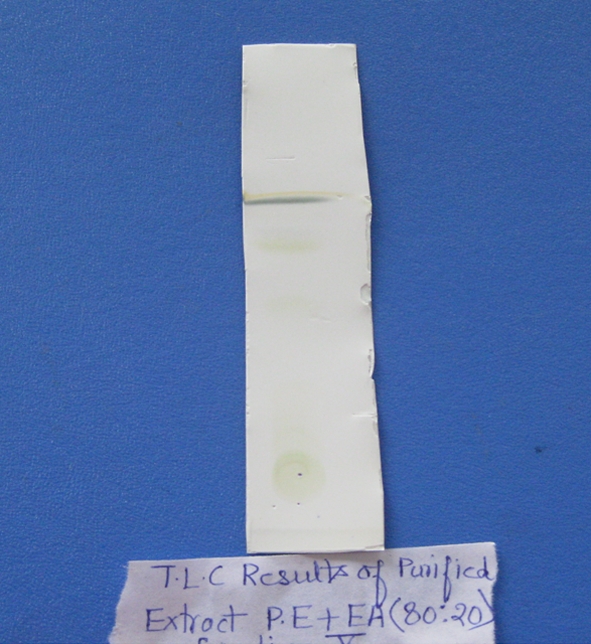
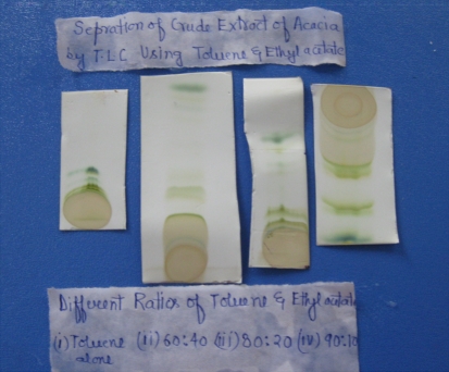
Acacia catechu extract prepared in methanol was subjected to separation by column chromatography with the combinations of solvents i.e. (a) petroleum ether + ethyl acetate, (b) toluene + ethyl acetate and TLC purification of these fractions indicated the Rf values in (a) and (b) solvents which were 0.65 and 0.83 respectively (Figs. 1, 2; Tables 3, 4).
Fig. 1.
TLC results of crude extract of A. catechu in different ratios of toluene and ethyl acetate
Fig. 2.

TLC results of purified extract of A. catechu in ratio 80:20 of toluene and ethyl acetate
Table 3.
TLC results of extract in different individual organic solvents
| Elution of organic solvent | TLC analysis of A. catechu leaves extract |
|---|---|
| Methanol | Serious tailing phenomena |
| Chloroform | No distinct phenomena |
| Toluene | One rather blur spot |
| Petroleum ether | Blur spot |
| Ethyl acetate | No distinct phenomena |
Table 4.
TLC results of extract in different combination of organic solvents
| Organic solvents | Ratio | TLC analysis of A. catechu leaves extract |
|---|---|---|
| PE + EA | 40:60 | Not so clear spot |
| PE + EA | 50:50 | Not so clear spot, on the edge |
| PE + EA | 70:30 | One spot |
| PE + EA | 80:20 | One single clear spot |
| PE + EA | 60:40 | ND |
| EA + DW | 50:50 | ND |
| BE + EA | 50:50 | ND |
| BE + EA | 70:30 | ND |
| TO + EA | 40:60 | One spot |
| TO + EA | 60:40 | One single clear spot |
| TO + EA | 80:20 | One spot |
| TO + EA | 70:30 | ND |
| PE + CH | 60:40 | ND |
| PE + CH | 40:60 | ND |
PE Petroleum ether, EA ethyl acetate, BE benzene, TO toluene, CH chloroform, DW distilled water, ND not detected
These purified fractions were subjected to GC/MS analysis. The results were summarized in Table 5 and the chromatogram was shown in Fig. S1 in supplementary material. The composition of A. catechu extract had shown major components of terpene i.e. camphor (76.40%) and phytol (27.56%) along with other terpenes in minor amounts which are related with their high antibacterial and antifungal properties.
Table 5.
Compounds present in the Acacia catechu leaf extract
| Compound | Retention time (min) | Amount (%) |
|---|---|---|
| Camphor | 5.382 | 76.40 |
| Phytol | 20.276 | 27.56 |
| Hexadecane | 19.440 | 5.82 |
| Vitamin E acetate | 40.906 | 11.85 |
Further the present studies showed that fractions obtained after purification have low antimicrobial activity for which the antimicrobial activity was determined by the agar well diffusion method. The zone of inhibition was found to have decreased by extract purification. It was noted during purification that some compounds are lost, which may explain the lesser antimicrobial properties observed with purified extracts than with crude extract, or may suggest the presence of more than one active compound.
The present study evaluated the antibacterial and antifungal potential of leaf extract of A. catechu so the use of this plant as an anti-infective agent in the ayurvedic medicine has been justified. The antibacterial and antifungal potential of leaf extracts of A. catechu is due to its high terpene contents. Terpenes are biologically active molecules and are considered to be part of plants defence systems and as such have been included in a large group of protective molecules found in plants named as ‘phytoprotectants’ [18]. The extract of A. catechu is effective both in Gram-positive and Gram-negative bacteria as well as against fungus C. albicans. The crude extract was more effective than its purified form as shown in Figs. S2, S3, S4 and S5 in supplementary material. Consequently the extracts obtained by the plant can potentially be used in the treatment of infectious diseases caused by microorganisms that showed emergence of resistance to currently available antibiotics.
Electronic supplementary material
Below is the link to the electronic supplementary material.
Acknowledgments
We are grateful to Prof. YK Agarwal for the corrections and valuable comments on the manuscript. We are also thankful to Central Salt and Marine Research Institute (CSMRI), Bhavnagar for carrying out GC–MS analysis.
References
- 1.Parekh J, Karathia N, Chanda S. Screening of some traditionally used medicinal plants for potential antibacterial activity. Indian J Pharm Sci. 2006;68(6):832–834. doi: 10.4103/0250-474X.31031. [DOI] [Google Scholar]
- 2.Chambers HF, Deleo FR. Waves of resistance: Staphylococcus aureus in the antibiotic era. Nat Rev Microbiol. 2009;7:629–641. doi: 10.1038/nrmicro2200. [DOI] [PMC free article] [PubMed] [Google Scholar]
- 3.Tenover F. Mechanisms of antimicrobial resistance in bacteria. Am J Med. 2006;9:3–10. doi: 10.1016/j.amjmed.2006.03.011. [DOI] [PubMed] [Google Scholar]
- 4.Zhou L, Lin Q, Li B, Li N, Zhang S. Expression and purification of the antimicrobial peptide CM4 in Escherichia coli. Biotechnol Lett. 2009;31:437–441. doi: 10.1007/s10529-008-9893-0. [DOI] [PubMed] [Google Scholar]
- 5.Livermore DM. Multiple mechanisms of antimicrobial resistance in Pseudomonas aeruginosa: our worst nightmare. Clin Infect Dis. 2002;34:634–640. doi: 10.1086/338782. [DOI] [PubMed] [Google Scholar]
- 6.Arunkumar S, Muthuselvam M. Analysis of phytochemical constituents and antimicrobial activities of Aloe vera L. against clinical pathogens. World J Agric Sci. 2009;5(5):572–576. [Google Scholar]
- 7.Samie A, Obi CL, Bessong PO, Namrita L. Activity profiles of fourteen selected medicinal plants from Rural Venda communities in South Africa against fifteen clinical bacterial species. Afr J Biotechnol. 2005;4(12):1443–1451. [Google Scholar]
- 8.Shen D, Wu Q, Wang M, Yang Y, Lavoie EJ, Simon JE. Determination of the predominant catechins in Acacia catechu by liquid chromatography/electrospray ionization-mass spectrometry. J Agric Food Chem. 2006;54(9):3219–3224. doi: 10.1021/jf0531499. [DOI] [PubMed] [Google Scholar]
- 9.Berezkin VG. A new approach to the determination of relative retention in thin-layer liquid chromatography. J Anal Chem. 2007;62(4):366–368. doi: 10.1134/S1061934807040120. [DOI] [Google Scholar]
- 10.Vardar-Unlu G, Silici S, Unlu M. Composition and in vitro antimicrobial activity of Populus buds and poplar-type propolis. World J Microbiol Biotechnol. 2008;24:1011–1017. doi: 10.1007/s11274-007-9566-5. [DOI] [Google Scholar]
- 11.Andrews JM. Determination of minimum inhibitory concentrations. J Antimicrob Chemother. 2001;48(suppl 1):5–16. doi: 10.1093/jac/48.suppl_1.5. [DOI] [PubMed] [Google Scholar]
- 12.Nostro A, Germano MP, D’Angelo V, Marino A, Cannatelli MA. Extraction methods and bioautography for evaluation of medicinal plant antimicrobial activity. Lett Appl Microbiol. 2000;30(1):379–384. doi: 10.1046/j.1472-765x.2000.00731.x. [DOI] [PubMed] [Google Scholar]
- 13.Clinical and Laboratory Standards Institute (CLSI) (2005) Methods for dilution antimicrobial susceptibility tests for bacteria that grow aerobically—sixth edition: approved standard M7-A6. CLSI, Wayne
- 14.Clinical and Laboratory Standards Institute (CLSI) (2008) Reference method for broth dilution antifungal susceptibility testing of yeasts: approved standard, 3rd edn. M27-A3. CLSI, Wayne
- 15.Gajera HP, Patel SV, Golakiya BA. Antioxidant properties of some therapeutically active medicinal plants—an overview. JMAPS. 2005;27:91–100. [Google Scholar]
- 16.Al-Bayati FA, AL-Mola HF. Antibacterial and antifungal activities of different parts of Tribulus terrestris L. growing in Iraq. J Zhejiang Univ Sci B. 2008;9(2):154–159. doi: 10.1631/jzus.B0720251. [DOI] [PMC free article] [PubMed] [Google Scholar]
- 17.Eloff JN. Which extractant should be used for the screening and isolation of antimicrobial compounds from plants? J Ethanopharmacol. 1998;60:1–8. doi: 10.1016/S0378-8741(97)00123-2. [DOI] [PubMed] [Google Scholar]
- 18.Morrissey JP, Osbourn AE. Fungal resistance to plant antibiotics as a mechanism of pathogenesis. Microbiol Mol Biol Rev. 1999;63:708–724. doi: 10.1128/mmbr.63.3.708-724.1999. [DOI] [PMC free article] [PubMed] [Google Scholar]
Associated Data
This section collects any data citations, data availability statements, or supplementary materials included in this article.
Supplementary Materials
Below is the link to the electronic supplementary material.


