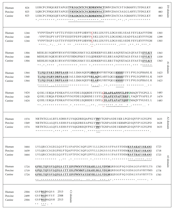
Figure 2.
Alignments of Human, Porcine, and Canine VWF. (a) Region of D′/D3 domain highlighting the F.VIII : C binding region (underlined). (b) Region of A1 domain showing the conserved 1272–1458 disulfide bonds (C1272 and C1458) in red and GP1b binding sites in black underlined. The D1472H human polymorphism site is highlighted in green. (c) Region of A2 domain showing the ADAMTS13 cleavage site (underlined). (d) Region of A3 domain highlighting Collagen binding site. (e) Region in C1 domain indicating the RGD binding site of integrin αIIb/βIII. The VWF amino acid sequences were analyzed by Clustal W multiple sequence alignment program [16] and derived from NCBI Accession NP_000543 (Human), AF052036 and AY004876 (Porcine), and NP_001002932 (Canine).

