

Diverticulitis of the appendix is an uncommon cause of right-lower-quadrant pain. Whether it presents symptomatically or is an incidental finding at surgery or barium enema, understanding its clinical behaviour is important for its management. We report here a case of appendiceal diverticulitis including what we believe to be the first reported case of this condition diagnosed preoperatively by CT imaging.
Case report
A large 38-year-old man came to the emergency department with right-lower-quadrant pain. His leukocyte count was elevated; his medical history was significant only for sleep apnea. Because his size made him difficult to assess, he was scanned with CT (Fig. 1). The scan confirmed appendicitis, but also showed suspected diverticular disease of the appendix. After laparoscopic appendectomy, the pathology report confirmed appendiceal diverticulosis complicated by diverticulitis, peridiverticular abscess and rupture (Fig. 2).
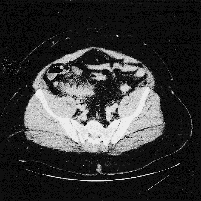
FIG. 1. Computed tomographic scan of abdomen showing diverticular disease of appendix.
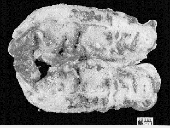
FIG. 2. Appendiceal diverticulosis.
Discussion
Appendiceal diverticulitis was first described in 1893 by Kelynack.1 As with all intestinal diverticula, appendiceal diverticula can be classified as congenital or acquired. The congenital form, which is very rare, is a true diverticulum; the more prevalent acquired form is a false diverticulum on the mesenteric border of the appendix. The incidence of diverticula found in appendectomy specimens ranges from 0.004% to 2.1%; from routine autopsies, 0.20% to 0.6%.2
Patients with appendiceal diverticulitis present at an average age of 38 years.3 It is more common in men and in patients with cystic fibrosis.2 Curiously, the patient in this case was also a 38-year-old man with a respiratory condition.
Appendiceal diverticulitis has been classified into 4 subtypes.1 Type 1 occurs when a normal-appearing appendix is found with an acutely inflamed diverticulum. Type 2 involves an acutely inflamed diverticulum with surrounding appendicitis, as seen in this case. Type 3 is conventional appendicitis with an incidental uninvolved diverticulum. Type 4 is an incidental appendiceal diverticulum with no evidence of appendicitis or diverticulitis.
Acute diverticulitis of the appendix has been shown to be more than 4 times as likely as acute appendicitis to perforate (occurring in 66% of cases), increasing mortality 30-fold compared with simple appendicitis.1 In addition, several cases of pseudomyxoma peritonei have been reported from appendiceal diverticuli.4 This may make removal of an appendix with diverticuli appropriate when found incidentally during surgery or upon barium enema.
A literature review by the authors found no case reference to appendiceal diverticulitis identifiable on CT. The scans in our case report allowed consideration of appendiceal diverticulitis before the operation. Although this is of interest, it should not change the management, as the appendix still should be removed.
Competing interests: None declared.
Correspondence to: Dr. Martin Friedlich, Ottawa Hospital—General Campus, 501 Smyth Rd. room 2003, Ottawa ON K1H 8L6; fax 613 739-6646; mfriedlich@ottawahospital.on.ca
Accepted for publication Aug. 12, 2003
References
- 1.Phillips BJ, Perry CW. Appendiceal diverticulitis. Mayo Clin Proc 1999;74:890-2. [DOI] [PubMed]
- 2.Place RJ, Simmang CL, Huber PJ. Appendiceal diverticulitis. South Med J 2000;93:76-9. [PubMed]
- 3.Lock JH, Wheeler WE. Diverticular disease of the appendix. South Med J 1990;83:350. [DOI] [PubMed]
- 4.Lin CH, Chen TC. Diverticulosis of the appendix with diverticulitis: case report. Chang Gung Med J 2000;23:711-4. [PubMed]