Abstract
The formation of insoluble amyloid fibrils is associated with an array of devastating human diseases. Dialysis-related amyloidosis (DRA) is a severe complication of hemodialysis that results in the progressive destruction of the bones and joints. Elevated concentrations of β2-microglobulin (β2m) in the serum of subjects on hemodialysis promote the formation of amyloid fibrils in the osteoarticular tissues, but the cellular basis for the destruction of these tissues in DRA is poorly understood. In this study we performed a systematic analysis of the interaction of monomeric and fibrillar β2m with primary human cells of the types present in the synovial joints of subjects with DRA. Building upon observations that macrophages infiltrate β2m amyloid deposits in vivo we demonstrate that monocytes, the precursors of macrophages, cannot degrade β2m fibrils, and that both monomeric β2m and fibrillar β2m are cytotoxic to these cells. β2m fibrils also impair the formation of bone resorbing osteoclasts from monocytes and reduce the viability of osteoblasts, the cell type that produces bone. As a consequence, we predict that β2m amyloid will disrupt the remodelling of the bone, which is critical for the maintenance of this tissue. Moreover, we show that β2m fibrils reduce the viability of chondrocytes, rationalizing the loss of cartilage in DRA. Together, our observations demonstrate that β2m cytotoxicity has multiple cellular targets in the osteoarticular tissues and is likely to be a key factor in the bone and joint destruction characteristic of DRA.
Introduction
The formation of insoluble amyloid fibrils is associated with a spectrum of devastating human diseases, many of which are characterised by tissue destruction [1]. One such disorder is dialysis-related amyloidosis (DRA), a debilitating complication of long-term hemodialysis [2], [3]. The culprit protein of DRA is β2-microglubulin (β2m) [4], [5], the non-covalent light-chain of cell surface major histocompatibility complex (MHC) class I molecules [6]. Upon dissociation from MHC molecules, β2m is normally removed from the bloodstream by the kidneys [6]. The normal serum concentration of β2m is 1–3 µg/ml but, in end stage renal disease, neither the kidney nor the dialysis membrane can efficiently remove β2m from the circulation and serum levels increase by up to 60 fold and can exceed 100 µg/ml [2], [3], [7]. At these elevated concentrations, β2m forms amyloid fibrils in the osteoarticular tissues resulting in arthropathy, cartilage destruction, bone cysts leading to pathologic fractures, carpal tunnel syndrome and spondyloarthropathy [2]–[6], [8], [9].
β2m fibril formation is promoted by collagen and the glycosaminoglycans (GAGs) chondroitin-sulfate and heparin [10]–[14]. Collagen and chondroitin-sulfate are abundant in osteoarticular tissues, rationalizing the deposition of β2m amyloid at these sites, whereas heparin is an anti-coagulant used in hemodialysis. β2m lacking the N-terminal six residues of the mature protein (ΔN6β2m) constitutes ≤30% of the β2m in ex vivo DRA amyloid [15]. Unlike full-length wild type (WT) β2m, ΔN6β2m forms amyloid fibrils de novo at neutral pH in vitro, a process that is enhanced by GAGs [12], [13], [16]–[18]. ΔN6β2m fibrils also cross-seed fibril formation with WT β2m and can convert WT β2m into an amyloidogenic state, suggesting that ΔN6β2m could initiate fibril formation in vivo [17], [18].
In contrast to the increasing knowledge of the mechanism of β2m fibril assembly in vitro [6], [19], how β2m amyloid causes skeletal morbidity in DRA is poorly understood. Macrophages infiltrate β2m amyloid deposits and have been implicated in the development of symptomatic DRA [20]–[22]. Cells of the monocyte/macrophage lineage are precursors of osteoclasts, which may be responsible for osteolytic lesions in DRA [23]–[27]. Indeed, human β2m has been shown to promote osteoclastogenesis from murine macrophages [28], but whether monomeric or fibrillar β2m species promote human osteoclast formation is not known. Since β2m fibrils generated in vitro are cytotoxic to some cultured cell lines [29], β2m amyloid formation could also cause bone and joint destruction via cytotoxicity to cell types that are responsible for the maintenance of the osteoarticular tissues.
Herein we perform a systematic comparison of the effects of monomeric and fibrillar β2m on primary human cells relevant to the pathology of DRA (monocytes, osteoblasts and chondrocytes). Our data do not support a role for monomeric or fibrillar β2m in osteoclast formation by human monocytes, but instead show that β2m monomer and fibril preparations are cytotoxic to monocytes, osteoblasts and chondrocytes and hence β2m cytotoxicity may be an important factor in the bone and joint destruction associated with DRA.
Results
Generation of β2m fibrils in vitro at a physiologically relevant pH
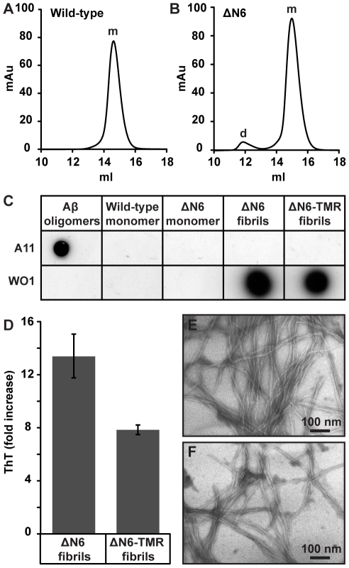
Recombinant human WT β2m and ΔN6β2m were expressed in E. coli and purified [12], [30], [31]. Size-exclusion chromatography (SEC) confirmed that both proteins are predominantly monomeric at pH 7.3 (≥98% of WT β2m; ∼96% of ΔN6β2m), although ∼4% of ΔN6β2m was dimeric (Fig. 1A and B). Neither protein was recognized by the A11 or WO1 antibodies (Fig. 1C), which bind generic epitopes in cytotoxic oligomers and amyloid fibrils, respectively [32], [33].
Figure 1. Analysis of monomeric and fibrillar β2-microglobulin (β2m).
Analytical size exclusion chromatography traces of 1 mg/ml full-length wild type (WT) β2m (a) and ΔN6β2m (ΔN6) (b), obtained in phosphate buffered saline pH 7.3 at 25°C. Monomer (m) and dimer (d) peaks are indicated. (c) Immunoblots of monomeric and fibrillar β2m probed with A11 anti-oligomer and WO1 anti-fibrillar antibodies. Aβ1–40 oligomers were used as a positive control for the A11 antibody. (d) Thioflavin-T fluorescence of unlabelled and TMR-labelled β2m fibrils, error bars represent one standard deviation over 60 readings. Negative stain transmission electron microscopy images of unlabelled (e) and TMR labelled (f) β2m fibrils.
To generate β2m fibrils at a physiologically relevant pH, and within an experimentally tractable timescale, fibrils generated at pH 2 from WT β2m were fragmented to produce seeds that were then extended with ΔN6β2m monomer at pH 7.3 in the presence of heparin [12]. The resultant fibrils were not recognized by the A11 antibody, were recognized by the WO1 antibody (Fig. 1C); bound the amyloid specific dye thioflavin-T (Fig. 1D) and exhibited a long straight morphology when visualized by negative stain transmission electron microscopy (TEM) (Fig. 1E) reminiscent of ex vivo β2m amyloid [34]–[36]. Moreover, we have shown previously with Fourier transform infrared spectroscopy that fibrils elongated from WT β2m seeds at pH 7 closely resemble ex vivo β2m amyloid [36]. Fibrils formed from seeds elongated with a 1∶9 mixture of ΔN6β2m and 5-(and -6)-carboxytetramethylrhodamine-SE (TMR) labelled ΔN6β2m also had the characteristic features of amyloid (Fig. 1C, D and F).
Human monocytes internalize, but do not degrade, β2m fibrils
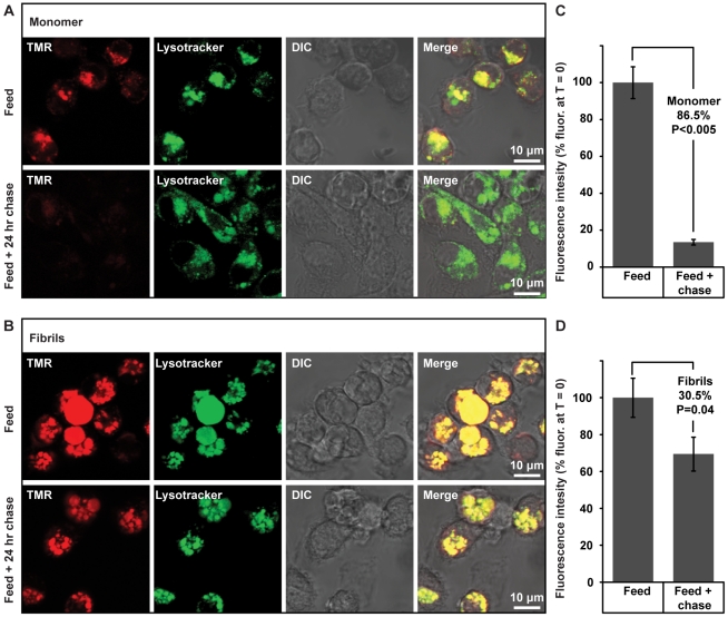
The macrophages that infiltrate β2m amyloid plaques are present around blood vessels consistent with these cells being derived from peripheral blood monocytes [20], [37]. We, therefore, investigated whether these cells could play a protective role in DRA via the degradation of β2m fibrils. CD14+ monocytes isolated from peripheral blood were cultured in the presence of macrophage colony stimulating factor (M-CSF), whereupon they become adherent and develop a macrophage-like morphology. These cells were incubated with either TMR-labelled monomeric ΔN6β2m or TMR-labelled β2m fibrils. Prior to imaging by confocal microscopy the cells were incubated with LysoTracker green, a fluorescent probe that accumulates in acidic intracellular compartments. After 24 hours, both monomer and fibrils were internalized and co-localized with Lysotracker green in punctate structures (Fig. 2A and B). The smaller puncta stained by Lysotracker green are characteristic of lysosomes, whereas the larger punctate structures that are most prominent in cells incubated with fibrils may correspond to acidified phagosomes. Indeed, this would be consistent with electron microscopy studies of ex vivo DRA amyloid, in which macrophages were shown to phagocytose β2m amyloid [38].
Figure 2. Internalization and degradation of monomeric and fibrillar β2-microglobulin (β2m) by primary human monocytes.
Monocytes were incubated with 10 µg/ml of ΔN6β2m monomer (90% unlabelled ΔN6β2m, 10% TMR-labelled ΔN6β2m) (a) or TMR-labelled fibrils formed from a 9∶1 mixture of unlabelled: labelled ΔN6β2m (red, feed) for 24 hours (b). Lysosomes were visualized by staining with LysoTracker green prior to confocal microscopy. Differential interferance contrast (DIC) images are also shown. Cells were then washed to remove non-cell associated protein and chased for a further 24 hours before imaging again (feed +24 hour chase). The mean intracellular fluorescence of 20 cells was quantified for cells incubated with monomeric β2m (c) and fibrillar β2m (d) before and after the 24 hour chase. Values are normalized to 100% for the intracellular fluoresence of cells before the chase and the percentage reduction after 24 hour chase is indicated. Error bars represent the SEM; P values differing from the PBS control were calculated by the Student's t-test and are stated.
After incubation for a further 24 hours in the absence of extracellular TMR-labelled monomeric ΔN6β2m or fibrils, a pronounced decrease in intracellular fluorescence was observed for cells that had internalized monomeric protein (Fig. 2A and C). In contrast only a modest reduction in intracellular fluorescence occurred in cells that had internalized β2m fibrils (Fig. 2B and D). Thus monocytes internalize and traffic both β2m monomer and fibrils to acidic vesicles, but only the monomeric protein is degraded efficiently. The inability of these phagocytic cells to degrade β2m fibrils would allow amyloid deposition to proceed unchecked in the synovial joints of subjects with DRA.
Monomeric and fibrillar β2m do not promote osteoclast formation
In addition to forming macrophages, monocytes can differentiate into bone resorbing osteoclasts [24]–[26]. Since primary human monocytes interact with both monomeric and fibrillar β2m (Fig. 2), and human β2m monomer has been shown to stimulate osteoclast formation from murine macrophages [28], we next examined whether either monomeric or fibrillar β2m can promote osteoclast formation from human monocytes. Osteoclasts are defined as large multinucleate cells, which express high levels of tartrate-resistant acid phosphatase (TRAP) and can resorb artificial bone substrates [24]–[26]; hence each of these characteristics were used to monitor osteoclast formation.
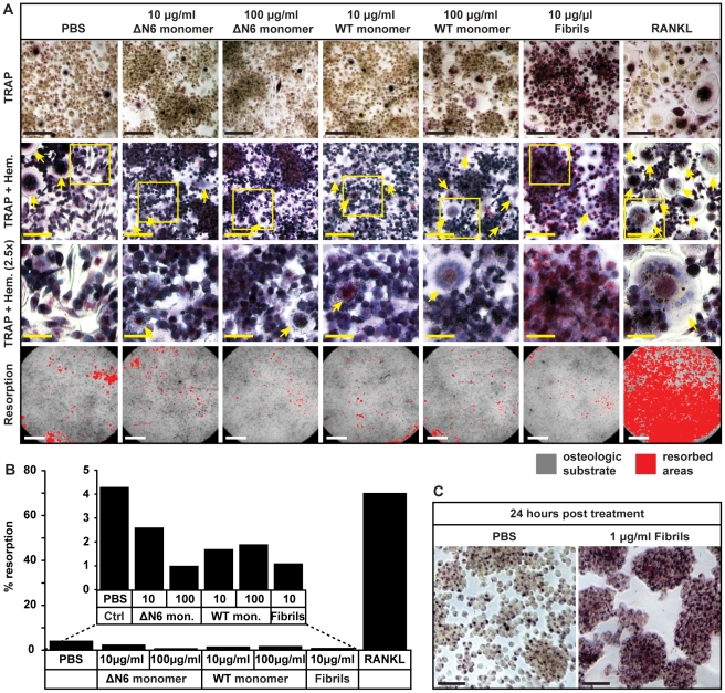
In a control experiment, monocytes were incubated with receptor activator of nuclear factor-Κ ligand (RANKL), a cytokine that promotes osteoclast formation [24]–[26]. After 14 days there were abundant large (≥20 µm) cells, which stained positive (dark pink) for TRAP in centrally located intracellular vesicles (Fig. 3A). Many of the cells were multinucleate, as visualised by hematoxylin staining, and they were able to resorb the majority (∼70%) of an artificial bone substrate (Fig. 3A and B). This confirmed that under the culture conditions used in this study osteoclasts form in 14 days. In contrast, when cells were incubated with buffer, ΔN6β2m monomer or WT β2m monomer, at concentrations (10 and 100 µg/ml) that span those observed in the serum of subjects with DRA [2], [3], [7], only a small number of multinucleate, TRAP positive cells were formed, and little of the artificial bone substrate was resorbed (<5%) (Fig. 3A and B) indicating that neither WT β2m nor ΔN6β2m monomeric proteins promote the differentiation of human monocytes into osteoclasts.
Figure 3. Effects of monomeric or fibrillar β2-microglobulin (β2m) on primary human monocyte differentiation into osteoclasts.
Monocytes were incubated with the indicated substances and macrophage colony-stimulating factor (M-CSF) for 14 days. RANKL induces osteoclast formation and was used as a positive control. (a) Top row (scale bar 50 µm): cells stained for the osteoclast marker, TRAP. 2nd row (scale bar 50 µm): TRAP stained cells were counter stained with hematoxylin to visualize cell morphology (nuclei, blue), arrows indicate large multinucleate cells. Regions within the yellow boxes are magnified in the 3rd row (scale bar 20 µm). Bottom row (scale bar 1 mm): resorption of an osteologic substrate by the cells treated as indicated. Resorbed areas are coloured red. (b) Resorption was quantified and results from a representative experiment are shown. The inset shows an expansion of the first six columns. (c) Monocytes were incubated with either PBS control or β2m fibrils for 24 hours, and then stained for TRAP (scale bar 50 µm).
When monocytes were incubated with 10 µg/ml β2m fibrils very few large multinucleate cells were formed and there was limited resorption of the artificial bone substrate (Fig. 3A and B), thus demonstrating that β2m fibrils do not promote osteoclast formation. Interestingly many cells did, however exhibit a dramatic increase in TRAP positive vesicles, giving them a dark pink appearance after staining for the enzyme (Fig. 3A). Elevated TRAP expression has also been observed in pulmonary alveolar macrophages that have phagocytosed microbes and other particles [39]; therefore TRAP could be increased in monocytes due to the internalization of β2m fibrils. Indeed after only 24 hours incubation with β2m fibrils, the level of TRAP expression was increased markedly (Fig. 3C).
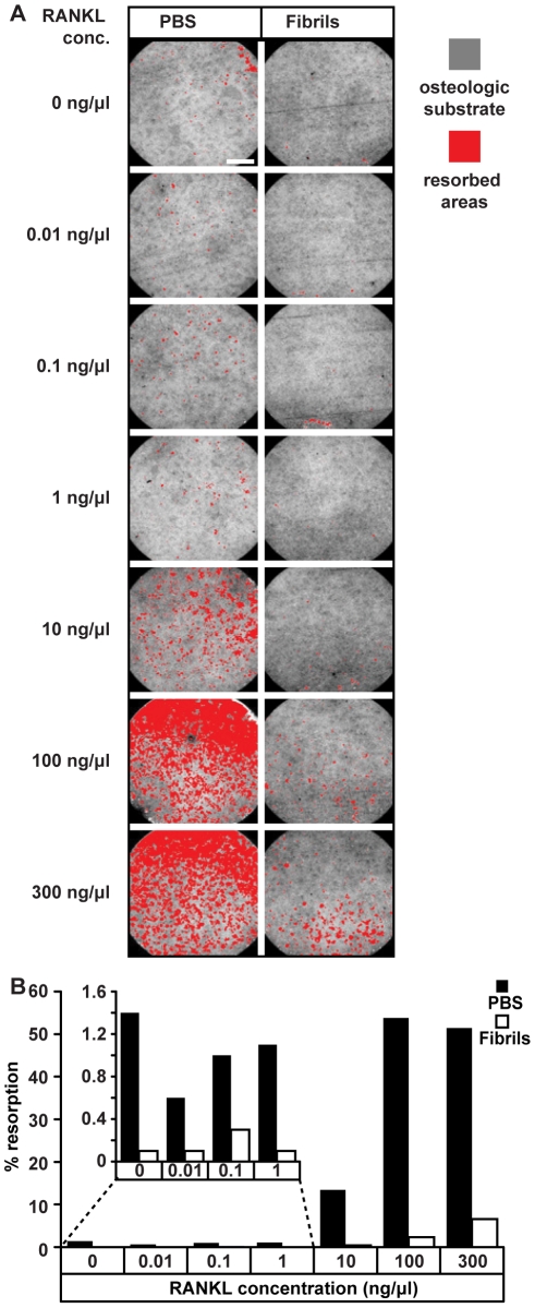
An alternative possibility for the observed increase in TRAP expression was that β2m fibrils may provide a partial osteoclastogenic signal that synergizes with other signalling molecules to promote the formation of mature osteoclasts. In vivo osteoclastogenesis is promoted by RANKL, a cytokine expressed on the surface of osteoblasts [24]–[26], this molecule was absent from all but the positive control cultures. We, therefore, examined whether β2m fibrils act in synergy with RANKL to promote osteoclast formation. Monocytes were incubated with increasing concentrations of RANKL in either the presence or absence of 10 µg/ml β2m fibrils and osteoclast function assessed by measuring resoprtion of the artificial bone substrate. As expected when monocytes were incubated with RANKL alone the number of osteoclasts formed and resorption of the artificial bone substrate increased as the concentration of this cytokine was increased (Fig. 4A and B). However, when cells were incubated with RANKL and β2m fibrils, a reduction in resorption of the artificial bone substrate was observed relative to cells incubated with RANKL alone (Fig. 4A and B). Thus although β2m fibrils increase TRAP expression they do not promote osteoclast formation, instead β2m fibrils reduce RANKL-dependent osteoclast formation.
Figure 4. Effects of β2-microglobulin (β2m) fibrils on RANKL-dependent osteoclast formation.
Primary human monocytes were incubated with M-CSF and the indicated concentrations of RANKL in the presence of PBS or 10 µg/ml β2m-fibrils for 14 days and their ability to resorb an osteologic substrate was then assessed. (a) Images of the osteologic substrate following the removal of cells. Resorbed regions are highlighted in red, scale bar 1 mm. (b) Resorption was quantified and results from a representative experiment are shown. The inset shows an expansion of the first four columns.
β2m fibrils are cytotoxic to monocytes, osteoblasts and chondrocytes
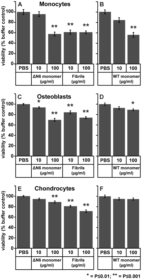
Previously we have shown that β2m fibrils are cytotoxic to the murine macrophage cell line RAW 264.7 [29]. We therefore investigated whether β2m fibrils are also cytotoxic to primary human monocytes. Using the 3-(4,5-dimethyl-2-thiazolyl)-2,5-diphenyl-2H-tetrazolium bromide (MTT) assay, a pronounced and significant reduction in cell viability was observed for monocytes incubated with 10 µg/ml β2m fibrils (Fig. 5A and S1A). By contrast to the fibrils, 10 µg/ml of ΔN6β2m and WT β2m had a minimal effect on the viability of monocytes (Fig. 5A, 5B, S1A and S1B). Thus fibrils are more cytotoxic than monomeric β2m, which is consistent with our previous study of the effect of monomeric and fibrillar β2m on the viability of cell lines [29]. β2m fibril cytotoxicity may contribute to the failure of monocytes to degrade β2m fibrils and could also be responsible for the observed reduction in ostoeclast formation, as monocyte death would prevent these cells from fusing together to form osteoclasts. However, at 100 µg/ml, β2m fibrils reduced cell viability by ∼40%, as did the monomeric forms of ΔN6β2m and WT β2m (Fig. 5A and S1A), demonstrating that monomeric forms of β2m are also cytotoxic at this concentration.
Figure 5. Effects of monomeric and fibrillar β2-microglobulin (β2m) on primary human monocytes, osteoblasts and chondrocyte viability.
Cells were incubated with the indicated substances for 24 hours and then their ability to reduce MTT, as a measure of viability, was measured. (a) Effects of β2m fibrils and ΔN6β2m (ΔN6) monomer on monocyte viability; the mean from three donors is plotted. (b) Effects of full-length wild type (WT) β2m monomer on monocyte viability; the mean from three donors is plotted. (c) The effects of β2m fibrils and ΔN6β2m monomer on osteoblast viability; the mean from four donors is plotted. (d) The effect of WT β2m monomer on osteoblast viability; the mean from one donor is plotted. (e) The effect of β2m fibrils and ΔN6β2m monomer on chondrocyte viability; the mean from four donors is plotted. (f) Effects of WT β2m monomer on chondrocyte viability, the mean from one donor is plotted. The data from individual donors for monocytes, osteoblasts and chondrocytes are presented in Figures S1, S2B and S3B, respectively. Error bars represent the SEM; P values differing from the PBS control were calculated by the Student's t-test, * = P≤0.01; ** = P≤0.001.
Next we investigated whether β2m could contribute to bone and joint destruction in DRA by reducing the viability of osteoblasts and chondrocytes, the cells that produce bone and cartilage respectively. The association of TMR-labelled ΔN6β2m monomer or β2m fibrils with primary human osteoblasts and chondrocytes was visualized by confocal microscopy. After 24 hours, monomeric TMR-labelled ΔN6β2m was present in punctate intracellular structures in osteoblasts and chondrocytes, consistent with internalisation of the protein (Fig. S2A and S3A). Similar internalization was seen when β2m fibrils were incubated with chondrocytes, while fibrils added to osteoblasts remained in close proximity to the cell surface as large aggregates, with some limited cell-associated punctate fluorescence indicative of cell association and internalization (Fig. S2A and S3A). Our observations suggest both cell types internalize monomeric β2m, although chondrocytes internalize β2m-fibrils more readily than osteoblasts.
The effect of β2m monomer and fibrils on osteoblast and chondrocyte viability was then assessed; both 10 and 100 µg/ml β2m fibrils caused a significant reduction in the viability of osteoblasts and chondrocytes (Fig. 5C, 5E, S2B and S3B). ΔN6β2m monomer also reduced cell viability of both cell types (Fig. 5C, 5E, S2B and S3B). In contrast to monomeric ΔN6β2m, 100 µg/ml of WT β2m monomer had no significant effect on chondrocyte viability and caused only a limited decrease in viability of osteoblasts, at the highest concentration used (Fig. 5D and F). Taken together, these data suggest that monomeric ΔN6β2m and fibrillar β2m cytotoxicity to chondrocytes and osteoblasts are important factors in the pathology of DRA. Chondrocyte cell death would be predicted to decrease formation of new cartilage, while the reduction in osteoclast formation in combination with the death of osteoblasts may impair bone turnover in subjects with DRA.
Discussion
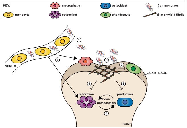
Despite the identification of β2m as the culprit protein of DRA in 1985 [4], [5], how β2m amyloid causes bone and joint destruction in DRA remains poorly understood. In order to elucidate the cellular basis for the pathological consequences in DRA we performed a systematic analysis of the effects of β2m fibrils and monomeric β2m on cell types that are present in the synovial joints of subjects with DRA. Our data suggest that the bone and joint destruction characteristic of DRA is a multifactorial process in which β2m amyloid fibrils and elevated concentrations of monomeric β2m are cytotoxic to multiple cellular targets (Fig. 6).
Figure 6. Model for a role of monomeric and fibrillar β2-microglobulin (β2m) cytotoxicity in dialysis related amyloidosis.
(1) Elevated levels of circulating β2m monomer form amyloid deposits in osteoarticular tissues. (2) Circulating monocytes are able to differentiate into both macrophages and osteoclasts. (3) Monocyte-derived macrophages infiltrate β2m amyloid deposits, but the resistance of the amyloid fold to proteolysis in combination with β2m amyloid cytotoxicity, prevents the clearance of the amyloid deposits. (4) β2m amyloid induced monocyte/macrophage cell death also contributes to reduced osteoclast formation, causing a decrease in the resorption of old bone. (5) Osteoblast cell death in the presence of β2m monomer and/or fibrils causes decreased bone formation. (6) Decreased bone resorption and formation contribute to pathological alterations to normal bone homeostasis. (7) Chondrocyte cell death in the presence of β2m monomer and/or fibrils causes decreased cartilage formation, which could allow gradual loss of this tissue and exposure of bone to cytotoxic β2m monomer and fibrils.
We demonstrate that primary human monocytes internalize, but do not degrade, β2m fibrils. This is consistent with the resistance of the β2m amyloid fold to lysosomal proteolysis [40], [41], but could be exacerbated by the cytotoxicity of β2m fibrils to the monocytic precursors of macrophages. The death of macrophages that infiltrate amyloid deposits in vivo [20]–[22], [37], would be predicted to prevent the clearance of β2m monomer and fibrils allowing the continued accumulation of β2m amyloid in the synovial joints.
Cells of the monocytic lineage are also the precursors of osteoclasts, which in combination with osteoblasts are responsible for bone remodelling [26]. Monomeric β2m did not promote osteoclast formation from human monocytes. This contrasts with a recent study in which monomeric β2m was shown to promote osteoclast formation from murine macrophages [28], and implies that β2m has differential effects on human and mouse cells. Similarly, β2m fibrils did not promote the formation of bone resorbing osteoclasts despite increased expression of TRAP, as there was no increase in the number of multinucleate cells nor did β2m fibrils promote the resorption of the artificial bone substrate. Once considered just as a marker of osteoclasts, it has become clear that TRAP has a number of important roles in both the skeleton and the immune system, and is expressed by a number of different cell types including macrophages [39], [42]. Thus the increase in TRAP expression in monocytes may be the result of the activation of these cells by β2m fibrils. Moreover, we found that β2m fibrils impaired RANKL-induced osteoclast formation. This was presumably as a consequence of cytotoxicity to monocytes, as fibril-induced cell death of the monocytic precursors would be predicted to prevent these cells from fusing together to form multinucleate osteoclasts. This reduction in osteoclast formation, in combination with the reduction in viability of osteoblasts caused by β2m fibrils and to a lesser extent monomeric β2m, suggests that bone remodelling will be perturbed in DRA. The formation of bone cysts in DRA may, therefore, be the consequence of a reduction in the turnover of damaged bone, rather than an increase in osteoclast activity. Moreover, the effect of β2m fibrils on osteoclast formation and the viability of osteoblasts may exacerbate the reduction in bone turnover that is a common feature of renal osteodystrophy associated with chronic kidney disease [43]. β2m fibrils were also shown to be cytotoxic to chondrocytes, rationalizing why the formation of β2m amyloid deposits in cartilage results in the loss of this tissue [2], [3], [8], [9], [22], which could in turn expose the bone surface to the cytotoxic effects of amyloid deposits.
The mechanism by which β2m fibrils kill cells is unknown, although other amyloid fibrils have been reported to cause both necrotic and apoptopic cell death [44]–[46]. β2m fibrils disrupt purified lipid membranes [29] suggesting a role for membrane disruption in fibril-associated cytotoxicity. Since β2m fibrils were visualized in association with monocytes, osteoblasts and chondrocytes in proximity to the plasma membrane and within intracellular punctate structures, β2m fibrils could kill these cells by disrupting the plasma membrane and/or intracellular membranes. No immunoreactivity was detected by the antibody A11, which recognises amyloid oligomers [32]. This suggests that cytotoxicity associated with β2m fibril preparation is mediated by the interaction of β2m fibrils with the cells.
Incubation of cells with monomeric WT β2m or ΔN6β2m also resulted in reduced viability, with the ΔN6β2m monomer exhibiting the greatest degree of cytotoxicity. While, no oligomers were detected in either protein preparation as determined by SEC and by immunoblotting with the oligomer specific antibody A11, WT β2m can form cytotoxic non-fibrillar aggregates [47], [48], whereas the cytotoxicity of ΔN6β2m could reflect the increased propensity of this truncation to aggregate or its different conformational properties [12], [13], [15]–[18]. Although elevated concentrations of WT β2m are characteristic of DRA [2], [3], [7], the relevance of non-fibrillar ΔN6β2m cytotoxicity is unclear, as ΔN6β2m has only been detected in amyloid deposits and is not found in the serum of hemodialysis patients [15], [49]. ΔN6β2m, however, has an increased affinity for collagen [50], which could lead to localized high concentrations of the soluble protein and resultant cytotoxicity.
The study outlined herein examined how monomeric and fibrillar β2m affect primary human cells present in the osteoarticular tissues of subjects with DRA. We have shown previously that β2m fibrils are cytotoxic to the murine macrophage RAW 264.7 and human neuroblastoma SH-SY5Y cell lines [29], demonstrating that β2m fibril-associated cytotoxicity is not unique to monocytes, osteoblasts and chondrocytes. Furthermore, since fibrillar preparations of other amyloidogenic precursors are also cytotoxic [29], [44]–[46], [51]–[54], it is likely that these would reduce viability of monocytes, osteoblasts and chondrocytes. Amyloid deposits in the osteoarticular tissues can be present in other amyloid diseases [55], [56] and may cause the death of osteoblasts and chondrocytes. Macrophages and macrophage-like cells such as microglia are found in association with other forms of amyloid [57], [58]. As such fibril-associated cytotoxicity to macrophages and macrophage-like cells could be a factor in other amyloid diseases.
In conclusion, our observations show that cytotoxicity of monomeric β2m and β2m fibrils to cell types in the joints of subjects with DRA could be important factors in the development of the osteoarticular pathology of this disorder (Fig. 6).
Materials and Methods
Ethics statement
The procedure for the isolation of monocytes from peripheral blood of healthy volunteers was approved by the Faculty of Biological Sciences Research Ethics Committee at the University of Leeds. Written informed consent was given by each donor.
Expression, purification and labelling of β2m
Recombinant WT β2m and ΔN6β2m were expressed in E. coli and purified as described previously [12], [30], [31]. Purification was confirmed by electrospray ionization mass spectrometry and size exclusion chromatography, as previously described [17]. Endotoxins were removed using an “EndoTrap Red” column (Hyglos, Bernreid, Germany) and Limulus amoebocyte lysate assays performed by Lonza (Verviers, Belgium) showed endotoxin concentrations to be ≤7.35 EU/mg protein. Purified proteins were labelled with TMR (Molecular Probes, Eugene, OR, USA) using methods described previously [40]. Further details are provided in File S1.
Generation of β2m fibrils at physiological pH
Fibrils formed at pH 2 [29] were fragmented by freezing in liquid nitrogen and thawing at 37°C three times to generate fibril seeds. 5% (v/v) seeds were elongated with 1 mg/ml ΔN6β2m in Dulbecco's Phosphate Buffered Saline (PBS) pH 7.3, at 37°C, with agitation at 200 rpm in the presence of 0.1 mg/ml low molecular weight heparin (Sigma-Aldrich H8537), 100 U/ml penicillin and 100 µg/ml streptomycin, for 21 days. To generate labelled fibrils, seeds were extended with 0.9 mg/ml unlabelled ΔN6β2m and 0.1 mg/ml TMR-ΔN6β2m. Fibril concentrations are stated as monomer equivalents.
TEM, thioflavin-T binding and immunoblotting
Negative stain TEM and thioflavin-T assays were performed as previously described [17], [30]. Immunoblotting with A11 and WO1 antibodies was performed as described previously [32], [33], [59], and in detail in File S1.
Isolation and culture of primary human monocytes
Mononuclear cells were isolated from peripheral blood by centrifugation over Lymphoprep (Axis-Shield, Oslo, Norway), monocytes were enriched using the MACS monocyte II kit (Miltenyi Biotec, Bergisch Gladbach, Germany) and enrichment confirmed by flow cytometric analysis of cells stained with anti-CD14-Phycoerythrin (Miltenyi Biotec) on a FACSCalibur instrument (BD Biosciences). Monocytes were cultured at 500 000 cells/cm2, in αMEM supplemented with 10% (v/v) fetal bovine serum (Biosera, Ringmer, UK), 2 mM L-glutamine, 100 U/ml penicillin, 100 µg/ml streptomycin and 750 ng/ml M-CSF (Peprotech, Rocky Hill, NJ, USA), at 37°C, 5% CO2.
Culture of primary human chondrocytes and osteoblasts
Primary human osteoblasts, chondrocytes, cell specific media and an optimized cell detach kit were obtained from PromoCell (Heidelberg, Germany). Cells were cultured at 10 000–20 000 cells/cm2, in cell specific media supplemented with 100 U/ml penicillin and 100 µg/ml streptomycin at 37°C, 5% CO2. Cells were passaged a maximum of three times.
MTT assay of cell viability
Cells were plated in 96 well plates (Corning Inc., Corning, NY, USA) and cultured overnight. MTT assays were then performed as described previously [29] and in File S1. For each cell type MTT assays were performed using the same culture conditions, enabling each experiment to be internally controlled to exclude any effects of the growth medium on the reduction of MTT.
Imaging of β2m internalization and degradation
Uptake of 10 µg/ml ΔN6β2m monomer (90% unlabelled, 10% TMR labelled) or TMR-ΔN6β2m fibrils over 24 hours, and degradation over a further 24 hours was analyzed using methods described previously [40] and in detail in File S1.
Osteoclast formation and characterization
Primary human monocytes were cultured in 96 well plates (Corning) or on 16 well osteologic slides (BD Biosciences). Osteoclast formation was assessed after 14 days by staining for TRAP and counterstaining with hematoxylin, using a leukocyte acid phosphatase (TRAP) kit (Sigma-Aldrich 387A) and manufacturer's protocol, and by quantifying the percentage resorption of the osteologic substrate. Further details are provided in File S1.
Supporting Information
Effects of monomeric and fibrillar β2-microglobulin (β2m) on monocyte viability. Primary human monocytes from three independent donors were treated with the indicated substances for 24 hours and then their ability to reduce MTT, as a measure of viability, was measured for each donor. (a) Effects of ΔN6β2m (ΔN6) monomer and β2m fibrils on viability. (b) Effect of full-length wild type (WT) β2m monomer on viability. Error bars represent the SEM; P values differing from the PBS control were calculated by the Student's t-test, * = P≤0.01; ** = P≤0.001.
(TIFF)
Monomeric and fibrillar β2-microglobulin (β2m) association with osteoblasts and their effects on cell viability. (a) Primary human osteoblasts were incubated with either TMR-labelled ΔN6β2m monomer or TMR-labelled β2m fibrils (red) for 24 hours and then imaged by confocal microscopy. Differential interferance contrast (DIC) images are also shown. The regions within the white boxes are magnified in the right hand panels. (b) Cells from four independent donors were treated with ΔN6β2m monomer or β2m fibrils for 24 hours and then their ability to reduce MTT, as a measure of viability, was measured for each donor. Error bars represent the SEM; P values differing from the PBS control were calculated by the Student's t-test, * = P≤0.01; ** = P≤0.001.
(TIFF)
Monomeric and fibrillar β2-microglobulin (β2m) association with chondrocytes and their effects on cell viability. (a) Primary human chondrocytes were treated with either TMR-labelled ΔN6β2m monomer or TMR-labelled fibrils (red) for 24 hours and then imaged by confocal microscopy. Differential interferance contrast (DIC) images are also shown. The regions within the white boxes are magnified in the in the right hand panels. (b) Chondrocytes from four independent donors were treated with ΔN6β2m monomer or β2m fibrils for 24 hours and then their ability to reduce MTT, as a measure of viability, was measured for each donor. Error bars represent the SEM; P values differing from the PBS control were calculated by the Student's t-test, * = P≤0.01; ** = P≤0.001.
(TIFF)
Acknowledgments
We thank Gareth Howell for advice on microscopy, James Ault for performing mass spectrometry, Charles Glabe (University of California, Irvine) for the A11 antibody and Aβ1–40 oligomers, Ron Wetzel (University of Pittsburg) for the WO1 antibody, and Andrew Hellewell, Toral Jakhria and Aneika Leney for WT β2m samples. We thank members of the Hewitt and Radford labs for helpful discussions.
Footnotes
Competing Interests: The authors have declared that no competing interests exist.
Funding: This work was funded by the Yorkshire Kidney Research Fund (http://www.ykrf.co.uk). The funder had no role in study design, data collection and analysis, decision to publish, or preparation of the manuscript.
References
- 1.Chiti F, Dobson CM. Protein misfolding, functional amyloid, and human disease. Annu Rev Biochem. 2006;75:333–366. doi: 10.1146/annurev.biochem.75.101304.123901. [DOI] [PubMed] [Google Scholar]
- 2.Floege J, Ehlerding G. β2-microglobulin-associated amyloidosis. Nephron. 1996;72:9–26. doi: 10.1159/000188801. [DOI] [PubMed] [Google Scholar]
- 3.Floege J, Ketteler M. β2-microglobulin-derived amyloidosis: an update. Kidney Int. 2001;Suppl 78:S164–171. doi: 10.1046/j.1523-1755.2001.59780164.x. [DOI] [PubMed] [Google Scholar]
- 4.Gorevic PD, Casey TT, Stone WJ, DiRaimondo CR, Prelli FC, et al. β2-microglobulin is an amyloidogenic protein in man. J Clin Invest. 1985;76:2425–2429. doi: 10.1172/JCI112257. [DOI] [PMC free article] [PubMed] [Google Scholar]
- 5.Gejyo F, Yamada T, Odani S, Nakagawa Y, Arakawa M, et al. A new form of amyloid protein associated with chronic hemodialysis was identified as β2-microglobulin. Biochem Biophys Res Commun. 1985;129:701–706. doi: 10.1016/0006-291x(85)91948-5. [DOI] [PubMed] [Google Scholar]
- 6.Heegaard NH. β2-microglobulin: from physiology to amyloidosis. Amyloid. 2009;16:151–173. doi: 10.1080/13506120903151775. [DOI] [PubMed] [Google Scholar]
- 7.Drueke TB, Massy ZA. β2-microglobulin. Semin Dial. 2009;22:378–380. doi: 10.1111/j.1525-139X.2009.00584.x. [DOI] [PubMed] [Google Scholar]
- 8.Danesh F, Ho LT. Dialysis-related amyloidosis: history and clinical manifestations. Semin Dial. 2001;14:80–85. doi: 10.1046/j.1525-139x.2001.00035.x. [DOI] [PubMed] [Google Scholar]
- 9.Ohashi K. Pathogenesis of β2-microglobulin amyloidosis. Pathol Int. 2001;51:1–10. doi: 10.1046/j.1440-1827.2001.01156.x. [DOI] [PubMed] [Google Scholar]
- 10.Relini A, Canale C, De Stefano S, Rolandi R, Giorgetti S, et al. Collagen plays an active role in the aggregation of β2-microglobulin under physiopathological conditions of dialysis-related amyloidosis. J Biol Chem. 2006;281:16521–16529. doi: 10.1074/jbc.M513827200. [DOI] [PubMed] [Google Scholar]
- 11.Relini A, De Stefano S, Torrassa S, Cavalleri O, Rolandi R, et al. Heparin strongly enhances the formation of β2-microglobulin amyloid fibrils in the presence of type I collagen. J Biol Chem. 2008;283:4912–4920. doi: 10.1074/jbc.M702712200. [DOI] [PubMed] [Google Scholar]
- 12.Myers SL, Jones S, Jahn TR, Morten IJ, Tennent GA, et al. A systematic study of the effect of physiological factors on β2-microglobulin amyloid formation at neutral pH. Biochemistry. 2006;45:2311–2321. doi: 10.1021/bi052434i. [DOI] [PubMed] [Google Scholar]
- 13.Borysik AJ, Morten IJ, Radford SE, Hewitt EW. Specific glycosaminoglycans promote unseeded amyloid formation from β2-microglobulin under physiological conditions. Kidney Int. 2007;72:174–181. doi: 10.1038/sj.ki.5002270. [DOI] [PubMed] [Google Scholar]
- 14.Yamamoto S, Yamaguchi I, Hasegawa K, Tsutsumi S, Goto Y, et al. Glycosaminoglycans enhance the trifluoroethanol-induced extension of β2-microglobulin-related amyloid fibrils at a neutral pH. J Am Soc Nephrol. 2004;15:126–133. doi: 10.1097/01.asn.0000103228.81623.c7. [DOI] [PubMed] [Google Scholar]
- 15.Bellotti V, Stoppini M, Mangione P, Sunde M, Robinson C, et al. β2-microglobulin can be refolded into a native state from ex vivo amyloid fibrils. Eur J Biochem. 1998;258:61–67. doi: 10.1046/j.1432-1327.1998.2580061.x. [DOI] [PubMed] [Google Scholar]
- 16.Esposito G, Michelutti R, Verdone G, Viglino P, Hernandez H, et al. Removal of the N-terminal hexapeptide from human β2-microglobulin facilitates protein aggregation and fibril formation. Protein Sci. 2000;9:831–845. doi: 10.1110/ps.9.5.831. [DOI] [PMC free article] [PubMed] [Google Scholar]
- 17.Eichner T, Radford SE. A generic mechanism of β2-microglobulin amyloid assembly at neutral pH involving a specific proline switch. J Mol Biol. 2009;386:1312–1326. doi: 10.1016/j.jmb.2009.01.013. [DOI] [PubMed] [Google Scholar]
- 18.Eichner T, Kalverda AP, Thompson GS, Homans SW, Radford SE. Conformational conversion during amyloid formation at atomic resolution. Mol Cell. 2011;41:161–172. doi: 10.1016/j.molcel.2010.11.028. [DOI] [PMC free article] [PubMed] [Google Scholar]
- 19.Eichner T, Radford SE. Understanding the complex mechanisms of β2-microglobulin amyloid assembly. FEBS J. 2011;278:3868–83. doi: 10.1111/j.1742-4658.2011.08186.x. [DOI] [PMC free article] [PubMed] [Google Scholar]
- 20.Inoue H, Saito I, Nakazawa R, Mukaida N, Matsushima K, et al. Expression of inflammatory cytokines and adhesion molecules in haemodialysis-associated amyloidosis. Nephrol Dial Transplant. 1995;10:2077–2082. [PubMed] [Google Scholar]
- 21.Argiles A, Mourad G, Kerr PG, Garcia M, Collins B, et al. Cells surrounding haemodialysis-associated amyloid deposits are mainly macrophages. Nephrol Dial Transplant. 1994;9:662–667. doi: 10.1093/ndt/9.6.662. [DOI] [PubMed] [Google Scholar]
- 22.Garbar C, Jadoul M, Noel H, van Ypersele de Strihou C. Histological characteristics of sternoclavicular β2-microglobulin amyloidosis and clues for its histogenesis. Kidney Int. 1999;55:1983–1990. doi: 10.1046/j.1523-1755.1999.00430.x. [DOI] [PubMed] [Google Scholar]
- 23.Kazama JJ, Maruyama H, Gejyo F. Osteoclastogenesis and osteoclast activation in dialysis-related amyloid osteopathy. Am Journal Kidney Dis. 2001;38:S156–160. doi: 10.1053/ajkd.2001.27427. [DOI] [PubMed] [Google Scholar]
- 24.Asagiri M, Takayanagi H. The molecular understanding of osteoclast differentiation. Bone. 2007;40:251–264. doi: 10.1016/j.bone.2006.09.023. [DOI] [PubMed] [Google Scholar]
- 25.Roodman GD. Regulation of osteoclast differentiation. Ann N Y Acad Sci. 2006;1068:100–109. doi: 10.1196/annals.1346.013. [DOI] [PubMed] [Google Scholar]
- 26.Raggatt LJ, Partridge NC. Cellular and molecular mechanisms of bone remodeling. J Biol Chem. 2010;285:25103–25108. doi: 10.1074/jbc.R109.041087. [DOI] [PMC free article] [PubMed] [Google Scholar]
- 27.Kazama JJ, Yamamoto S, Takahashi N, Ito Y, Maruyama H, et al. Aβ-2M-amyloidosis and related bone diseases. J Bone Miner Metab. 2006;24:182–184. doi: 10.1007/s00774-005-0669-5. [DOI] [PubMed] [Google Scholar]
- 28.Menaa C, Esser E, Sprague SM. β2-microglobulin stimulates osteoclast formation. Kidney Int. 2008;73:1275–1281. doi: 10.1038/ki.2008.100. [DOI] [PubMed] [Google Scholar]
- 29.Xue WF, Hellewell AL, Gosal WS, Homans SW, Hewitt EW, et al. Fibril fragmentation enhances amyloid cytotoxicity. J Biol Chem. 2009;284:34272–34282. doi: 10.1074/jbc.M109.049809. [DOI] [PMC free article] [PubMed] [Google Scholar]
- 30.Kad NM, Thomson NH, Smith DP, Smith DA, Radford SE. β2-microglobulin and its deamidated variant, N17D form amyloid fibrils with a range of morphologies in vitro. J Mol Biol. 2001;313:559–571. doi: 10.1006/jmbi.2001.5071. [DOI] [PubMed] [Google Scholar]
- 31.Ladner CL, Chen M, Smith DP, Platt GW, Radford SE, et al. Stacked sets of parallel, in-register β-strands of β2-microglobulin in amyloid fibrils revealed by site-directed spin labeling and chemical labeling. J Biol Chem. 2010;285:17137–17147. doi: 10.1074/jbc.M110.117234. [DOI] [PMC free article] [PubMed] [Google Scholar]
- 32.Kayed R, Head E, Thompson JL, McIntire TM, Milton SC, et al. Common structure of soluble amyloid oligomers implies common mechanism of pathogenesis. Science. 2003;300:486–489. doi: 10.1126/science.1079469. [DOI] [PubMed] [Google Scholar]
- 33.O'Nuallain B, Wetzel R. Conformational Abs recognizing a generic amyloid fibril epitope. Proc Natl Acad Sci U S A. 2002;99:1485–1490. doi: 10.1073/pnas.022662599. [DOI] [PMC free article] [PubMed] [Google Scholar]
- 34.Nishi S, Ogino S, Maruyama Y, Honma N, Gejyo F, et al. Electron-microscopic and immunohistochemical study of β2-microglobulin-related amyloidosis. Nephron. 1990;56:357–363. doi: 10.1159/000186175. [DOI] [PubMed] [Google Scholar]
- 35.Inoue S, Kuroiwa M, Ohashi K, Hara M, Kisilevsky R. Ultrastructural organization of hemodialysis-associated β2-microglobulin amyloid fibrils. Kidney Int. 1997;52:1543–1549. doi: 10.1038/ki.1997.484. [DOI] [PubMed] [Google Scholar]
- 36.Jahn TR, Tennent GA, Radford SE. A common beta-sheet architecture underlies in vitro and in vivo β2-microglobulin amyloid fibrils. J Biol Chem. 2008;283:17279–17286. doi: 10.1074/jbc.M710351200. [DOI] [PMC free article] [PubMed] [Google Scholar]
- 37.Gordon S, Taylor PR. Monocyte and macrophage heterogeneity. Nat Rev Immunol. 2005;5:953–964. doi: 10.1038/nri1733. [DOI] [PubMed] [Google Scholar]
- 38.Argiles A, Garcia Garcia M, Mourad G. Phagocytosis of dialysis-related amyloid deposits by macrophages. Nephrol Dial Transplant. 2002;17:1136–1138. doi: 10.1093/ndt/17.6.1136. [DOI] [PubMed] [Google Scholar]
- 39.Efstratiadis T, Moss DW. Tartrate-resistant acid phosphatase in human alveolar macrophages. Enzyme. 1985;34:140–143. doi: 10.1159/000469376. [DOI] [PubMed] [Google Scholar]
- 40.Morten IJ, Gosal WS, Radford SE, Hewitt EW. Investigation into the role of macrophages in the formation and degradation of β2-microglobulin amyloid fibrils. J Biol Chem. 2007;282:29691–29700. doi: 10.1074/jbc.M705004200. [DOI] [PubMed] [Google Scholar]
- 41.Garcia-Garcia M, Argiles Gouin-Charnet A, Durfort M, Garcia-Valero J, et al. Impaired lysosomal processing of β2-microglobulin by infiltrating macrophages in dialysis amyloidosis. Kidney Int. 1999;55:899–906. doi: 10.1046/j.1523-1755.1999.055003899.x. [DOI] [PubMed] [Google Scholar]
- 42.Hayman AR. Tartrate-resistant acid phosphatase (TRAP) and the osteoclast/immune cell dichotomy. Autoimmunity. 2008;41:218–223. doi: 10.1080/08916930701694667. [DOI] [PubMed] [Google Scholar]
- 43.Malluche HH, Mawad HW, Monier-Faugere MC. Renal osteodystrophy in the first decade of the new millennium: analysis of 630 bone biopsies in black and white patients. J Bone Miner Res. 2011;26:1368–1376. doi: 10.1002/jbmr.309. [DOI] [PMC free article] [PubMed] [Google Scholar]
- 44.Gharibyan AL, Zamotin V, Yanamandra K, Moskaleva OS, Margulis BA, et al. Lysozyme amyloid oligomers and fibrils induce cellular death via different apoptotic/necrotic pathways. J Mol Biol. 2007;365:1337–1349. doi: 10.1016/j.jmb.2006.10.101. [DOI] [PubMed] [Google Scholar]
- 45.Rigacci S, Bucciantini M, Relini A, Pesce A, Gliozzi A, et al. The (1–63) region of the p53 transactivation domain aggregates in vitro into cytotoxic amyloid assemblies. Biophys J. 2008;94:3635–3646. doi: 10.1529/biophysj.107.122283. [DOI] [PMC free article] [PubMed] [Google Scholar]
- 46.Novitskaya V, Bocharova OV, Bronstein I, Baskakov IV. Amyloid fibrils of mammalian prion protein are highly toxic to cultured cells and primary neurons. J Biol Chem. 2006;281:13828–13836. doi: 10.1074/jbc.M511174200. [DOI] [PubMed] [Google Scholar]
- 47.Giorgetti S, Raimondi S, Cassinelli S, Bucciantini M, Stefani M, et al. β2-microglobulin is potentially neurotoxic, but the blood brain barrier is likely to protect the brain from its toxicity. Nephrol Dial Transplant. 2009;24:1176–1181. doi: 10.1093/ndt/gfn623. [DOI] [PubMed] [Google Scholar]
- 48.Giorgetti S, Raimondi S, Pagano K, Relini A, Bucciantini M, et al. Effect of tetracyclines on the dynamics of formation and destructuration of β2-microglobulin amyloid fibrils. J Biol Chem. 2011;286:2121–2131. doi: 10.1074/jbc.M110.178376. [DOI] [PMC free article] [PubMed] [Google Scholar]
- 49.Corlin DB, Sen JW, Ladefoged S, Lund GB, Nissen MH, et al. Quantification of cleaved β2-microglobulin in serum from patients undergoing chronic hemodialysis. Clin Chem. 2005;51:1177–1184. doi: 10.1373/clinchem.2005.049544. [DOI] [PubMed] [Google Scholar]
- 50.Giorgetti S, Rossi A, Mangione P, Raimondi S, Marini S, et al. β2-microglobulin isoforms display an heterogeneous affinity for type I collagen. Protein Sci. 2005;14:696–702. doi: 10.1110/ps.041194005. [DOI] [PMC free article] [PubMed] [Google Scholar]
- 51.Dehle FC, Ecroyd H, Musgrave IF, Carver JA. αB-Crystallin inhibits the cell toxicity associated with amyloid fibril formation by κ-casein and the amyloid-β peptide. Cell stress & chaperones. 2010;15:1013–1026. doi: 10.1007/s12192-010-0212-z. [DOI] [PMC free article] [PubMed] [Google Scholar]
- 52.Lee YJ, Savtchenko R, Ostapchenko VG, Makarava N, Baskakov IV. Molecular structure of amyloid fibrils controls the relationship between fibrillar size and toxicity. PloS One. 2011;6:e20244. doi: 10.1371/journal.pone.0020244. [DOI] [PMC free article] [PubMed] [Google Scholar]
- 53.Petkova AT, Leapman RD, Guo Z, Yau WM, Mattson MP, et al. Self-propagating, molecular-level polymorphism in Alzheimer's β-amyloid fibrils. Science. 2005;307:262–265. doi: 10.1126/science.1105850. [DOI] [PubMed] [Google Scholar]
- 54.Mossuto MF, Dhulesia A, Devlin G, Frare E, Kumita JR, et al. The non-core regions of human lysozyme amyloid fibrils influence cytotoxicity. J Mol Biol. 2010;402:783–796. doi: 10.1016/j.jmb.2010.07.005. [DOI] [PMC free article] [PubMed] [Google Scholar]
- 55.Schonland SO, Hansmann J, Mechtersheimer G, Goldschmidt H, Ho AD, et al. Bone involvement in patients with systemic AL amyloidosis mimics lytic myeloma bone disease. Haematologica. 2008;93:955–956. doi: 10.3324/haematol.12497. [DOI] [PubMed] [Google Scholar]
- 56.Shiraishi M, Ando Y, Mizuta H, Nakamura E, Takagi K, et al. Charcot knee arthropathy with articular amyloid deposition in familial amyloidotic polyneuropathy. Scand J rheumatol. 1997;26:61–64. doi: 10.3109/03009749709065667. [DOI] [PubMed] [Google Scholar]
- 57.Cameron B, Landreth GE. Inflammation, microglia, and Alzheimer's disease. Neurobiol Dis. 2010;37:503–509. doi: 10.1016/j.nbd.2009.10.006. [DOI] [PMC free article] [PubMed] [Google Scholar]
- 58.de Koning EJ, van den Brand JJ, Mott VL, Charge SB, Hansen BC, et al. Macrophages and pancreatic islet amyloidosis. Amyloid. 1998;5:247–254. doi: 10.3109/13506129809007297. [DOI] [PubMed] [Google Scholar]
- 59.Gosal WS, Morten IJ, Hewitt EW, Smith DA, Thomson NH, et al. Competing pathways determine fibril morphology in the self-assembly of β2-microglobulin into amyloid. J Mol Biol. 2005;351:850–864. doi: 10.1016/j.jmb.2005.06.040. [DOI] [PubMed] [Google Scholar]
Associated Data
This section collects any data citations, data availability statements, or supplementary materials included in this article.
Supplementary Materials
Effects of monomeric and fibrillar β2-microglobulin (β2m) on monocyte viability. Primary human monocytes from three independent donors were treated with the indicated substances for 24 hours and then their ability to reduce MTT, as a measure of viability, was measured for each donor. (a) Effects of ΔN6β2m (ΔN6) monomer and β2m fibrils on viability. (b) Effect of full-length wild type (WT) β2m monomer on viability. Error bars represent the SEM; P values differing from the PBS control were calculated by the Student's t-test, * = P≤0.01; ** = P≤0.001.
(TIFF)
Monomeric and fibrillar β2-microglobulin (β2m) association with osteoblasts and their effects on cell viability. (a) Primary human osteoblasts were incubated with either TMR-labelled ΔN6β2m monomer or TMR-labelled β2m fibrils (red) for 24 hours and then imaged by confocal microscopy. Differential interferance contrast (DIC) images are also shown. The regions within the white boxes are magnified in the right hand panels. (b) Cells from four independent donors were treated with ΔN6β2m monomer or β2m fibrils for 24 hours and then their ability to reduce MTT, as a measure of viability, was measured for each donor. Error bars represent the SEM; P values differing from the PBS control were calculated by the Student's t-test, * = P≤0.01; ** = P≤0.001.
(TIFF)
Monomeric and fibrillar β2-microglobulin (β2m) association with chondrocytes and their effects on cell viability. (a) Primary human chondrocytes were treated with either TMR-labelled ΔN6β2m monomer or TMR-labelled fibrils (red) for 24 hours and then imaged by confocal microscopy. Differential interferance contrast (DIC) images are also shown. The regions within the white boxes are magnified in the in the right hand panels. (b) Chondrocytes from four independent donors were treated with ΔN6β2m monomer or β2m fibrils for 24 hours and then their ability to reduce MTT, as a measure of viability, was measured for each donor. Error bars represent the SEM; P values differing from the PBS control were calculated by the Student's t-test, * = P≤0.01; ** = P≤0.001.
(TIFF)


