

Introduction
Optimal hand function requires good thumb movement and compromise of thumb motion may result in substantial incapacity. Rupture of the flexor pollicis longus (FPL) tendon may occur following distal radius fracture and there has been an increase in the number of reported cases in the literature. Authors have reported cases of flexor tendon ruptures attributed to improper plate placement, screw back-out, or chronic use of steroids [2, 3, 5, 8, 16, 17, 20, 21, 24, 26]. Despite the increased number of FPL ruptures reported in the literature, rehabilitation protocols used following rupture and subsequent tendon repair are lacking. FPL ruptures associated with distal radius fracture can be more challenging due to the FPL tendon shortening following the repair because the tendon first attenuates and shreds before it ruptures. To restore optimum hand function, consideration must be given to the loss of passive motion of the thumb joint to avoid an interphalangeal (IP) flexion joint contracture and possible tendon rupture by overstretching the newly repaired tendon.
The use of an early motion protocol is advocated to promote intrinsic healing of the tendon and to minimize extrinsic scarring and adhesion formation [16]. Many different protocols for rehabilitation have been advocated in the literature [4, 7, 9, 18, 25]. Published clinical series typically advocate one protocol without allowances for individual physiologic tissue or biologic responses. The hand therapist is often challenged with the task of matching the appropriate protocol to the injured patient. This task is made more difficult because no single mobilization strategy has yet to be accepted as the gold standard [26]. Time-based protocols progress rehabilitation based upon the time lapsed from the date of surgery when prescribing therapeutic exercise, rather than on individual tissue response. Groth reported that the most commonly reported clinical reasoning strategy used by hand therapists are the use of established protocols to make clinical decisions (such as the Modified Kleinert or Duran protocols) [9, 10, 18]. Conversely, Groth reported that advanced clinical reasoning strategies which include knowledge of suture technique, compliance issues, and range of motion (ROM) measurements were infrequently reported [13]. The purpose of this case series is to demonstrate the use of early active motion following rupture of the FPL. Secondarily, the purpose is also to document the clinical reasoning process underlying the decisions that were made to modify protocols and treatment plans to meet specific patient needs, which are a vital part of evidence-based practice.
Methods
Participants
The four patients in this case series were treated by one surgeon between 2008 and 2010 for hardware removal and tenorrhaphy of the ruptured flexor tendons (Table 1). The suture repair used was an eight-strand modified Kessler technique in all cases. Tendon repair with the strengthened modified Kessler technique provides the highest resistance to both 3-mm separation and rupture [22]. Knowing the biomechanical properties of the suture repair permitted the use of an early active motion protocol. All radiographs demonstrated anatomic alignment of the distal radius without evidence of implant fatigue/failure. All patients underwent surgical repair of the FPL and hardware removal a few days after tendon rupture. The surgeon reported that the plate fixation was unchanged from the time of initial placement and there was no screw back-out in all cases. There was thickened and mildly proliferative tenosynovium surrounding the FPL tendon. No gross metallic debris was evident. Because the mechanism involved attritional rupture of the tendon, there was a loss of tendon substance, and end-to-end repair resulted in shortening of the FPL muscle–tendon unit.
Table 1.
Patient demographics
| Case | Age (years) | Gender | Hand dominance | Affected hand | Year of ORIF | Date of tenorrhaphy 8 strand modified Kessler | Maximum Passive extension of combined thumb MP and IP at initial visit | Approximate time from ORIF to rupture (years) | Digits involved | Secondary diagnoses | Return to work |
|---|---|---|---|---|---|---|---|---|---|---|---|
| 1 | 71 | F | R | R | 2008 | 2009 | −70° | 2 | FPL | Hand OA | Retired |
| 2 | 70 | F | R | R | 2005 | 2010 | −110° | 5 | FPL partial FDP of index | NA | Retired |
| 3 | 62 | F | R | R | 2005 | 2010 | −70° | 5 | FPL FDP of index | Hand OA pan-trapezial arthritis of the wrist | Full duty |
| 4 | 59 | M | R | R | 2005 | 2008 | −20° | 3 | FPL partial FDP of index | NA | Full duty |
F female, M male, R right, L left, NA not applicable, OA osteoarthritis, ORIF open reduction internal fixation, FPL flexor pollicis longus, FDP flexor digitorum profundus
Therapy treatment was provided in one outpatient therapy facility by one certified hand therapist (CHT). The course of treatment was discussed with all patients to ensure compliance and patient understanding of the treatment that would be provided. The average time postoperatively that the patient was initiated in therapy was 3.8 days. The variability of the initiation of treatment was dependent upon the physician’s referral to therapy. Baseline, monthly, and discharge ROM measurements were assessed using a goniometer (see Table 2). Initial treatment plan for all cases included sterile dressing change, wound assessment, home exercise program provision, and edema control techniques. Home program exercises included hand exercises ten times/session, three sessions per day: active extension of thumb to the dorsal block, active flexion of involved digits without forceful effort, and passive flexion of the involved digits at IP joints with the help of the other hand. Thumb active flexion exercises were initiated under the direction of the therapist. Edema and pain control techniques (pumping, elevation, ROM, manual mobilization), mobilization of unaffected joints, and functional activities to maintain digit and shoulder ROM were also incorporated into early treatment. A variety of scar management techniques, such as silicone gel sheeting, scar mobilization with movement, cross-friction, and circular massage were used in all cases. At all subsequent therapy visits, flexor lag was assessed to determine if tendon gliding was restricted and determine when more forceful exercises could be initiated.
Table 2.
Patient ROM, grip measurements, and ULFI scores at specified time points
| Initial evaluation | 4 weeks | 8 weeks | 12 weeks | Discharge | ||
|---|---|---|---|---|---|---|
| MP A motion (all fingers) | Case 1 | −25–90 | 0–90 | 0–90 | 0–90 | 0–90 |
| Other digits WNL | Case 2 | Index −40−90 | 0–90 | 0–90 | 0–90 | 0–90 |
| Case 3 | Index −45−90 | 0–90 | 0–90 | 0–90 | ||
| Case 4 | Index −45−90 | 0–90 | 0–90 | 0–90 | ||
| PIP A motion (all fingers) | Case 1 | −15–90 | 0–100 | 0–110 | 0–110 | 0–110 |
| Case 2 | Index −30−90 | 0–90 | 0–100 | 0–100 | 0–100 | |
| Case 3 | Index −30−90 | 0–100 | 0–100 | 0–100 | ||
| Case 4 | Index −30−90 | 0–80 | 0–110 | 0–110 | ||
| Thumb flexion | Case 1 | A | A | A | A | A |
| IP 50 MP 40 | IP 55 MP 40 | IP 55 MP 45 | IP 65 MP 60 | IP 65 MP 60 | ||
| Case 2 | IP 55 MP 55 | IP 75 MP 40 | IP 75 MP 45 | IP 80 MP 55 | IP 90 MP 55 | |
| Case 3 | IP 65 MP45 | IP 70 MP 45 | IP 70 MP 55 | IP 70 MP 55 | ||
| Case 4 | IP 50 MP 45 | IP 60 MP 50 | IP 70 MP 50 | IP 80 MP 50 | ||
| Thumb extension MP joint | Case 1 | P | A | A | A | A |
| −35 | −20 | 0 | 0 | 0 | ||
| Case 2 | −55 | −20 | 0 | 0 | 0 | |
| Case 3 | −35 | 0 | 0 | 0 | ||
| Case 4 | −10 | 0 | 0 | 0 | ||
| Thumb extension IP joint | Case 1 | P | A | A | A | A |
| −35 | −20 | −5 | 0 | 0 | ||
| Case 2 | −55 | −45 | −40 | −37 | 0 | |
| Case 3 | −35 | −25 | 0 | 0 | ||
| Case 4 | −10 | 0 | 0 | 0 | ||
| Grip strength | Case 1 | 13 R 34 L | 35 R 34 L | 35 R 34 L | ||
| Case 2 | 22 R 54 L | 27 R 54 L | 48 R 54 L | |||
| Case 3 | 41 R 33 L | 41 R 33 L | ||||
| Case 4 | 45 R 69 L | 57 R 69 L | ||||
| ULFI score | Case 1 | 88 | 52 | 32 | 0 | 0 |
| Case 2 | 48 | 28 | 16 | 8 | 4 | |
| Case 3 | 40 | 20 | 4 | 4 | ||
| Case 4 | 64 | 28 | 4 | 0 |
ROM range of motion, ULFI Upper Limb Functional Index, MP metacarpal phalangeal, IP interphalangeal, CMC carpometacarpal. A active, P passive, R right, L left
Each potential subject was adequately informed of the aims, methods, the anticipated benefits, and potential risks of the study. All subjects signed a consent letter to allow the researcher to use their health information for research. Every precaution was taken to protect the privacy of research subjects and the confidentiality of their personal information to minimize the impact of the study on their physical, mental, and social integrity.
Outcome Measures
Functional outcomes were evaluated using the Upper Limb Functional Index (ULFI). The patient-rated ULFI score was recorded at the initiation of therapy, at monthly re-evaluation periods, and at the time of discharge for all cases. The ULFI is a standardized survey, which assesses the patients’ perception of their functional status [11]. Scores produced from the ULFI range from 0 to 100; the lowest score indicates no functional disability and the highest score indicates severe disability. A change in ULFI score of 10.5% or 2.6 ULFI points indicates that the observed change is real and not a measurement error. Grip strength measurements were taken using the Jamar dynamometer on the second setting. Mathiowetz et al. found the highest test–retest reliability with the use of the Jamar dynamometer, as well as it having the highest calibration accuracy [19, 20]. Lateral and pinch strength were tested using the B&L pinch gauge. Measuring grip and pinch strength in people with hand injuries has also been shown to be reliable and valid [19, 20, 23]. Active ROM measurements of the thumb and digits were measured with a standard goniometer [14]. Standardized methods as described by the American Society of Hand Therapists for the measurement of grip strength, pinch strength, and range of motion were used, and the same techniques were used for each side and completed consistently between all patients [1, 10]. The 10-cm visual analog scale (VAS) was used to evaluate subjective pain. Descriptive statistics and analyses were used to determine the statistical significance of the results of the treatment provided.
Postoperative Protocol
The standard protocol used was an early active motion protocol for use after flexor tendon repair as described by Klein [15] (Table 3). The protocol uses a simple dorsal blocking splint with the wrist in neutral and the fingers in rubber band traction for the first 5 weeks, and then gradually advances the patient over the next 7 weeks. The patient is able to perform the exercises without changing the splint at home during the first 5 weeks of the protocol.
Table 3.
Klein early active motion protocol
| Deviations from protocol | ||
|---|---|---|
| First postop visit | A dorsal blocking splint is fabricated, wrist in neutral, MCPs 50° to 70° of flexion, and IPs allowed full extension. The hood of the dorsal blocking splint extends to the fingertips, allowing IPs to be strapped loosely in extension at night. Rubber band traction is applied to all fingertips and attached at the proximal forearm strap | Cases 1, 2, and 3. Due to shortening of the FPL tendon, a volar orthotic was fabricated. The thumb was positioned in maximum active extension under a dorsal block. Cases 2, 3, and 4. The index finger was not immobilized due to the strength of the suture used to repair the FDP combined with the fact that the thumb was positioned directly beneath the index finger because of FPL shortening |
| The patient is instructed in initial edema control, neck, shoulder, and elbow AROM, and light wound bandaging | ||
| Instruction is given for passive flexion of all joints and full active IP extension to dorsal hood. Maximal passive MP flexion using the contralateral hand is instructed while performing active IP extension against the resistance of the rubber bands, to minimize PIP flexion contractures | Cases 1, 2, and 3. Active thumb extension performed against the block following passive thumb flexion. When not performing exercises, the thumb was held in maximum IP extension with a strap to regain tendon length | |
| Rubber bands are allowed to be disconnected proximally during active IP extension. Place-active hold in flexion is then performed, with passive flexion of all digits by the contralateral hand followed by gentle active hold of the fingers in flexed position for 2 to 3 s when released by the contralateral hand. The rubber band traction is detached from the forearm attachment for place-active hold exercises | All patients performed active flexion of involved digits without forceful effort | |
| In therapy, the splint is removed for cleansing, wrist tenodesis exercises, passive flexion, active IP extension, and place-active hold with the wrist in 20° to 30° of extension. Patients are seen in therapy 1–3 times per week, depending on their level of swelling, pain, passive range of motion, ability to hold in flexion with minimal effort, and ability to perform the exercises independently | Gentle active flexion of involved digits was performed in therapy under therapist supervision | |
| 5 weeks | The dorsal blocking splint is continued, with fingers strapped to dorsal hood between exercises. The patient is instructed to remove the splint at home for exercises. Active wrist tenodesis exercises are performed at home | All patients removed their orthotic at 6 weeks. Case 2. Static progressive IP extension splinting was initiated because of ROM limitations |
| 6 weeks | Exercises continue with the addition of composite extension of all fingers with wrist in neutral | |
| 8 weeks | Passive IP extension exercises are added to home program if flexion contractures present. Dorsal blocking splint is cut to free wrist | |
| 12 weeks | All protective splinting is discontinued. Hand is allowed normal use, avoiding strong tip prehension for another 2 weeks |
MCP metacarpal phalangeal, IP interphalangeal, FPL flexor pollicis longus, FDP flexor digitorum profundus, AROM active range of motion, PIP proximal interphalangeal
Results
The average time from the open reduction internal fixation (ORIF) of the distal radius fracture to tendon rupture of all cases was 3.8 years. The specifics of each particular case are as follows.
Case 1
C.P. is a retired 71-year-old right hand dominant female who sustained a right distal radius fracture in 2008. In 2009, she was attempting to fasten a necklace when she incurred a FPL rupture of the right hand. She was referred for orthotic fabrication 4 days postop. Because of the amount of tendon loss, the thumb could not be positioned in neutral between the thumb carpometacarpal (CMC) flexion and extension. The maximum passive thumb extension allowed the thumb to be positioned below the index and long finger across the palm. It was determined that the patient would be able to apply a volar orthotic with less difficulty than a dorsal based orthotic that covered approximately one half of her palm and the dorsum of the hand (see Fig. 1). The thumb was positioned at maximum active extension (−35 of metacarpal phalangeal (MP) and IP joints), protected with a dorsal hood; the wrist was positioned in neutral; and the fingers were left free.
Fig. 1.
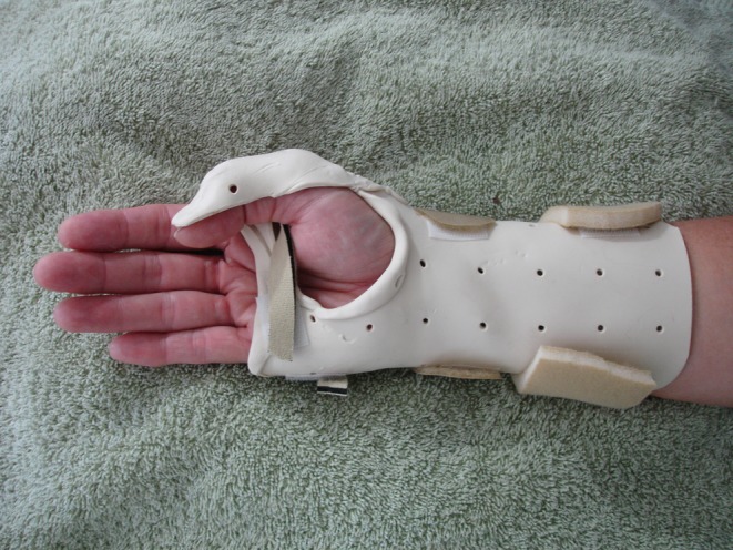
Modified thumb orthoses
The patient rated her initial pain level at 7/10 using VAS. Thumb extension gains were measured on a weekly basis to allow progressive adjustment of the orthotic to allow maximum thumb extension. The patient was able to achieve 50° of flexion at the IP joint and 40° of MP flexion at the time of the initial evaluation. No lag of motion was noted.
At 4 weeks postop, the orthotic was again adjusted to allow additional thumb extension. She had regained 30° of thumb extension from the time of the initial evaluation. Prehension training was initiated at 1 month postop, to regain thumb function and prehension skills. Her pain level had decreased from 7/10 to 4/10. At 6 weeks postop, the protective orthotic was removed per protocol and the patient was instructed to start using the hand for light activities of daily living (ADL) tasks. The patient was unable to place her hand around a glass due to loss of combined thumb extension and abduction. Hand therapy continued two times weekly and consisted of PROM to the thumb to achieve full extension and abduction of the thumb. At 8 weeks, strengthening exercises (putty, gripper, and Baltimore Therapeutic Equipment (BTE) functional strengthening tasks) were performed. CP was discharged from therapy after 3 months of therapy (24 visits). Thumb CMC extension measured 0–70°. Her ULFI score was 0. She reported 0/10 pain on the VAS scale at the time of discharge.
Case 2
M.U. is a retired 72-year-old right hand dominant female who sustained a right distal radius fracture in 2005. In May of 2010, she presented with a chief complaint of clumsiness of her right hand. Examination by her physician demonstrated no active and full passive flexion of the thumb IP joint and mild discomfort with resisted flexion of the index finger distal joint. He scheduled her for surgery to repair her ruptured FPL and the partial rupture of the flexor digitorum profundus (FDP) to the index finger and for hardware removal. She was referred for orthotic fabrication 3 days postop. The thumb was positioned at maximum extension (−55 IP and MP) with a dorsal blocking hood, the wrist was positioned in 0° of extension, and the fingers were left free. The index finger was not immobilized.
The patient rated her initial pain level at 5/10 using VAS. Thumb passive extension gains were measured on a weekly basis to allow progressive adjustment of the orthotic to allow maximum thumb extension. She was able to achieve thumb flexion of 55° at the IP and MP joints at the time of the initial evaluation. No lag of flexion motion was present at the time of the initial evaluation. Full active flexion of index finger was also present.
At 4 weeks postop, the orthotic was adjusted to allow additional thumb extension. She had regained 45° of thumb extension from the time of the initial evaluation. Prolonged thumb IP extension stretch was performed in therapy to regain motion. At 4 weeks, full active finger extension was present. It was also determined that full composite wrist extension and digit extension was not present. Wrist prolonged weighted stretch over a bolster was performed to regain composite motion. At 6 weeks postop, the protective orthotic was removed and the patient was instructed to start using the hand for light ADL tasks. Static progressive IP extension splinting was initiated because of thumb ROM limitations (Fig. 2). The patient was instructed to wear the splint for 8 h at night and two times a day for a period of 30 min to 1 h as tolerated (Fig. 3). Hand therapy continued two times weekly and consisted of PROM to the thumb to achieve full extension and abduction and strengthening exercises (putty, gripper, and BTE functional strengthening tasks). At 12 weeks postop, her grip measured 27 lbs. Active thumb CMC extension measured 20°. The patient was discharged from therapy after 4 months of therapy (28 visits). Her ULFI score was 4. She reported 0/10 pain on the VAS scale.
Fig. 2.
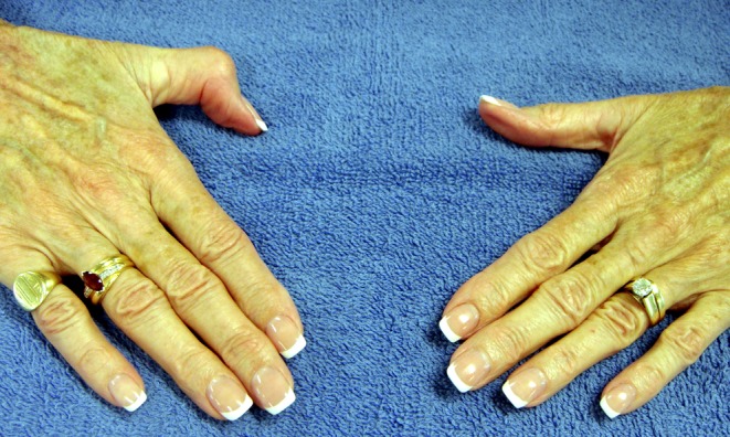
Case 2: range of motion comparison right and left
Fig. 3.
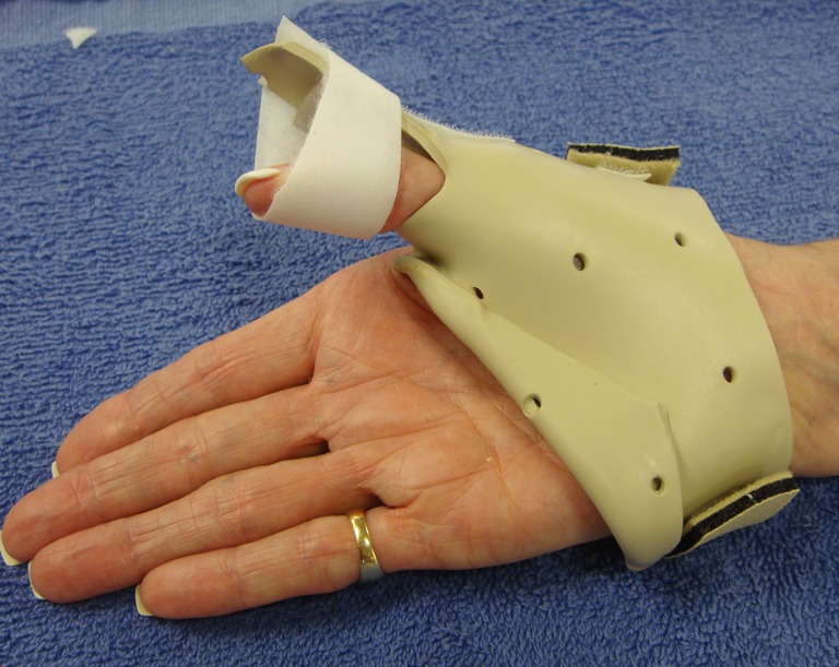
Case 2: static progressive thumb IP extension splint
Case 3
M.T. is 62-year-old female who is employed as a nurse. The patient has co-existing diagnosis of osteoarthritis of the digits and pan-trapezial arthritis of the wrist. She sustained a right distal radius fracture in 2005. She reported onset of sharp wrist pain and localized edema and made an appointment with her physician. She had her surgical procedure scheduled to remove hardware when her FPL and FDP of the index finger ruptured 1 week prior to scheduled surgery. Hand therapy was initiated 6 days postop for orthotic fabrication and initiation of therapeutic exercises. The orthoses positioned the wrist in neutral. The thumb was positioned, under a dorsal hood, in palmar adduction and 35° of both MP and IP thumb flexion, as further extension was not possible. The index finger was not immobilized because the thumb was positioned directly beneath the index finger due to shortened surgical repair of the FPL.
During her initial evaluation in therapy, she reported that she had experienced mild pain with resistive pinch tasks during the 5 years post injury, but she had not reported this to her physician. Index finger passive flexion was within normal limits (WNL). The patient rated her initial pain level at 4/10 using the VAS scale. The patient was able to achieve 65° of flexion of the IP joint and 45° of MP flexion at the time of the initial evaluation. No lag of motion was noted as passive motion and active flexion range of motion were equal. Full active flexion of index finger was also present.
At 4 weeks postop, the orthotic was adjusted to allow additional thumb extension. She had regained 45° of thumb extension from the time of the initial evaluation. Therapy frequency was decreased to one time per week at the patient’s request. Full finger extension was present. The patient was able to oppose thumb to all fingertips. Her pain level decreased from 4/10 to 2/10. At 6 weeks postop, the protective orthotic was removed and the patient was instructed to start using her hand for light ADL tasks. At 8 weeks postop, active thumb CMC extension measured 45°. She reported pain 1/10 on the VAS pain scale. Hand therapy was discontinued and the patient returned to full-time work as a nurse without restrictions. A written home program of strengthening exercises was provided to the patient. She did not perform strengthening exercises in therapy. She received a total of ten therapy visits. The patient opted out of therapy at this point because she had regained functional status and had financial constraints.
Case 4
R.S. is a 59-year-old male who was self-employed. He suffered a right distal radius fracture in 2005 as a result of a motorcycle accident. He sustained a complete rupture of his FPL tendon and partial rupture of the FDP tendon of his index finger. Fracture fixation was performed by a different surgeon than the surgeon who performed the tenorrhaphy. Because the amount of tendon loss would allow active thumb extension against a dorsal block, a dynamic dorsal splint with rubber band traction on the fingernail was fabricated. The orthoses positioned the wrist in 20° of extension. The thumb was maintained in a neutral position between thumb CMC flexion and extension and 20° of MP flexion. The index finger was not immobilized. Hand therapy was initiated 2 days postop for orthotic fabrication and initiation of therapeutic exercises.
He demonstrated full active and passive extension of long, ring, and small fingers. Index finger passive flexion was WNL. The patient rated his initial pain level at 2/10 using the VAS scale. He performed active thumb extension exercises to the dorsal block of the splint ten times per waking hour. The patient was able to achieve 50° of thumb flexion at the IP joint and 45° of MP flexion at the initial evaluation. No lag of FPL and FDP motion was noted. Active index finger flexion was to 1.5 cm to the palm. At 4 weeks postop, full finger extension was present. At 1 month postop, the patient was able to oppose thumb to all fingertips. Lag of index FDP tendon was noted at 1 month. Passive index finger flexion was to the palm, but active motion was to 1 cm to the palm. The patient was instructed to remove his orthotic and perform composite fist exercises three to five times per day. The rubber band traction device was removed from the orthotic. His pain level decreased from 2/10 to a 1/10. At 6 weeks postop, the protective orthotic was removed and the patient was instructed to start using the hand for light ADL tasks. Thumb active motion measured 0–70° at the IP joint and 0–50° at the MP joint. Active index finger flexion was to 0.5 cm to the palm. At 8 weeks postop, thumb CMC abduction measured 75°. He reported pain 0/10 on the VAS pain scale. Hand therapy continued two times weekly for one more week and consisted of PROM to the thumb to achieve full extension and abduction and strengthening exercises (putty, gripper, and BTE functional strengthening tasks). Hand therapy was discontinued 9 weeks postoperatively and he received a total of 11 therapy visits. A written home program of strengthening exercises was provided to the patient. The patient opted out of therapy at this point because he had regained functional status and had financial constraints.
Statistical Analysis of Outcome Measures
Descriptive statistics and analyses were used to evaluate the data (Table 4). The two patients who were employed returned back to work full duty between 8 and 9 weeks postoperatively. Paired t tests were used to compare initial VAS and ULFI scores to discharge scores. The mean ULFI score at the initial evaluation was 60 and at discharge the mean score was 2. A statistically significant difference (p < 0.01) (95% CI 23.99–94.00) was present between ULFI scores initially to the score at time of discharge. ULFI effect size (ES) and standardized response mean (SRM) values were determined to be greater than 0.80 which demonstrate a large treatment effect. When comparing VAS scores at the initial evaluation to discharge, a statistically significant difference (p < 0.03) (95% CI 0.721–7.777) was present. VAS pain ES and SRM values were determined to be greater than 0.80 which demonstrate a large treatment effect. The mean VAS score at the initial evaluation was 4.5/10. At discharge, the mean VAS score for pain was 0.25/10. Excellent results were observed in 100% of cases when using the Buck-Gramcko assessment [6] (Table 5). The Buck-Gramcko assessment qualifies the ROM results achieved with scores ranging between (0–6) poor and (13–15) excellent.
Table 4.
Results
| Case | 1 | 2 | 3 | 4 | ES | SRM | Mean | Standard deviation |
|---|---|---|---|---|---|---|---|---|
| Thumb TAM (IP and MP) | ||||||||
| Operated | 125 | 145 | 125 | 130 | 131 | 9.46 | ||
| Nonoperated | 125 | 155 | 135 | 135 | 138 | 12.58 | ||
| Percentage of nonoperated side | 100% | 94% | 93% | 96% | 96% | 0.03 | ||
| Index finger TAM | ||||||||
| Operated | 265 | 255 | 235 | 265 | 255 | 14.14 | ||
| Nonoperated | 265 | 255 | 265 | 265 | 263 | 5 | ||
| Percentage of nonoperated side | 100% | 100% | 89% | 100% | 97% | 5.5 | ||
| Buck-Gramcko score | 15 | 15 | 15 | 15 | 15 | 0 | ||
| Grip strength | ||||||||
| Operated | 35 | 48 | 41 | 57 | 43 | 9.46 | ||
| Nonoperated | 34 | 54 | 33 | 69 | 47.5 | 17.29 | ||
| Percentage of nonoperated side | 103% | 91% | 124% | 83% | 100% | 17.84 | ||
| Lateral pinch | ||||||||
| Operated | 20 | 19 | 22 | 15 | 19 | 2.94 | ||
| Nonoperated | 22 | 22 | 20 | 17 | 20 | 2.06 | ||
| Percentage of nonoperated side | 91% | 86% | 110% | 88% | 94% | 11.03 | ||
| Tripod pinch | ||||||||
| Operated | 18 | 20 | 18 | 16 | 18 | 1.63 | ||
| Nonoperated | 18 | 22 | 16 | 18 | 19 | 2.52 | ||
| Percentage of nonoperated side | 100% | 91% | 113% | 89% | 98% | 10.94 | ||
| ULFI | 0.89 | 0.87 | ||||||
| Initial score | 88 | 48 | 40 | 64 | 60 | 21.16 | ||
| Final score | 0 | 4 | 4 | 0 | 2 | 2.31 | ||
| VAS pain | 0.84 | 0.83 | ||||||
| Initial score | 7/10 | 5/10 | 4/10 | 2/10 | 4.5/10 | 2.08 | ||
| Final score | 0/10 | 0/10 | 1/10 | 0/10 | .25/10 | 0.5 | ||
TAM total active motion, IP interphalangeal, MP metacarpal phalangeal, ULFI Upper Limb Functional Index, VAS visual analog scale, ES effect size, SRM standardized response means
Table 5.
Buck-Gramcko method of assessment of tendon outcomes for the thumb
| Degrees | Points | |
|---|---|---|
| Flexion of interphalangeal joint | 50–90 | 6 |
| 30–49 | 4 | |
| 10–29 | 2 | |
| <10 | 0 | |
| Extension lag | 0–10 | 3 |
| 11–20 | 2 | |
| 21–30 | 1 | |
| >30 | 0 | |
| Total active motion | >40 | 6 |
| 30–39 | 4 | |
| 20–29 | 2 | |
| <20 | 0 | |
| Evaluation | Excellent | 14–15 |
| Good | 11–13 | |
| Fair | 7–10 | |
| Poor | 0–6 |
Discussion
When the range of motion data was compared with the data found in the literature for early active mobilization of FPL repairs, similar results were obtained when using the Buck-Gramcko assessment. Sirotakova and his colleagues reported on 48 FPL repairs that were rehabilitated by early active mobilization [24]. Excellent or good results were observed in 77% of cases determined by the Buck-Gramcko assessments. Giesen and his colleagues reported on 50 flexor pollicis longus repairs which were rehabilitated by early active motion [12]. The authors reported excellent or good results in 82% of cases using the Buck-Gramcko assessments with no reports of tendon rupture as a result of early active mobilization [12]. The early active protocol used in the studies of Sirotakova et al. and Giesen et al. restricted active flexion of the thumb to touch the middle finger tip during the first week and touch the ring finger tip during the second week. During the third week, the patients were encouraged to actively flex the thumb as far as possible.
All four cases experienced FPL rupture after volar plating of a distal radius fracture. The case similarities included FPL tendon shortening which was initially demonstrated by loss of thumb extension and delayed rupture of the tendon, all cases received tenorrhaphy surgery by the same hand surgeon who used an eight-strand modified Kessler technique, and all cases received hand therapy from the same CHT. The most significant differences between cases were the degree of thumb extension loss and whether or not the FDP of the index finger was also ruptured. The differences in the cases are important as they demonstrate how clinical reasoning differs when the therapist is faced with situations that are less than optimal during the course of rehabilitation. Some of the challenges that were faced during the treatment of these four cases included lag of active flexion of FDP of the index finger, loss of thumb extension that prevented fabrication of protocol specified orthotic, and financial constraints that limited the patient’s ability to attend recommended therapy visits. The standard postoperative rehabilitation regimens do not clearly define how deviations from the based protocols can occur. Knowledge of suture technique and strength of the repair permitted early active motion and dictated the initiation of exercises throughout the patients’ treatments. All of the patients were compliant and their ability to correctly follow directions enabled the patients to remove their orthotic for exercises and start and an early active motion program. When case 4 demonstrated flexion lag determined by ROM measurements, his program was modified to start composite fist exercises.
One of the limitations of the study is the information in retrospective data, recollections of past events, and is therefore subject to the problems inherent to memory. This case series uses descriptive method, not an explanatory one. That is, without the controlled conditions an experiment, conclusions about cause-and-effect relationships cannot be drawn. Outcomes can only be described, not explained. The series also involves only a few individuals and therefore may not be representative of the general group or population.
Conclusion
Flexor tendon rupture of the FPL and FDP of the index finger following volar plate fixation of the distal radius is a condition that has been reported in the literature in increased numbers and requires some modifications of the usual rehabilitation program due to the shortened tendon length. The case examples outlined here illustrate the variability in presentation and challenges associated when rehabilitating patients with delayed tendon rupture following volar plate fixation of the distal radius. A tailored regimen of exercise, education, and equipment is a distinctive feature of quality rehabilitation. Advanced clinical reasoning strategies which include knowledge of suture technique, compliance issues, and ROM measurements are useful tools that the clinician can use when making decisions that deviate from standard protocols. In all of the reported cases, each patient returned to independent ADL status as demonstrated by ULFI scores ranging between 0 and 4. Excellent range of motion and functional strength were achieved by all patients. Average total active motion of thumb measured 131°, 96% of noninjured side. Injured hand grip regained an average of 100% of uninjured grip strength. These cases demonstrate the importance of hand therapy and the necessity of individualizing treatment plans to restore patients to their highest level of function.
References
- 1.Adams LS, Greene LW, Toppoozian E. Range of motion. In: ASHT clinical recommendations, 2nd ed. Chicago: American Society of Hand Therapists; 1992, pp 55–69.
- 2.Adham MN, Porembski M, Adham C. Flexor tendon problems after volar plate fixation of distal radius fractures. Hand. 2009;4:406–409. doi: 10.1007/s11552-009-9180-0. [DOI] [PMC free article] [PubMed] [Google Scholar]
- 3.Arora R, Lutz M, Hennerbichler A, et al. Complications following internal fixation of unstable distal radius fracture with palmar locking plate. Jour Ortho Trau. 2007;21:316–321. doi: 10.1097/BOT.0b013e318059b993. [DOI] [PubMed] [Google Scholar]
- 4.Bainbridge LC, Robertson C, Gillies D, et al. A comparison of post-operative mobilization of flexor tendon repairs with “passive flexion-active extension” and “controlled active motion” techniques. J Hand Surg Br. 1994;19:517–521. doi: 10.1016/0266-7681(94)90219-4. [DOI] [PubMed] [Google Scholar]
- 5.Bell JS, Wollstein R, Citron ND. Rupture of flexor pollicis longus tendon: a complication of volar plating of the distal radius. J Bone Joint Surg Br. 1998;80:225–226. doi: 10.1302/0301-620X.80B2.8351. [DOI] [PubMed] [Google Scholar]
- 6.Buck-Gramcko D, Dietrich FE, Gogge S. Evaluation criteria in follow-up studies of flexor tendon therapy. Handchirurgie. 1976;8:65–69. [PubMed] [Google Scholar]
- 7.Chow JA, Thomes LJ, Dovelle S, et al. A combined regimen of controlled motion following flexor tendon repair in “no man’s land”. Plast Reconstr Surg. 1987;79:447–455. doi: 10.1097/00006534-198703000-00025. [DOI] [PubMed] [Google Scholar]
- 8.Douthit JD. Volar plating of dorsally comminuted fractures of the distal radius: a 6 year study. Am J Orthop. 2005;34:140–147. [PubMed] [Google Scholar]
- 9.Duran RJ, House RG. Controlled passive motion following tendon repairs in zone 2 and 3. In: American Academy of Orthopedic Surgeons: symposium on tendon surgery in the hand. St. Louis: CV Mosby Co; 1975. p. 105–14.
- 10.Fess EE. Grip strength. In: Casanova JS, editor. ASHT clinical recommendations. 2. Chicago: American Society of Hand Therapists; 1992. pp. 41–44. [Google Scholar]
- 11.Gabel CP, Michener LA, Burkett B, et al. The Upper Limb Functional Index: development and determination of reliability, validity, and responsiveness. J Hand Ther. 2006;19:328–349. doi: 10.1197/j.jht.2006.04.001. [DOI] [PubMed] [Google Scholar]
- 12.Giesen T, Sirotakova M, Copsey AJ, et al. Flexor pollicis longus primary repair: further experience with the Tang technique and controlled active mobilization. J Hand Surg Eu. 2009;34:758–761. doi: 10.1177/1753193408096025. [DOI] [PubMed] [Google Scholar]
- 13.Groth GN. Clinical decision making and therapists’ autonomy in the context of flexor tendon rehabilitation. J Hand Ther. 2008;21:254–260. doi: 10.1197/j.jht.2007.10.022. [DOI] [PubMed] [Google Scholar]
- 14.Horger MM. The reliability of goniometric measurements of active and passive wrist motion. Am J Occup Ther. 1990;44:342–348. doi: 10.5014/ajot.44.4.342. [DOI] [PubMed] [Google Scholar]
- 15.Klein L. Early active motion flexor tendon protocol using one splint. J Hand Ther. 2003;16:199–206. doi: 10.1016/S0894-1130(03)00035-8. [DOI] [PubMed] [Google Scholar]
- 16.Lee H. Double loop locking suture: a technique of tendon repair for early active mobilization. Part II: clinical experience. J Hand Surg Am. 1990;15:953–958. doi: 10.1016/0363-5023(90)90022-J. [DOI] [PubMed] [Google Scholar]
- 17.Lifchez SD. Flexor pollicis longus rupture after volar plating of a distal radius fracture. Plast Reconstr Surg. 2010;125:21e–23e. doi: 10.1097/PRS.0b013e3181c2a353. [DOI] [PubMed] [Google Scholar]
- 18.Lister GD, Kleinert HE, Kutz JE, et al. Primary flexor tendon repair followed by immediate controlled mobilization. J Hand Surg Am. 1977;2:441–451. doi: 10.1016/s0363-5023(77)80025-7. [DOI] [PubMed] [Google Scholar]
- 19.Mathiowetz V, Kashman N, Volland G, et al. Grip and pinch strength: normative data for adults. Arch Phys Med Rehabil. 1985;66:69–74. [PubMed] [Google Scholar]
- 20.Mathiowetz V, Weber K, Volland G, et al. Reliability and validity of grip and pinch strength evaluations. J Hand Surg [Am] 1984;9:222–226. doi: 10.1016/s0363-5023(84)80146-x. [DOI] [PubMed] [Google Scholar]
- 21.Nunley JA, Rowan PR. Delayed rupture of the flexor pollicis longus tendon after inappropriate placement of the pi plate on the volar surface of the distal radius. J Hand Surg. 1999;24:1279–1280. doi: 10.1053/jhsu.1999.1279. [DOI] [PubMed] [Google Scholar]
- 22.Piskin A, Yuceturk A, Tomak Y, Ozer M, Gulman B, Ataman A, Kangal M, Sahin Y, Desteli E, Alic T. Tendon repair with the strengthened modified Kessler, modified Kessler, and Savage suture techniques: a biomechanical comparison. Acta Orthop Traumatol Turc. 2007;41:238–243. [PubMed] [Google Scholar]
- 23.Schreuders T, Roebroeck ME, Goumans J, et al. Measurement error in grip and pinch force measurements in patients with hand injuries. Phys Ther. 2003;83:806–815. [PubMed] [Google Scholar]
- 24.Sirotakova M, Elliot D. Early active mobilization of primary repairs of the flexor flexor pollicis longus tendon with two Kessler two-strand core sutures and a strengthened circumferential suture. J Hand Surg. 2004;29:531–535. doi: 10.1016/j.jhsb.2004.07.002. [DOI] [PubMed] [Google Scholar]
- 25.Siu CK. Delayed rupture of flexor pollicis longus tendon after volar plating of the distal radius. Hand Surg. 2006;11:67–70. doi: 10.1142/S0218810406003048. [DOI] [PubMed] [Google Scholar]
- 26.Thien TB, Becker JH, Theis JC. Rehabilitation after flexor tendon injuries in the hand. Cochrane Database Syst Rev 2004; 4:CD003979. [DOI] [PubMed]