
Fig. 6.
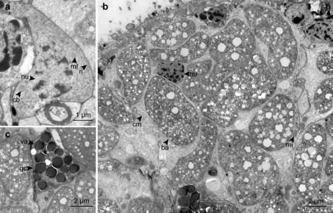
TEM micrographs of different cell types on a cross-section. a Neoblast. b Bacteriocyte, dashed line indicates bacteriocyte, dotted line indicates single symbiont. c Gland cell. ba bacteria, cb chromatoid body, cm cell membrane, gc gland cell, mi mitochondrion, nu nucleus, ri free ribosome, va vacuole