Sir,
Tadayoni et al1 reported that the retina acquired a particular appearance, featuring ‘arcuate striae', after pars plana vitrectomy (PPV) for treatment of a macular hole. They considered that the feature was attributable to the presence of a dissociated optic nerve fiber layer (DONFL). Ito et al2 found that a DONFL resulted in the development of numerous arcuate retinal striae mimicking a retinal nerve fiber layer (RNFL) defect. Earlier conventional time-domain (TD) OCT showed only focal dimples on two-dimensional imaging.
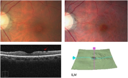
We performed a PPV with internal limiting membrane (ILM) peeling to treat macular hole. No frank retinal trauma was observed during PPV or the ILM peeling procedure. Preoperative fundus photograph showed no RNFL defect (Figure 1). On 12-months postoperative review, multiple dark round lesions were detected in the superior-temporal perimacular area. We performed high-resolution spectral-domain (SD) OCT with three-dimensional imaging (Cirrus OCT, Carl Zeiss Meditec, Inc., Dublin, CA, USA). SD OCT showed deep focal dimples, with clear margins, in the perimacular area, compatible with the presence of a DONFL. Three-dimensional SD OCT images, which reveal the inner retinal surface, confirmed the presence of multiple round, cobblestone-shaped, ‘beaten bronze' dimples in the superior temporal perimacular area. DONFL did not present as the arcuate striae described in many previous reports.2, 3, 4 Many reports have described RNFL morphological changes after ILM peeling during vitrectomy with TD OCT.1, 2, 3, 4 Removal of the ILM is a common procedure in macular hole surgery, as it increases the probability that the macular hole will close. In our case, we used SD OCT, which provides images of greater clarity and higher resolution than afforded by TD OCT. SD OCT clearly revealed the dimple margins and the exact depth of each DONFL. The shape and size of a DONFL could be visualized using the new three-dimensional SD OCT imaging facility. Our patient had DONFLs with a ‘beaten bronze' cobblestone-like appearance. DONFLs can, thus, present in various forms, including the cobblestones of our patients and the arcuate striae described in many reports.
Figure 1.
Upper left: preoperative fundus photography. No retinal nerve fiber layer defect in the perimacular area. Upper right: 12-months follow-up images after vitrectomy. Multiple dark round lesions in the superior temporal perimacular area. Lower left: well-demarcated DONFL (arrow) margin on OCT (vertical axis). Lower right: three-dimensional OCT mapping shows multiple round DONFLs, with a ‘beaten bronze' cobblestone-like appearance, in the superior temporal perimacular area.
The authors declare no conflict of interest.
References
- Tadayoni R, Paques M, Massin P, Mouki-Benani S, Mikol K, Gaudric A. Dissociated optic nerve fiber layer appearance of the fundus after idiopathic epiretinal membrane removal. Ophthalmology. 2001;108:2279–2283. doi: 10.1016/s0161-6420(01)00856-9. [DOI] [PubMed] [Google Scholar]
- Ito Y, Terasaki H, Takahashi A, Yamakoshi T, Kondo M, Nakamura M. Dissociated optic nerve fiber layer appearance after internal limiting membrane peeling for idiopathic macular holes. Ophthalmology. 2005;112:1415–1420. doi: 10.1016/j.ophtha.2005.02.023. [DOI] [PubMed] [Google Scholar]
- Mitamura Y, Ohtsuka K. Relationship of dissociated optic nerve fiber layer appearance to internal limiting membrane peeling. Ophthalmology. 2005;112:1766–1770. doi: 10.1016/j.ophtha.2005.04.026. [DOI] [PubMed] [Google Scholar]
- Mitamura Y, Suzuki T, Kinoshita T, Miyano N, Tashimo A, Ohtsuka K. Optical coherence tomographic findings of dissociated optic nerve fiber layer appearance. Am J Ophthalmol. 2004;137:1155–1156. doi: 10.1016/j.ajo.2004.01.052. [DOI] [PubMed] [Google Scholar]


