Sir,
Penetrating keratoplasty performed for perforated corneal ulcers is associated with a high incidence of secondary glaucoma.1, 2 Anterior segment optical coherence tomography (ASOCT) provides non-contact, non-invasive, high-resolution, real-time cross-sectional images of the anterior segment of the eye.3, 4
Case report
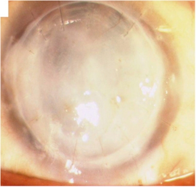
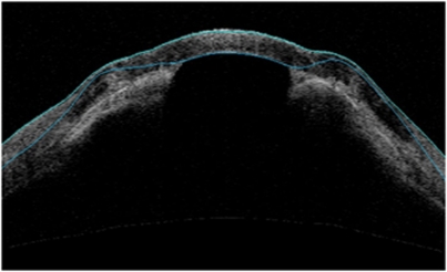
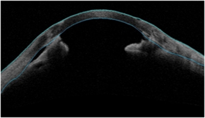
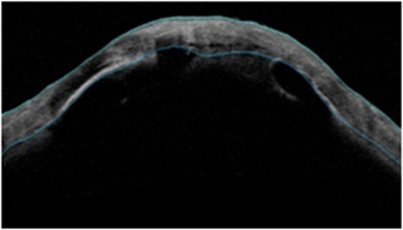
We conducted a prospective study to evaluate the role of ASOCT in the evaluation of the anterior segment in opaque grafts with secondary glaucoma following penetrating keratoplasty performed for perforated corneal ulcers. Eyes with opaque corneal grafts (Figure 1) and with an intraocular pressure (IOP)>25 mm of Hg on two separate occasions presenting three or more months post surgery were included in the study. Twenty five eyes of twenty-five patients with opaque corneal grafts and secondary glaucoma following therapeutic penetrating keratoplasty were enrolled in the present study. The mean IOP at presentation was 37.4±8.9 mm Hg (range, 26–61 mm Hg). ASOCT examination revealed peripheral anterior synechiae in 5 eyes (Figure 2) (20%), a combination of peripheral anterior synechiae+graft-host junction synechiae (Figure 3) in 14 eyes (56%) and a combination of peripheral anterior synechiae+graft-host junction+central synechiae (Figure 4) in 6 eyes (24%). The extent of synechial angle closure was 90–180° in 2 eyes (8%), 180–270° in 4 eyes (16%) and 270–360° in the remaining 19 eyes (76%).
Figure 1.
Post-therapeutic penetrating keratoplasty glaucoma with opaque graft.
Figure 2.
ASOCT image showing peripheral anterior synechiae approaching paracentral area.
Figure 3.
ASOCT image showing both peripheral anterior synechiae and graft-host junction synechiae.
Figure 4.
ASOCT image showing combined peripheral anterior synechiae, graft-host junction and central synechiae.
Comment
ASOCT enables detailed visualization of the anterior segment and angle anatomy in eyes with opaque corneal grafts and secondary glaucoma. Secondary angle closure due to peripheral anterior synechiae formation is an important mechanism for intraocular pressure elevation in these eyes. Knowledge of the exact sites of peripheral anterior synechiae pre-operatively in such eyes may be of help in planning the site of trabeculectomy or placement of aqueous drainage devices. Being a non-contact procedure, ASOCT helps in the immediate post-operative follow up of these patients. It provides rapid and high-resolution images of the entire circumference of the anterior chamber angle. Angle evaluation using ASOCT is recommended in all eyes with post-penetrating keratoplasty secondary glaucoma before planning any corneal or glaucoma intervention.
The authors declare no conflict of interest.
References
- Ayyala RS. Penetrating keratoplasty and glaucoma. Surv Ophthalmol. 2000;45:91–105. doi: 10.1016/s0039-6257(00)00141-7. [DOI] [PubMed] [Google Scholar]
- Dada T, Aggarwal A, Minudath KB, et al. Post-Penetrating Keratoplasty Glaucoma. Indian J Ophthalmol. 2008;56:269–277. doi: 10.4103/0301-4738.41410. [DOI] [PMC free article] [PubMed] [Google Scholar]
- Radhakrishnan S, Rollins AM, Roth JE, Yazdanfar S, Westphal V, Bardenstein DS, et al. Real-time optical coherence tomography of the anterior segment at 1310 nm. Arch Ophthalmol. 2001;119:1179–1185. doi: 10.1001/archopht.119.8.1179. [DOI] [PubMed] [Google Scholar]
- Müller M, Dahmen G, Pörksen E, Geerling G, Laqua H, Ziegler A, et al. Anterior chamber angle measurement with optical coherence tomography: intraobserver and interobserver variability. J Cataract Refr Surg. 2006;32:1803–1808. doi: 10.1016/j.jcrs.2006.07.014. [DOI] [PubMed] [Google Scholar]


