Abstract
As a consequence of the Earth's rotation, almost all organisms experience day and night cycles within a 24-hr period. To adapt and synchronize biological rhythms to external daily cycles, organisms have evolved an internal time-keeping system. In mammals, the master circadian pacemaker residing in the suprachiasmatic nucleus (SCN) of the anterior hypothalamus generates circadian rhythmicity and orchestrates numerous subsidiary local clocks in other regions of the brain and peripheral tissues. Regardless of their locations, these circadian clocks are cell-autonomous and self-sustainable, implicating rhythmic oscillations in a variety of biochemical and metabolic processes. A group of core clock genes provides interlocking molecular feedback loops that drive the circadian rhythm even at the single-cell level. In addition to the core transcription/translation feedback loops, post-translational modifications also contribute to the fine regulation of molecular circadian clocks. In this article, we briefly review the molecular mechanisms and post-translational modifications of mammalian circadian clock regulation. We also discuss the organization of and communication between central and peripheral circadian oscillators of the mammalian circadian clock.
Keywords: circadian pacemaker, SCN, feedback loop, mammalian circadian clock
CIRCADIAN TIMING SYSTEM
Regardless of disparate phylogenetic origins and huge differences in complexity among species, organisms have evolved internal timing systems to adapt to the external day and night cycles. This daily time-keeping system is referred to as the 'circadian clock' from the Latin circa diem, literally meaning 'approximately one day'. Circadian clocks from all organisms have many similar properties. Circadian rhythms are entrained by an external cue called a 'zeitgeber'. Any exogenous cue can synchronize the endogenous time-keeping system. The primary zeitgeber is light for organisms on Earth, and specialized photoreceptive and phototransductive mechanisms have evolved in almost all biological clock systems (Moore, 1997). Consequently, changes in the zeitgeber time (e.g., traveling to a different time zone) result in the resetting and phase shifting of internal rhythmicity. Such an internal rhythm is generated intrinsically and is self-sustainable (Reppert and Weaver, 2002). In other words, the circadian rhythm persists even in the absence of cyclic environmental zeitgebers. However, the free-running period in the absence of an external time cue differs from the period under light-dark cycles. Thus, free-running periodicity under constant conditions is often described by circadian time (CT), a standardized 24-hour phase in a circadian cycle that represents an estimation of the organism's subjective time. Circadian rhythm is temperature compensated (Hastings et al., 2008), meaning that organisms can maintain their circadian periodicity over a range of environmental temperatures. Circadian periodicity is not affected by changes in temperature, even though the rates of most physiological processes are easily susceptible to changes in temperature. Therefore, unresolved mechanisms are likely participating in the circadian clock machinery and can compensate for the temperature-related changes in biophysical and biochemical processes.
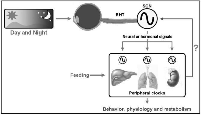
The mammalian circadian clock system is composed of three basic components: input signals (environmental timing cue), a circadian oscillator (rhythm generator) and output signals (overt rhythm). In opaque animals, including mammals, the light signal is mainly detected by eyes. Behavioral studies using laboratory animals, such as mice, rats and hamsters, revealed that intact animals exhibit approximately 24 hr of circadian rhythm even in the absence of external time cues (e.g., constant darkness). These findings strongly support the existence of a 'master circadian oscillator', which generates intrinsic circadian rhythmicity (Fig. 1). In an effort to determine the master circadian pacemaker of mammals, the suprachiasmatic nucleus (SCN) of the hypothalamus was anatomically found to be a direct target of the retinal fibers (Reppert and Weaver, 2002). More importantly, selective disruption of the SCN revealed a complete loss of circadian rhythmicity, whereas transplantation of an intact SCN to the mutant animal restored circadian rhythmicity (Stephan and Zucker, 1972; Ralph et al., 1990). Thus, the SCN is currently regarded as the master circadian oscillator where the circadian rhythm is generated. The circadian rhythm generated in the SCN is likely converted into neuronal or hormonal signals that affect the behavior, physiology and metabolic processes of entire animals (Schibler and Sassone-corsi, 2002).
Fig. 1.
Components of the mammalian circadian clock system. Changes in light due to the day/night cycle are directly detected by eyes. The light information is transported to the suprachiasmatic nucleus (SCN) in the anterior hypothalamus by the retinohypothalamic tract (RHT). The SCN functions as a master circadian oscillator where circadian rhythmicity is generated. The generated circadian rhythmicity is converted into output pathways that control the behavior, physiology and metabolism of the organisms. These environmental signals, core circadian oscillator and output rhythms are three basic components of circadian clock system.
In addition to the central pacemaker, numerous subsidiary circadian clocks exist outside the SCN in peripheral tissues (including the liver, heart, lung and muscle) and even in immortalized cell lines, which are kept ex vivo for long periods of time (Yamazaki et al., 2000). The presence of mammalian peripheral clocks has been demonstrated by measuring circadian gene expression in cultured fibroblasts or tissue explants. Schibler and colleagues showed that immortalized fibroblasts could generate rhythmic gene expression when cells are exposed to a serum shock (a brief treatment with 50% horse serum for two hours) (Balsalobre et al., 1998). These peripheral clocks are now believed to have tissue- or organ-specific roles in controlling circadian rhythms. Unlike the central pacemaker in the SCN, these peripheral oscillators are not directly entrained by light. Instead, output signals from the SCN or other external stimuli, such as feeding, can control the circadian rhythmicity of the peripheral local clocks (Fig. 1). Despite such differences, the underlying molecular bases of the peripheral circadian clocks are very similar to those of the central oscillator.
MOLECULAR CIRCADIAN CLOCKWORK
Most recent chronobiological studies have focused on the molecular basis of the circadian clock. Intensive studies have revealed that at least one internal autonomous circadian oscillator consisting of positive and negative elements of autoregulatory feedback loops is at the center of all examined circadian clocks. For example, the circadian clock of cyanobacteria is regulated by a cluster of three genes: kaiA, kaiB and kaiC (Ishiura et al., 1998). This clock sustains a 22-hr rhythm over several days upon the addition of ATP. In Neurospora, the molecular clock is composed of the positive regulator, White collar complex (Wcc), and the negative regulator, Frequency (Frq) (Dunlap, 1999). In Drosophila, Clock (Clk) and Cycle (Cyc) activate the transcription of several circadian clock genes. Among these genes, Period (Per) and Timeless (Tim) form heterodimers in the cytoplasm and then translocate to the nucleus, where PER inhibits the transcriptional activity of the CLK/CYC complex (Gallego and Virshup, 2007).
The Drosophila Per gene is the first identified molecular circadian clock component. In 1971, Konopka and Benzer identified a genetic component of the biological clock for the first time using the fruit fly as a model system (Konopka and Benzer, 1971). They found three mutant lines of flies displaying aberrant circadian behaviors; one had a shorter period, the second had a longer one and the third had no apparent periodicity. All three mutations mapped to the same gene, which was named Period. Thirty years later, a human ortholog of this gene was found to be defective in patients with the sleep disorder familial advanced sleep phase syndrome (FASPS) (Vanselow et al., 2006). The first circadian mutant in mammals was identified in 1988 and designated as tau (τ). Ralph and Menaker found a single male hamster exhibiting an abnormally short period in constant darkness (DD). Whereas the normal circadian period for golden hamsters averages about 24.1 hr and is rarely shorter than 23.5 hr, the free-running period of the abnormal male was 22.0 hr (Ralph and Menaker, 1988). About 10 years later, Takahashi and colleagues identified tau as an allele of casein kinase 1ε (CK1ε), a protein kinase that can phosphorylate mammalian PER proteins (Lowrey et al., 2000).
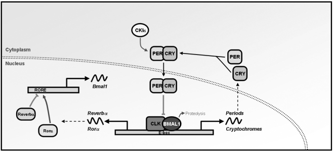
The mammalian circadian oscillator consists of a network of interlocking transcriptional-translational feedback loops that drive rhythmic expression of core clock components (Reppert and Weaver, 2002). The core clock components are established by genes whose protein products are necessary for the generation and maintenance of circadian rhythms within individual cells throughout the organism. As core components of the mammalian circadian molecular clock, CLOCK and BMAL1, which belong to the family of bHLH-PAS-containing transcription factors, constitute a positive feedback loop. CLOCK and BMAL1 form a heterodimer, which binds to the E-box cis-regulatory enhancer elements of their target genes, including Periods (Per1, Per2 and Per3) and Cryptochromes (Cry1 and Cry2; Fig. 2). A negative feedback loop is achieved when the PERs and CRYs form heterocomplexes that translocate back to the nucleus and inhibit their own transcription (Gallego and Virshup, 2007). In addition to the primary feedback loops, another regulatory feedback loop is formed by the orphan nuclear receptors REV-ERBα and RORα (Gallego and Virshup, 2007). Expression of these nuclear receptors is also controlled by the CLOCK/BMAL1 heterodimer. In the nucleus, REV-ERBα competes with RORα for binding to the ROR-responsive element (RORE) in the Bmal1 promoter. Whereas RORα activates transcription of Bmal1, REV-ERBα represses it. Consequently, the cyclic expression of Bmal1 is achieved by both positive and negative regulation of RORs and REV-ERBs, respectively. This secondary feedback loop is called the 'stabilizing loop'.
Fig. 2.
Mammalian molecular circadian clock. Several core clock genes constitute interlocking molecular feedback loops. In this molecular clock, the bHLH-PAS (basic helix-loop-helix, Per-Arnt-Sim)-containing transcription factors BMAL1 and CLOCK function as positive regulators. Upon heterodimerization, they translocate into the nucleus, where they bind to the E-box enhancer element in the promoter region of downstream clock genes such as Periods (Pers) and Cryptochromes (Crys). Rhythmically expressed PER and CRY form a heterocomplex. In the nucleus, PER and CRY constitute a negative feedback loop by repressing their own transcriptional activation by CLOCK/BMAL1 heterodimer. The CLOCK/BMAL1 heterodimer also induces the transcription of the nuclear receptors Rev-erbα and Rorα. Rev-erbα and Rorα constitute another feedback loop by repressing and activating Bmal1 transcription, respectively.
In addition to the feedback loops of transcription and translation, various post-translational modifications are also involved in the normal functioning of the circadian clockwork. A few hours would be sufficient for a molecular feedback loop to run a cycle by only transcriptional activation and following feedback repression. Therefore, if not for the significant delay between transcriptional activation and repression, 24-hr circadian periodicity would not be achieved. Increasing evidence supports the involvement of post-translational modifications for the required delay (Gallego and Virshup, 2007). Studies in molecular circadian clock machinery in various phyla have found several protein kinases involved in circadian regulation. The double-time (DBT) kinase was the first enzyme identified as an essential component of the Drosophila circadian clock. Mutations in DBT result in abnormal locomotor activity periods and eclosion (Price et al., 1998). Mammalian orthologs of DBT, casein kinase members CK1δ and CK1ε were also identified as clock components. Mutation in CK1ε resulted in a shorter free-running period of hamsters (Lowrey et al., 2000). In humans, mutations in the CK1δ and CK1ε phosphorylation sites in PER2 have been found in families with FASPS (Toh et al., 2001). More recently, inhibition of glycogen synthase kinase 3 (GSK-3) was reported to shorten the mammalian circadian period (Hirota et al., 2008). Moreover, the cAMP-dependent protein kinase (PKA) and mitogen-activated protein kinase (MAPK) pathways are also implicated in the CRE-mediated induction of Per1 (Travnickova-Bendova et al., 2002). In addition to the CREB-mediated induction, the Ca2+-dependent protein kinase C (PKC)-mediated phosphorylation of CLOCK seems to be important for phase resetting of the mammalian circadian clock (Shim et al., 2007). Treatment with PMA, a PKC-activating phorbol ester, induces the phosphorylation of CLOCK and rapid induction of Per1 mRNA expression in fibroblasts cultured in vitro.
Phosphorylation is a prerequisite step for the recruitment of ubiquitin ligases and the subsequent degradation of PERs. In Drosophila, DBT kinase reduces the stability of PER; in the absence of DBT kinase activity, the protein levels of PER remain constitutively high (Price et al., 1998). In mammals, PER proteins bind to and are phosphorylated by CK1s. The half-lives of PER1 and PER2 proteins are moderately shortened when CK1δ or CK1ε is overexpressed (Akashi et al., 2002). Phosphorylation of PERs creates binding sites for β-transducin repeat-containing protein (βTrCP), an F-box-containing E3 ubiquitin ligase (Eide et al., 2005; Shirogane et al., 2005). Another F-box protein, Fbxl3, is an E3 ligase of CRYs (Busino et al., 2007). Mutation in Fbxl3 results in impaired ubiquitination and subsequent degradation of CRYs. Consequently, prolonged stability of the CRY proteins leads to an extended negative phase and period lengthening. The post-translational modifications of BMAL1 have also been extensively studied. BMAL1 is rhythmically sumoylated in vivo, and the interaction with CLOCK is necessary for proper sumoylation (Cardone et al., 2005). More recently, we showed that BMAL1 sumoylation is closely related to protein turnover of BMAL1 itself (Lee et al., 2008). Sumoylation localizes BMAL1 exclusively to promyelocytic leukemia (PML) nuclear bodies and simultaneously promotes its transactivation and ubiquitin-dependent degradation (Kwon et al., 2006; Lee et al., 2008). BMAL1 also can be acetylated, a process essential for the maintenance of circadian rhythmicity (Hirayama et al., 2007). Phosphorylation of BMAL1 by casein kinase 2 alpha (CK2α) is known to be important for nuclear accumulation of BMAL1 and circadian clock function (Tamaru et al., 2009). Thus, these BMAL1 modifications are required for proper functioning of BMAL1 in the circadian clockwork.
CENTRAL VS. PERIPHERAL CLOCKS
Although the peripheral oscillators share the same basic molecular components with the central pacemaker, the peripheral clocks are thought to be less self-sustainable while the master pacemaker is indispensible for rhythm generation in peripheral clocks. Indeed, circadian rhythms in peripheral clocks of liver, lung and skeletal muscle are damped in two to seven days, whereas the SCN exhibits robust rhythmicity up to 30 days in vitro (Yamazaki et al., 2000). These findings strongly support the hierarchical organization of circadian oscillators, where the self-sustainable SCN clock entrains other easily dampened peripheral oscillators. However, Takahashi and coworkers reported in 2004 that the explants of peripheral tissues, such as liver and lung, could also sustain the circadian rhythmicity of PER2 expression for more than 20 days (Yoo et al., 2004). Moreover, SCN lesions did not abolish circadian rhythmicity in peripheral tissues of mPer2-luciferase transgenic mice. Instead, SCN lesions resulted in dispersed phases in peripheral tissues within and among animals. These results contradict other reports in that the SCN lesions caused dampening and abrogation of circadian gene expression (Sakamoto et al., 1998; Akhtar et al., 2002; Terazono et al., 2003). Therefore, the SCN is a master synchronizer rather than a driver for persistent circadian rhythmicity in the peripheral tissues. Considering the possibility of differential regulation of Per1 and Per2 in peripheral tissues, the peripheral tissues and cells seem to possess self-sustainable circadian clocks that can generate overt rhythms in the absence of the central pacemaker (Dibner et al., 2010).
The roles of such self-sustainable peripheral oscillators are still unknown. The adrenal local clock is a good example of a peripheral clock. The adrenal cortex is well known for synthesis and secretion of the steroid hormone glucocorticoid (GC). This hormone is secreted in a circadian rhythmic pattern and is implicated in a wide variety of biological processes, including stress response, growth, reproduction and immune response (Buckingham, 2006). Interestingly, injection of a synthetic GC, dexamethasone (DEX), induced a phase shift of circadian rhythmicity in mouse liver, while the central clock in the SCN was barely affected (Balsalobre et al., 2000), suggesting that GC is also important for the synchronization of circadian rhythmicity. The SCN has been reported to directly modulate GC secretion via the splanchnic nerve system (Ishida et al., 2005; Ulrich-Lai et al., 2006). Thus, the rhythmic GC signaling evoked by either the hypothalamus-pituitary-adrenal gland (HPA) axis or sympathetic nervous system is believed to be a possible mediator of cues from the SCN to peripheral local clocks. However, neither hypophysectomy nor DEX suppression abolished circadian secretion of GC (Meier, 1976; Torres-Farfan et al., 2008). In addition, denervation of the splanchnic nerve did not completely blunt rhythmic GC secretion (Ulrich-Lai et al., 2006). These reports raised the possibility of additional regulatory mechanisms of GC synthesis and/or secretion intrinsic to the adrenal gland. Recently, we demonstrated the functional importance of the adrenal local clock (Son et al., 2008); the clock-controlled adrenal steroidogenic acute regulatory protein (StAR) expression mediates circadian production of GC, and the adrenal-specific knock-down of a key clock protein, BMAL1, significantly dampened the circadian rhythmicity of GC in vivo. Given that GC is one of the key mediators between the central and peripheral circadian clocks, the adrenal local clock appears to have pivotal roles in harmonizing the central and other peripheral clocks by generating a robust GC rhythm. Moreover, as previously reported, GC receptor (GR) is expressed in almost all cells of an organism except for the SCN (Rosenfeld et al., 1993; Balsalobre et al., 2000). Thus, regulation of the rhythmicity of GC secretion by the adrenal local clock as well as the central clock can be crucial for understanding the connection between the master clock in the SCN and other local clocks in both peripheral and extra-SCN brain tissues.
The food-entrainable oscillator (FEO) is another good example of a SCN-independent local clock. Unlike the central circadian oscillator, the mammalian peripheral circadian oscillators are not entrained by light. Instead, feeding time and/or hormone stimulation can be a dominant zeitgeber for several peripheral circadian clocks (Balsalobre et al., 2000; Stokkan et al., 2001). SCN-disrupted animals were reported to still be able to respond to rhythmic food availability (Krieger et al., 1977). In addition, SCN lesions do not abolish food-anticipatory behaviors in rats (Stephan et al., 1979). When the feeding was restricted to a certain time, laboratory animals showed a food anticipatory activity (FAA), namely increased locomotor activity prior to food presentation. Thus, mammals surprisingly can exhibit FAA despite the presence of SCN lesions. Another local clock in stomach gland oxyntic cells that rhythmically secrete ghrelin, a hormone stimulating hunger, was reported recently to play a role in mouse FAA (LeSauter et al., 2009). In these cells, rhythmic expression of ghrelin was controlled by circadian clock machinery, and loss of ghrelin receptors resulted in diminished FAA. However, the mechanisms by which the gastric local clocks and their products act on the central nervous system to generate food-anticipation behaviors remain unclear.
EXTRA-SCN CLOCKS IN THE CENTRAL NERVOUS SYSTEM (CNS)
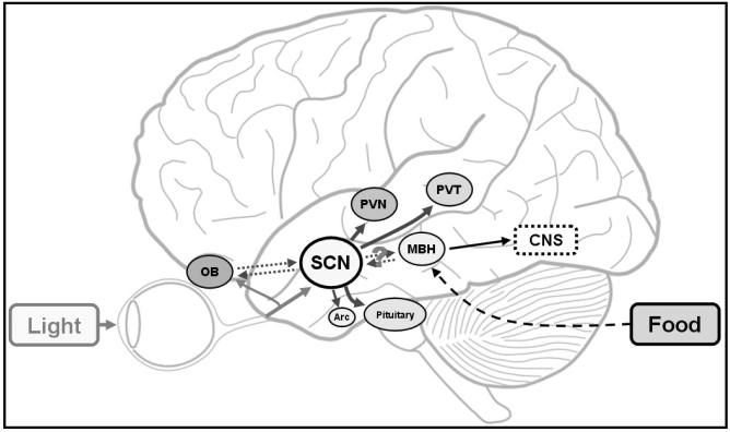
Circadian oscillators also exist in various areas of the brain outside of the SCN. Several transgenic animal models have been developed whose circadian rhythmicity can be measured in a real time-enabled evaluation of the molecular properties of these extra-SCN brain clocks. For example, 14 of 27 Per1-luciferase transgenic animals exhibited circadian rhythmicities in tissue cultures, and brain regions other than the SCN exhibited dampened rhythmicities (Abe et al., 2002). Cellular circadian rhythm was especially robust in the olfactory bulb. Other brain tissues such as the pineal gland, pituitary and arcuate nucleus also exhibited apparent circadian rhythms, but the amplitudes were attenuated within approximately six days (Abraham et al., 2005). Interestingly, the peak expression of Per1-luciferase occurred at dusk or at night in the olfactory bulb and other brain tissues, including the pituitary, pineal gland, and certain nuclei of the hypothalamus and thalamus, while the SCN exhibited peak expression during the daytime. In addition, the circadian rhythm phase was quite variable among individual neurons of the olfactory bulb although the rhythmic nature of Per1-luciferase expression was not abolished in the absence of the SCN (Granados-Fuentes et al., 2004; Abraham et al., 2005). Therefore the SCN appears to coordinate and synchronize the individual brain oscillators like local clocks in peripheral tissues rather than drive the circadian rhythmicity itself (Fig. 3; Dibner et al., 2010). The communication of the SCN with other oscillators in several brain regions remains to be elucidated; a number of neurochemical messengers, including transforming growth factor alpha (TGF-α), cardiotrophin-like cytokine and vasopression, have been proposed as possible output signals from the SCN (Kramer et al., 2001; Kalsbeek and Buijs, 2002; Kraves and Weitz, 2006). Prokineticin 2, a cysteine-rich secreted protein, is another strong candidate (PK2; Cheng et al., 2002). In the SCN, PK2 mRNA is rhythmically expressed, and the phase is entrained by light. Moreover, PK2 receptor (PKR2) is widely expressed in discrete brain target regions of SCN pathways, such as the paraventricular nucleus, dorsal medial hypothalamic nucleus, thalamic paraventricular nucleus, paracentral thalamic nuclei, lateral habenular nucleus, lateral septal nucleus, lateral globus pallidus and amygdala (Cheng et al., 2002). The intracerebroventricular injection of recombinant PK2 alters the locomotor activity of rats (Cheng et al., 2002). However, additional factors that mediate the signal output of the SCN are likely because mice lacking PK2 or PKR2 exhibited reduced, but not completely abolished circadian rhythms (Li et al., 2006; Prosser et al., 2007). Taken together, several self-sustainable circadian oscillators may exist in discrete brain regions, independent of the SCN central pacemaker. Therefore, the physiological roles of brain clocks outside the SCN need to be studied. Piggins and coworkers recently revealed a role for the brain clock residing in the mediobasal hypothalamus (MBH) in the regulation of metabolic homeostasis (Guilding et al., 2009). The MBH is crucial for maintenance of internal homeostasis and encompasses the arcuate, dorsomedial hypothalamus, ventromedial hypothalamus, median eminence and pars tuberalis. Using Per2-luciferase knock-in mice, they showed the presence of extra-SCN circadian oscillators in the MBH region. Circadian rhythmicities in median eminence and pars tuberalis were maintained even when synaptic transmission was inhibited. Moreover, the circadian oscillators residing in the median eminence and pars tuberalis were reset by food deprivation, while the SCN was not affected. These results can provide new insights into the relationship between metabolic cues and circadian oscillators in the CNS outside the SCN. However, the mechanism of integration of metabolic changes into circadian homeostasis by circadian oscillators in MBH and other brain regions remains unclear.
Fig. 3.
Circadian clocks in the SCN and other regions of the brain. In addition to the central pacemaker residing in the suprachiasmatic nucleus (SCN), other clocks exist in various regions of the brain. Light induces the generation of circadian rhythm in the master circadian clock, SCN. The SCN master clock seems to function as a synchronizer of the brain clocks. A metabolic signal, such as feeding, can be delivered to the CNS via a local clock in the DMH region. OB: olfactory bulb, SCN: suprachiasmatic nucleus, PVN: paraventricular nucleus, PVT: paraventricular nuclei of the thalamus, MBH: mediobasal hypothalamus, Arc: arcuate nucleus, CNS: central nervous system.
The extra-SCN brain clocks are also associated with a reward system (Dibner et al., 2010). This reward system is closely related to the survival of individual organisms, and accessibility to food and water can be a primary reward. Rewards can influence the higher brain functions of an organism, including behaviors, learning capability and mood. Several groups recently described the relationship between the brain reward system and the circadian clock. For example, mice lacking Per1 or Per2 exhibited diffent responses to repeated cocaine injection: the behavioral sensitization was absent in the Per1 KO mice, whereas Per2 KO mice showed a hypersensitized response to cocaine (Abarca et al., 2002). Moreover, Per2 mutant animals exhibited an increased alcohol preference compared with wild-type animals (Spanagel et al., 2005), which seems to result from an alteration in glutamatergic transmission. In Per2 mutant mice, expression of the glutamate transporter Eaat1 was significantly decreased, resulting in reduced glutamate uptake by astrocytes and thus increased glutamate concentrations in the extracellular space. Interestingly, treatment with acamprosate, a drug used with alcoholic patients, decreased extracellular glutamate levels and subsequently reduced the alcohol consumption of Per2 mutant mice. Therefore, the circadian timing system and components of the molecular clockwork are related to diverse CNS functions.
PERSPECTIVES
Circadian clock research during recent decades has focused on the elucidation of the molecular mechanisms involved in regulating these processes. However, studies have become more diverse, concentrating on the understanding of functional roles of molecular circadian clocks in the regulation of physiology, behaviors and metabolism. In particular, the circadian clock is associated with various metabolic processes. In unicellular organisms such as cyanobacteria, the circadian clock system segregates two essential metabolic processes, nitrogen fixation and photosynthesis, which are incompatible with each other (Tu and McKnight, 2006). By separating nitrogen fixation and photosynthesis between night and day, respectively, cyanobacteria can resolve the incompatibility of these two key processes. Even in mammals, expression of many metabolic genes is tissue-specifically regulated by circadian clockwork (Panda et al., 2002). In addition, nuclear receptors involved in lipid and carbohydrate metabolism are expressed in a circadian manner in various tissues, including liver, skeletal muscle and white and brown adipose tissues (Yang et al., 2006). In a more recent study, the core clock gene Per2 was reported to interact directly and indirectly with several nuclear receptors implicated in various biological processes (Schmutz et al., 2010). Mice lacking functional Per2 and Rev-erbα exhibit abnormal glucose metabolism in the liver. The interaction between nuclear receptors and core clock components may confer another phase of the circadian clock system controlling cellular metabolism.
The circadian clock is also implicated in mental disorders, such as bipolar disorder and depression. The mania-like behavior is defined as a state of abnormal mood, arousal and energy levels. Recently, mice bearing a mutant form of CLOCK were reported to exhibit mania-like behavior (Roybal et al., 2007). The Clock mutant mice displayed a lower level of anxiety- and depression-like behaviors mimicking symptoms of human bipolar disorder, and restoration of functional CLOCK rescued these abnormal behaviors. Moreover, Cry2 mRNA expression tends to be significantly decreased in depressed patients, but a known antidepressant did not increase Cry2 mRNA levels in depressed patients (Lavebratt et al., 2010). Therefore, the precise molecular mechanisms associated with the core clock components and mood regulation need to be further explored.
The most important development in the history of circadian clock research was the recognition of the development of animal models harboring mutations in core clock genes. Using these animals, investigators have identified the functions and implications of specific core clock components in various biological processes and diseases. Although most of the components of molecular clock loops and their interlocking networks are relatively well established using these animal models, several unsolved questions, particularly in terms of the human circadian clock system, still remain. In fact, the circadian clock is closely related to the everyday lives of humans. For example, travel to a different time zone results in fatigue, mild depression and sleep disorders. These symptoms are generally called jet lag and are caused by desynchronization of the internal body clock to day/night cycles. Moreover, tumorsuppressive actions of the molecular clock component Pers have been reported in mouse models (Fu et al., 2002). Interestingly, metastatic colorectal cancer patients with marked 24-hr rest/activity rhythm exhibited higher survival rates compared with the patients with altered daily rhythms (Mormont et al., 2000). We expect that more knowledge about the human circadian clock will contribute to a further understanding of circadian disturbances and lead to improved well-being.
ACKNOWLEDGEMENTS
This work was supported by the Brain Research Center for the 21st Century Frontier Program in Neuroscience, MEST, Korea.
References
- 1.Abarca C, Albrecht U, Spanagel R. Cocaine sensitization and reward are under the influence of circadian genes and rhythm. Proc Natl Acad Sci USA. 2002;99:9026–9030. doi: 10.1073/pnas.142039099. [DOI] [PMC free article] [PubMed] [Google Scholar]
- 2.Abe M, Herzog ED, Yamazaki S, Straume M, Tei H, Sakaki Y, Menaker M, Block GD. Circadian rhythms in isolated brain regions. J Neurosci. 2002;22:350–356. doi: 10.1523/JNEUROSCI.22-01-00350.2002. [DOI] [PMC free article] [PubMed] [Google Scholar]
- 3.Abraham U, Prior JL, Granados-Fuentes D, Piwnica-Worms DR, Herzog ED. Independent circadian oscillations of Period1 in specific brain areas in vivo and in vitro. J Neurosci. 2005;25:8620–8626. doi: 10.1523/JNEUROSCI.2225-05.2005. [DOI] [PMC free article] [PubMed] [Google Scholar]
- 4.Akashi M, Tsuchiya Y, Yoshino T, Nishida E. Control of intracellular dynamics of mammalian Period proteins by Casein kinase Iepsilon (CKIε) and CKIdelta in cultured cells. Mol Cell Biol. 2002;22:1693–1703. doi: 10.1128/MCB.22.6.1693-1703.2002. [DOI] [PMC free article] [PubMed] [Google Scholar]
- 5.Akhtar RA, Reddy AB, Maywood ES, Clayton JD, King VM, Smith AG, Gant TW, Hastings MH, Kyriacou CP. Circadian cycling of the mouse liver transcriptome, as revealed by cDNA microarray, is driven by the suprachiasmatic nucleus. Curr Biol. 2002;12:540–550. doi: 10.1016/s0960-9822(02)00759-5. [DOI] [PubMed] [Google Scholar]
- 6.Balsalobre A, Brown SA, Marcacci L, Tronche F, Kellendonk C, Reichardt HM, Schütz G, Schibler U. Resetting of circadian time in peripheral tissues by glucocorticoid signaling. Science. 2000;289:2344–2347. doi: 10.1126/science.289.5488.2344. [DOI] [PubMed] [Google Scholar]
- 7.Balsalobre A, Damiola F, Schibler U. A serum shock induces circadian gene expression in mammalian tissue culture cells. Cell. 1998;93:929–937. doi: 10.1016/s0092-8674(00)81199-x. [DOI] [PubMed] [Google Scholar]
- 8.Buckingham JC. Glucocorticoids: exemplars of multitasking. Br J Pharmacol. 2006;147:S258–S268. doi: 10.1038/sj.bjp.0706456. [DOI] [PMC free article] [PubMed] [Google Scholar]
- 9.Busino L, Bassermann F, Maiolica A, Lee C, Nolan PM, Godinho SIH, Draetta GF, Pagano M. SCFFbxl3 controls the oscillation of the circadian clock by directing thedegradation of Cryptochrome proteins. Science. 2007;316:900–904. doi: 10.1126/science.1141194. [DOI] [PubMed] [Google Scholar]
- 10.Cardone L, Hirayama J, Giordano F, Tamaru T, Palvimo JJ, Sassone-Corsi P. Circadian clock control by SUMOylation of BMAL1. Science. 2005;309:1390–1394. doi: 10.1126/science.1110689. [DOI] [PubMed] [Google Scholar]
- 11.Cheng MY, Bullock CM, Li C, Lee AG, Bermak JC, Belluzzi J, Weaver DR, Leslie FM, Zhou QY. Prokineticin 2 transmits the behavioural circadian rhythm of the suprachiasmatic nucleus. Nature. 2002;417:405–410. doi: 10.1038/417405a. [DOI] [PubMed] [Google Scholar]
- 12.Dibner C, Schibler U, Albrecht U. The mammalian circadian timing system: organization and coordination ofcentral and peripheral clocks. Annu Rev Physiol. 2010;72:517–549. doi: 10.1146/annurev-physiol-021909-135821. [DOI] [PubMed] [Google Scholar]
- 13.Dunlap JC. Molecular bases for circadian clocks. Cell. 1999;96:271–290. doi: 10.1016/s0092-8674(00)80566-8. [DOI] [PubMed] [Google Scholar]
- 14.Eide EJ, Woolf MF, Kang H, Woolf P, Hurst W, Camacho F, Vielhalber EL, Giovanni A, Virshup DM. Control of mammalian circadian rhythm by CKIε-regulated proteasome-mediated PER2 degradation. Mol Cell Biol. 2005;25:2795–2807. doi: 10.1128/MCB.25.7.2795-2807.2005. [DOI] [PMC free article] [PubMed] [Google Scholar]
- 15.Fu L, Pelicano H, Liu J, Huang P, Lee C. The circadian gene Period2 plays an important role in tumor suppression and DNA damage response in vivo. Cell. 2002;111:41–50. doi: 10.1016/s0092-8674(02)00961-3. [DOI] [PubMed] [Google Scholar]
- 16.Gallego M, Virshup DM. Post-translational modifications regulate the ticking of the circadian clock. Nat Rev Mol Cell Biol. 2007;8:139–148. doi: 10.1038/nrm2106. [DOI] [PubMed] [Google Scholar]
- 17.Granados-Fuentes D, Prolo LM, Abraham U, Herzog ED. The suprachiasmatic nucleus entrains, but does not sustain, circadian rhythmicity in the olfactory bulb. J Neurosci. 2004;24:615–619. doi: 10.1523/JNEUROSCI.4002-03.2004. [DOI] [PMC free article] [PubMed] [Google Scholar]
- 18.Guilding C, Hughes AT, Brown TM, Namvar S, Piggins HD. A riot of rhythms: neuronal and glial circadian oscillators in the mediobasal hypothalamus. Mol Brain. 2009;2:28. doi: 10.1186/1756-6606-2-28. [DOI] [PMC free article] [PubMed] [Google Scholar]
- 19.Hastings MH, Maywood ES, O'Neill JS. Cellular circadian pacemaking and the role of cytosolic rhythms. Curr Biol. 2008;18:R805–R815. doi: 10.1016/j.cub.2008.07.021. [DOI] [PubMed] [Google Scholar]
- 20.Hirayama J, Sahar S, Grimaldi B, Tamaru T, Takamatsu K, Nakahata Y, Sassone-Corsi P. CLOCK-mediated acetylation of BMAL1 controls circadian function. Nature. 2007;450:1086–1090. doi: 10.1038/nature06394. [DOI] [PubMed] [Google Scholar]
- 21.Hirota T, Lewis WG, Liu AC, Lee JW, Schultz PG, Kay SA. A chemical biology approach reveals period shortening of the mammalian circadian clock by specific inhibition of GSK-3beta. Proc Natl Acad Sci USA. 2008;105:20746–20751. doi: 10.1073/pnas.0811410106. [DOI] [PMC free article] [PubMed] [Google Scholar]
- 22.Ishida A, Mutoh T, Ueyama T, Bando H, Masubuchi S, Nakahara D, Tsujimoto G, Okamura H. Light activates the adrenal gland: timing of gene expression and glucocorticoid release. Cell Metab. 2005;2:297–307. doi: 10.1016/j.cmet.2005.09.009. [DOI] [PubMed] [Google Scholar]
- 23.Ishiura M, Kutsuna S, Aoki S, Iwasaki H, Andersson CR, Tanabe A, Golden SS, Johnson CH, Kondo T. Expression of a gene cluster kaiABC as a circadian feedback process in cyanobacteria. Science. 1998;281:1519–1523. doi: 10.1126/science.281.5382.1519. [DOI] [PubMed] [Google Scholar]
- 24.Kalsbeek A, Buijs RM. Output pathways of the mammalian suprachiasmatic nucleus: coding circadian time by transmitter selection and specific targeting. Cell Tissue Res. 2002;309:109–118. doi: 10.1007/s00441-002-0577-0. [DOI] [PubMed] [Google Scholar]
- 25.Konopka RJ, Benzer S. Clock mutants of Drosophila melanogaster. Proc Natl Acad Sci USA. 1971;68:2112–2116. doi: 10.1073/pnas.68.9.2112. [DOI] [PMC free article] [PubMed] [Google Scholar]
- 26.Kramer A, Yang FC, Snodgrass P, Li X, Scammell TE, Davis FC, Weitz CJ. Regulation of daily locomotor activity and sleep by hypothalamic EGF receptor signaling. Science. 2001;294:2511–2515. doi: 10.1126/science.1067716. [DOI] [PubMed] [Google Scholar]
- 27.Kraves S, Weitz CJ. A role for cardiotrophin-like cytokine in the circadian control of mammalian locomotor activity. Nat Neurosci. 2006;9:212–219. doi: 10.1038/nn1633. [DOI] [PubMed] [Google Scholar]
- 28.Krieger DT, Hauser H, Krey LC. Suprachiasmatic nuclear lesions do not abolish food-shifted circadian adrenal and temperature rhythmicity. Science. 1977;197:398–399. doi: 10.1126/science.877566. [DOI] [PubMed] [Google Scholar]
- 29.Kwon I, Lee J, Chang SH, Jung NC, Lee BJ, Son GH, Kim K, Lee KH. BMAL1 shuttling controls transactivation and degradation of the CLOCK/BMAL1 heterodimer. Mol Cell Biol. 2006;26:7318–7330. doi: 10.1128/MCB.00337-06. [DOI] [PMC free article] [PubMed] [Google Scholar]
- 30.Lavebratt C, Sjöholm LK, Soronen P, Paunio T, Vawter MP, Bunney WE, Adolfsson R, Forsell Y, Wu JC, Kelsoe JR, Partonen T, Schalling M. CRY2 is associated with depression. PLoS One. 2010;5:e9407. doi: 10.1371/journal.pone.0009407. [DOI] [PMC free article] [PubMed] [Google Scholar]
- 31.Lee J, Lee Y, Lee MJ, Park E, Kang SH, Chung CH, Lee KH, Kim K. Dual modification of BMAL1 by SUMO2/3 and ubiquitin promotes circadian activation of the CLOCK/BMAL1 complex. Mol Cell Biol. 2008;28:6056–6065. doi: 10.1128/MCB.00583-08. [DOI] [PMC free article] [PubMed] [Google Scholar]
- 32.LeSauter J, Hoque N, Weintraub M, Pfaff DW, Silver R. Stomach ghrelin-secreting cells as food-entrainable circadian clocks. Proc Natl Acad Sci USA. 2009;106:13582–13587. doi: 10.1073/pnas.0906426106. [DOI] [PMC free article] [PubMed] [Google Scholar]
- 33.Li JD, Hu WP, Boehmer L, Cheng MY, Lee AG, Jilek A, Siegel JM, Zhou QY. Attenuated circadian rhythms in mice lacking the prokineticin 2 gene. J Neurosci. 2006;26:11615–11623. doi: 10.1523/JNEUROSCI.3679-06.2006. [DOI] [PMC free article] [PubMed] [Google Scholar]
- 34.Lowrey PL, Shimomura K, Antoch MP, Yamazaki S, Zemenides PD, Ralph MR, Menaker M, Takahashi JS. Positional syntenic cloning and functional characterization of the mammalian circadian mutation tau. Science. 2000;288:483–492. doi: 10.1126/science.288.5465.483. [DOI] [PMC free article] [PubMed] [Google Scholar]
- 35.Meier AH. Daily variation in concentration of plasma corticosteroid in hypophysectomized rats. Endocrinology. 1976;98:1475–1479. doi: 10.1210/endo-98-6-1475. [DOI] [PubMed] [Google Scholar]
- 36.Moore RY. Circadian rhythms: basic neurobiology and clinical applications. Annu Rev Med. 1997;48:253–266. doi: 10.1146/annurev.med.48.1.253. [DOI] [PubMed] [Google Scholar]
- 37.Mormont MC, Waterhouse J, Bleuzen P, Giacchetti S, Jami A, Bogdan A, Lellouch J, Misset JL, Touitou Y, Lévi F. Marked 24-h rest/activity rhythms are associated with better quality of life, better response, and longer survival in patients with metastatic colorectal cancer and good performance status. Clin Cancer Res. 2000;6:3038–3045. [PubMed] [Google Scholar]
- 38.Pando MP, Morse D, Cermakian N, Sassone-Corsi P. Phenotypic rescue of a peripheral clock genetic defect via SCN hierarchical dominance. Cell. 2002;110:107–117. doi: 10.1016/s0092-8674(02)00803-6. [DOI] [PubMed] [Google Scholar]
- 39.Price JL, Blau J, Rothenfluh A, Abodeely M, Kloss B, Young MW. double-time is a novel Drosophila clock gene that regulates PERIOD protein accumulation. Cell. 1998;94:83–95. doi: 10.1016/s0092-8674(00)81224-6. [DOI] [PubMed] [Google Scholar]
- 40.Prosser HM, Bradley A, Chesham JE, Ebling FJ, Hastings MH, Maywood ES. Prokineticin receptor 2 (Prokr2) is essential for the regulation of circadian behavior by the suprachiasmatic nuclei. Proc Natl Acad Sci USA. 2007;104:648–653. doi: 10.1073/pnas.0606884104. [DOI] [PMC free article] [PubMed] [Google Scholar]
- 41.Ralph MR, Foster RG, Davis FC, Menaker M. Transplanted suprachiasmatic nucleus determines circadian period. Science. 1990;247:975–978. doi: 10.1126/science.2305266. [DOI] [PubMed] [Google Scholar]
- 42.Ralph MR, Menaker M. A mutation of the circadian system in golden hamsters. Science. 1988;241:1225–1227. doi: 10.1126/science.3413487. [DOI] [PubMed] [Google Scholar]
- 43.Reppert SM, Weaver DR. Coordination of circadian timing in mammals. Nature. 2002;418:935–941. doi: 10.1038/nature00965. [DOI] [PubMed] [Google Scholar]
- 44.Rosenfeld P, van Eekelen JA, Levine S, de Kloet ER. Ontogeny of corticosteroid receptors in the brain. Cell Mol Neurobiol. 1993;13:295–319. doi: 10.1007/BF00711575. [DOI] [PubMed] [Google Scholar]
- 45.Roybal K, Theobold D, Graham A, DiNieri JA, Russo SJ, Krishnan V, Chakravarty S, Peevey J, Oehrlein N, Birnbaum S, Vitaterna MH, Orsulak P, Takahashi JS, Nestler EJ, Carlezon WA, Jr, McClung CA. Mania-like behavior induced by disruption of CLOCK. Proc Natl Acad Sci USA. 2007;104:6406–6411. doi: 10.1073/pnas.0609625104. [DOI] [PMC free article] [PubMed] [Google Scholar]
- 46.Sakamoto K, Nagase T, Fukui H, Horikawa K, Okada T, Tanaka H, Sato K, Miyake Y, Ohara O, Kako K, Ishida N. Multitissue circadian expression of rat period homolog (rPer2) mRNA is governed by the mammalian circadian clock, the suprachiasmatic nucleus in the brain. J Biol Chem. 1998;273:27039–27042. doi: 10.1074/jbc.273.42.27039. [DOI] [PubMed] [Google Scholar]
- 47.Schibler U, Sassone-Corsi P. A web of circadian pacemakers. Cell. 2002;111:919–922. doi: 10.1016/s0092-8674(02)01225-4. [DOI] [PubMed] [Google Scholar]
- 48.Schmutz I, Ripperger JA, Baeriswyl-Aebischer S, Albrecht U. The mammalian clock component PERIOD2 coordinates circadian output by interaction with nuclear receptors. Genes Dev. 2010;24:345–357. doi: 10.1101/gad.564110. [DOI] [PMC free article] [PubMed] [Google Scholar]
- 49.Shim HS, Kim H, Lee J, Son GH, Cho S, Oh TH, Kang SH, Seen DS, Lee KH, Kim K. Rapid activation of CLOCK by Ca2+-dependent protein kinase C mediates resetting of the mammalian circadian clock. EMBO Reports. 2007;8:366–371. doi: 10.1038/sj.embor.7400920. [DOI] [PMC free article] [PubMed] [Google Scholar]
- 50.Shirogane T, Jin J, Ang XL, Harper JW. SCFbeta-TRCP controls clock-dependent transcription via casein kinase 1-dependent degradation of the mammalian period-1 (Per1) protein. J Biol Chem. 2005;280:26863–26872. doi: 10.1074/jbc.M502862200. [DOI] [PubMed] [Google Scholar]
- 51.Son GH, Chung S, Choe HK, Kim HD, Baik SM, Lee H, Lee HW, Choi S, Sun W, Kim H, Cho S, Lee KH, Kim K. Adrenal peripheral clock controls the autonomous circadian rhythm of glucocorticoid by causing rhythmic steroid production. Proc Natl Acad Sci USA. 2008;105:20970–10975. doi: 10.1073/pnas.0806962106. [DOI] [PMC free article] [PubMed] [Google Scholar]
- 52.Spanagel R, Pendyala G, Abarca C, Zghoul T, Sanchis-Segura C, Magnone MC, Lascorz J, Depner M, Holzberg D, Soyka M, Schreiber S, Matsuda F, Lathrop M, Schumann G, Albrecht U. The clock gene Per2 influences the glutamatergic system and modulates alcohol consumption. Nat Med. 2005;11:35–42. doi: 10.1038/nm1163. [DOI] [PubMed] [Google Scholar]
- 53.Stephan FK, Swann JM, Sisk CL. Anticipation of 24-hr feeding schedules in rats with lesions of the suprachiasmatic nucleus. Behav Neural Biol. 1979;25:346–363. doi: 10.1016/s0163-1047(79)90415-1. [DOI] [PubMed] [Google Scholar]
- 54.Stephan FK, Zucker I. Circadian rhythms in drinking behavior and locomotor activity of rats are eliminated by hypothalamic lesions. Proc Natl Acad Sci USA. 1972;69:1583–1586. doi: 10.1073/pnas.69.6.1583. [DOI] [PMC free article] [PubMed] [Google Scholar]
- 55.Stokkan KA, Yamazaki S, Tei H, Sakaki Y, Menaker M. Entrainment of the circadian clock in the liver by feeding. Science. 2001;291:490–493. doi: 10.1126/science.291.5503.490. [DOI] [PubMed] [Google Scholar]
- 56.Tamaru T, Hirayama J, Isojima Y, Nagai K, Norioka S, Takamatsu K, Sassone-Corsi P. CK2alpha phosphorylates BMAL1 to regulate the mammalian clock. Nat Struct Mol Biol. 2009;16:446–448. doi: 10.1038/nsmb.1578. [DOI] [PMC free article] [PubMed] [Google Scholar]
- 57.Terazono H, Mutoh T, Yamaguchi S, Kobayashi M, Akiyama M, Udo R, Ohdo S, Okamura H, Shibata S. Adrenergic regulation of clock gene expression in mouse liver. Proc Natl Acad Sci USA. 2003;100:6795–6800. doi: 10.1073/pnas.0936797100. [DOI] [PMC free article] [PubMed] [Google Scholar]
- 58.Toh KL, Jones CR, He Y, Eide EJ, Hinz WA, Virshup DM, Ptácek LJ, Fu YH. An hPer2 phosphorylation site mutation in familial advanced sleep phase syndrome. Science. 2001;291:1040–1043. doi: 10.1126/science.1057499. [DOI] [PubMed] [Google Scholar]
- 59.Torres-Farfan C, Valenzuela FJ, Ebensperger R, Méndez N, Campino C, Richter HG, Valenzuela GJ, Serón-Ferré M. Circadian cortisol secretion and circadian adrenal responses to ACTH are maintained in dexamethasone suppressed capuchin monkeys (Cebus apella) Am J Primatol. 2008;70:93–100. doi: 10.1002/ajp.20461. [DOI] [PubMed] [Google Scholar]
- 60.Travnickova-Bendova Z, Cermakian N, Reppert SM, Sassone-Corsi P. Bimodal regulation of mPeriod promoters by CREB-dependent signaling and CLOCK/BMAL1 activity. Proc Natl Acad Sci USA. 2002;99:7728–7733. doi: 10.1073/pnas.102075599. [DOI] [PMC free article] [PubMed] [Google Scholar]
- 61.Tu BP, McKnight SL. Metabolic cycles as an underlying basis of biological oscillations. Nat Rev Mol Cell Biol. 2006;7:696–701. doi: 10.1038/nrm1980. [DOI] [PubMed] [Google Scholar]
- 62.Ulrich-Lai YM, Arnhold MM, Engeland WC. Adrenal splanchnic innervation contributes to the diurnal rhythm of plasma corticosterone in rats by modulating adrenal sensitivity to ACTH. Am J Physiol Regul Integr Comp Physiol. 2006;290:R1128–R1135. doi: 10.1152/ajpregu.00042.2003. [DOI] [PubMed] [Google Scholar]
- 63.Vanselow K, Vanselow JT, Westermark PO, Reischl S, Maier B, Korte T, Herrmann A, Herzel H, Schlosser A, Kramer A. Differential effects of PER2 phosphorylation: molecular basis for the human familial advanced sleep phase syndrome (FASPS) Genes Dev. 2006;20:2660–2672. doi: 10.1101/gad.397006. [DOI] [PMC free article] [PubMed] [Google Scholar]
- 64.Yamazaki S, Numano R, Abe M, Hida A, Takahashi R, Ueda M, Block GD, Sakaki Y, Menaker M, Tei H. Resetting central and peripheral circadian oscillators in transgenic rats. Science. 2000;288:682–685. doi: 10.1126/science.288.5466.682. [DOI] [PubMed] [Google Scholar]
- 65.Yang X, Downes M, Yu RT, Bookout AL, He W, Straume M, Mangelsdorf DJ, Evans RM. Nuclear receptor expression links the circadian clock to metabolism. Cell. 2006;126:801–810. doi: 10.1016/j.cell.2006.06.050. [DOI] [PubMed] [Google Scholar]
- 66.Yoo SH, Yamazaki S, Lowrey PL, Shimomura K, Ko CH, Buhr ED, Siepka SM, Hong HK, Oh WJ, Yoo OJ, Menaker M, Takahashi JS. PERIOD2::LUCIFERASE real-time reporting of circadian dynamics reveals persistent circadian oscillations in mouse peripheral tissues. Proc Natl Acad Sci USA. 2004;101:5339–5346. doi: 10.1073/pnas.0308709101. [DOI] [PMC free article] [PubMed] [Google Scholar]


