Abstract
Retroperitoneal lipomas have remained the essentially rare tumors seen in clinical practice. The tumors are rarer in children, with very few reported cases in surgical literature worldwide. We are reporting the case of a six-month-old child who presented with a giant retroperitoneal lipoma that was successfully managed by complete excision. There has been no recurrence noticed during follow-up.
Keywords: Emergency, giant lipoma, infant, retroperitoneum
INTRODUCTION
Lipomas are the most common soft tissue tumors that are encountered in clinical practice. Lipoma of the abdominal cavity, a benign neoplasm of mature fat cells usually presents as an asymptomatic abdominal mass or progressive abdominal distention. Lipomas are sometimes detected incidentally as an intraperitoneal radiolucent fat density mass on a CT scan.[1] Extrahepatic, fat-containing masses of the abdomen and pelvis represent a broad spectrum of congenital, metabolic, inflammatory, traumatic, degenerative, and neoplastic processes.[2] We present a rare case of giant retroperitoneal lipoma in an infant, mimicking Hirschprung's disease, and presenting as an emergency. At laparotomy, a giant lipoma weighing about 1.7 kg was excised.
CASE REPORT
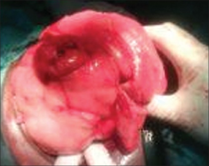
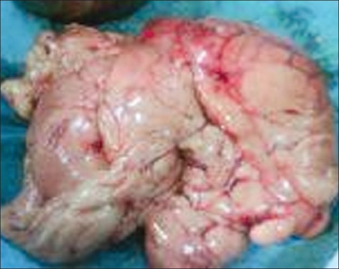
A six-month-old baby girl presented to our Accident and Emergency Department with progressive abdominal distention and recurrent constipation, with episodes lasting four to nine days, for all of the four-month duration, and significant wasting. There was no history of delayed passage of the meconeum, no vomiting, and no fever. The child was in respiratory distress, the abdomen was grossly distended, tensed, and nontender, but with uniform dullness to percussion [Figure 1]. The above-mentioned features were in-keeping with subacute intestinal obstruction. A plain X-ray of the abdomen revealed a soft tissue mass displacing and compressing the bowel loops, with the dome of the diaphragm at the level of the fourth rib anteriorly. An ultrasound (US) examination of the abdomen showed a huge heterogeneously hyperechoic mass occupying the whole abdomen, with the bowel loops displaced toward the periphery. The liver and spleen were also compressed and displaced upward. No obvious calcification was seen. Thin fibrous septations were also noted within the lesion. On Doppler scanning of the abdomen the vessels were noted to be traversing the lesion. Further evaluation with CT and/or MRI was not done. The patient was optimized and the nasogastric tube (NGT) drained, with scanty, clear effluent and no reduction of the abdominal girth. An emergency laparotomy was performed, which revealed a huge yellowish tumor arising from the retroperitoneal space occupying the whole abdomen [Figure 2]. By careful dissection the tumor was removed, It measured 26 × 19 × 13 cm and weighed 1.7 kg [Figure 3]. The patient had an uneventful recovery. The histological examination of the excised specimen confirmed lipoma.
Figure 1.

Gross abdominal distention due to retroperitoneal lipoma
Figure 2.

Retroperitoneal lipoma being delivered at the laparotomy
Figure 3.

The lipoma weighing 1.7 kg
DISCUSSION
Retroperitoneal lipoma is an unusual entity that is most often found in adults between 40 and 60 years of age and rarely occurs in the first decade of life. Retroperitoneal benign lipomas are extremely rare and represent about 2.9% of all primary retroperitoneal tumors and about 80% of them are malignant neoplasms.[3] In a series of 190 retroperitoneal tumors in infants and children only two were lipomata.[4] Till 1979, only 12 cases of retroperitoneal lipoma in children, diagnosed in the first decade of life, were reported in the literature.[4,5]
Clinically, these lipomas produce few symptoms and therefore tend to become large before being discovered.[6] The only report of malignant degeneration was by Kretschmer, who removed a large lipofibrosarcoma from a two-yearr-old girl.[5] Lipoma of the abdominal cavity usually presents as an asymptomatic abdominal mass or progressive abdominal distention. Lipomas are sometimes detected incidentally as an intraperitoneal radiolucent fat density mass, on a CT scan. Ultrasonograpy may present a confusing picture in some cases of retroperitoneal lipoma. Two cases of histologically proven giant retroperitoneal lipomas, evaluated by CT and sonography, appear to be similar on CT, but they exhibit different echographic patterns sonographically.[7] Weather the lipoma is congenital in infants is uncertain, as abdominal masses are not seen at birth in most cases,[4] but the presence of a huge abdominal mass in early infancy, in this patient, indicates that this retroperitoneal lipoma is most probably congenital. The lipoma may be too small at birth to be recognized. The long-term behavior of retroperitoneal lipoma in children is not well-defined as compared to adults, due to the inadequate number of cases,[4] therefore, a long-term follow-up is essential in children.
CONCLUSION
Retroperitoneal lipoma is rare in children, more especially in infancy; the emergency presentation was due to pressure symptoms. The characteristic behavior in children is yet to be defined; therefore a long-term follow-up is recommended.
Footnotes
Source of Support: Nil
Conflict of Interest: None declared.
REFERENCES
- 1.Srinivasan KG, Gaikwad A, Ritesh K, Ushanandini KP. Giant omental and mesenteric lipoma in an infant. Afr J Paediatr Surg. 2009;6:68–9. doi: 10.4103/0189-6725.48585. [DOI] [PubMed] [Google Scholar]
- 2.Pereira JM, Sirlin CB, Pinto PS, Casola G. CT and MR imaging of extrahepatic fatty masses of the abdomen and pelvis: techniques, diagnosis, differential diagnosis, and pitfalls. Radiographics. 2005;25:69–85. doi: 10.1148/rg.251045074. [DOI] [PubMed] [Google Scholar]
- 3.Shingo U, Masafumi K, Megumi O, Naoyuki M, Yukiyo Y. Retroperitoneal lipoma arising from the urinary bladder. Rare Tumors. 2009;1:12–3. doi: 10.4081/rt.2009.e13. [DOI] [PMC free article] [PubMed] [Google Scholar]
- 4.Weitzner S, Blumenthal IB, Moynihan PC. Retroperitoneal lipoma in children. J Pediatr Surg. 1979;14:88–90. doi: 10.1016/s0022-3468(79)80585-0. [DOI] [PubMed] [Google Scholar]
- 5.Wolk DP, Schuster KM. Retroperitoneal lipoma in a child. Arch Surg. 2000;136:343–4. doi: 10.1016/0022-3468(72)90143-1. [DOI] [PubMed] [Google Scholar]
- 6.Bowen A, Gaisie G, Bron K. Retroperitoneal lipoma in children choosing among the diagnostic imaging modalities. Paediatr Radiol. 1982;12:221–5. doi: 10.1007/BF00971768. [DOI] [PubMed] [Google Scholar]
- 7.Brazilai M, Biterman A. Two sonographic presentations of giant retroperitoneal lipomas. J Diagn Med Sonogr. 1997;13:140–2. [Google Scholar]


