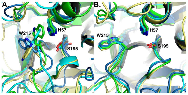
Figure 3. E* and E in the trypsin fold.
Active site conformations of relevant trypsin-like proteases in the E* form (A) or the E form (B). Note how the active site is fully open in the E form (B), but occluded to various degrees in the E* form (A). Structures are colored as follows: (A) prostasin (3DFJ, yellow), thrombin mutant N143P (3JZ2, cyan), complement factor D mutant S215W (1DST, green), prophenoloxidase activating factor II (2B9L, blue), (B) complement factor C1r (1MD8, yellow), thrombin mutant N143P (cyan, 3QGN), neuropsin (1NPM, green), trypsin (2G51, blue).

