Abstract
In order to the neuroprotective effect of Lycium chinense fruit (LCF), the present study examined the effects of Lycium chinense fruit on learning and memory in Morris water maze task and the choline acetyltransferase (ChAT) and cyclic adenosine monophosphate (cAMP) of rats with trimethyltin (TMT)-induced neuronal and cognitive impairments. The rats were randomly divided into the following groups: naïve rat (Normal), TMT injection+saline administered rat (control) and TMT injection+LCF administered rat (LCF). Rats were administered with saline or LCF (100 mg/kg, p.o.) daily for 2 weeks, followed by their training to the tasks. In the water maze test, the animals were trained to find a platform in a fixed position during 6d and then received 60s probe trial on the 7th day following removal of platform from the pool. Rats with TMT injection showed impaired learning and memory of the tasks and treatment with LCF (p<0.01) produced a significant improvement in escape latency to find the platform in the Morris water maze at the 2nd day. Consistent with behavioral data, treatment with LCF also slightly reduced the loss of ChAT and cAMP in the hippocampus compared to the control group. These results demonstrated that LCF has a protective effect against TMT-induced neuronal and cognitive impairments. The present study suggests that LCF might be useful in the treatment of TMT-induced learning and memory deficit.
Keywords: trimethyltin (TMT), choline acetyltransferase (ChAT), cyclic adenosine monophosphate (cAMP), Lycium chinense fruit (LCF)
INTRODUCTION
Trimethyltin (TMT) is a potent neurotoxicant which produces a dose-dependent degeneration of neurons in the limbic system (Dyer et al., 1982a, b; Chang and Dyer, 1983a; Earley et al., 1992), particularly the hippocampus, amygdale and entorhinal cortex (Dyer et al., 1982a, b; McMillan and Wenger, 1985; Balaban et al., 1988). The molecular basis of selective vulnerability of specific neuronal populations to neuronal insults has been a key focus in neurology and neuropathology (Brabeck et al., 2002). TMT-induced neurodegeneration is characterized by massive neuronal death, mainly localized in the limbic system, especially in the hippocampus, accompanied by reactive gliosis, epilepsy and marked neurobehavioral alterations, and is considered a useful model of neurodegeneration and selective neuronal death (Balaban et al., 1988; Koczyk et al., 1996b; Ishida et al., 1997; Brabeck et al., 2002; Geloso et al., 2002, 2004).
In the rat, TMT impairs hippocampally-dependent behaviors, including passive avoidance (Walsh et al., 1982a), water maze (Hagan et al., 1988; Earley et al., 1992) and working and/or reference memory measured in the radial arm maze (Walsh et al., 1982b; Miller and O'Callaghan, 1984; Bushnell and Angell, 1992; Alessandri et al., 1994). Furthermore, behavioral studies have shown increased locomotor activity, disruption in self-grooming and learning deficits in TMT-intoxicated rats (Swartzwelder et al., 1981, 1982; Cohen et al., 1987; Hagan et al., 1988; Segal, 1988; Cannon et al., 1991, 1994 a, b; Woodruff et al., 1991; Earley et al., 1992; Messing et al., 1992; Alessandri et al., 1994). TMT intoxication impairs the acquisition of water maze and Biel maze (water avoidance) task as well as Hebb-Williams maze and radial arm maze performance (Walsh et al., 1982a, b; O'Connell et al., 1994a, b, 1996; Ishida et al., 1997). Moreover, TMT has been shown to produce effects on operant behavior, since TMT-intoxicated rats had higher lever pressing rats under a fixed-ratio schedule of food presentation (Swartzwelder et al., 1981) and TMT impaired the performance of differential reinforcement at low response rates in an operant schedule (Woodruff et al., 1991). These anatomical and behavioral findings have made TMT-intoxicated rats an attractive model for degenerative diseases such as Alzheimer's disease, the most common cause of dementia (Woodruff et al., 1994).
Herbs have recently become attractive as health-beneficial foods (physiologically functional foods) and as a source material for the development of drugs. Herbal medicines derived from plant extracts are being utilized increasingly to treat a wide variety of clinical diseases, with relatively little knowledge regarding their modes of action (Matthews et al., 1999). Lycium chinense fruit is widely distributed in East Asia and has been used traditionally for anti-aging purpose (Xiao et al., 1999). Recently, the neuro-protective effect of Lycium chinense fruit has been reported by Geloso et al. (2002, 2004), but the underlying mechanism of protective action is not fully understood.
The present study was undertaken to evaluate the neuroprotective effect of Lycium chinense fruit on TMT-induced learning and memory deficits in the rats and to elucidate the mechanism underlying these protective effects in rats. Rats were tested on Morris water maze for the spatial learning and memory. The analyzed parameters included choline acetyltransferase (ChAT) and cyclic adenine monophosphate (cAMP) in the hippocampus.
MATERIALS AND METHODS
Animals and experimental design
Male Sprague-Dawley rats weighting 250~280 g each were purchased from Samtaco Animal Corp. (Kyungki-do, Korea). The animals were allowed to acclimatize themselves for at least 7 days prior to the experimentation. The animals were housed in individual cages under light-controlled conditions (12/12-hr light/dark cycle) and at 23℃ room temperature. Food and water were available ad libitum. All experiments were approved by the catholic university institutional animal care and use committee. They were allowed at least 1week to adapt to their environment before the experiments.
The rats were injected intraperitoneally (i.p.) with TMT (6.0 mg/kg, body weight) dissolved in 0.9% saline and then returned to their home cages. The rats were randomly divided into the following groups (n=8 per group): the naïve rat (Normal), TMT injection+Saline administered rat (Control), and TMT injection+LCF administered rat (LCF). Drug (400 mg/kg) was dissolved in saline and orally administered for two weeks after TMT injection. From the 15th after the injection of drug, the water maze test was performed for one week.
Morris water maze test
The swimming pool of the Morris water maze was a circular water tank 200 cm in diameter and 35 cm deep. It was filled to a depth of 21 cm with water at 23±2℃. A platform 15 cm in diameter and 20 cm in height was placed inside the tank with its top surface being 1.5cm below the surface of the water. The pool was surrounded by many cues that were external to the maze (D'Hooge and De deyn, 2001). A CCD camera was equipped with a personal computer for the behavioral analysis. Each rat was received four daily trials. For 6 consecutive days, the rats were tested with three acquisition tests. They also received retention tests on the 7th day. For the acquisition test, the rat was allowed to search for the hidden platform for 180s and the latency to escape onto the platform was recorded. The animals were trained to find the platform that was in a fixed position during 6 days for the acquisition test, and then for the retention test (at the 7th day), they received a 1 min probe trial in which the platform was removed from the pool. The intertrial interval time was 1 min. Performance of the test animals in each water maze trial was assessed by a personal computer for the behavioral analysis (S-mart program, Spain).
ChAT, cAMP immunohistochemistry
At the end of the behavioral observation, the animals were deeply anesthetized with sodium pentobarbital (100 mg/kg, i.p.) and then perfused transcardially with 100 ml of saline, followed by 500 ml of a 4% solution of formaldehyde prepared in phosphate buffer. The brains were then removed, postfixed in the same fixative for two to three hours at 4℃ and then placed overnight at 4℃ in PBS containing 20% sucrose. On the following day, the brain was cut into coronal sections that were sliced to 30 µm-thicknesses. The primary rabbit polyclonal antibodies against the following specific antigen were used: Cholinacetyl transferase (ChAT, concentration 1 : 2,000; Cambridge Research Biochemicals, Wilmington, DE), cAMP (concentration 1 : 200; Cambridge Research Biochemicals, Wilmington, DE). The primary antibody was prepared at a dilution of 2,000x in 0.3% PBST, 2% normal rabbit serum and 0.001% kehole limpit hemocyanin (Sigma, CA, USA). The sections were incubated in the primary antiserum for 72 h at 4℃. After three more rinses in PBST, the sections were placed in Vectastain Elite ABC reagent (Vector laboratories, Burlingame, CA) for 2 h at room temperature. Following a further rinsing in PBS, the tissue was developed using diaminobenzadine (Sigma, CA, USA) as the chromogen. Images were captured using an Axio Vision 3.0 imaging systems (Zeiss, Oberkochen, Germany) and processed in Adobe Photoshop. For measuring cells of ChAT, the grid was placed on CA1 and CA3 in the hippocampus areas according to the method of Paxinos et al. (1985). The number of cells was counted at 100x magnification using a microscope rectangle grid measuring 200×200 µm.
Statistical analysis
Statistical comparisons were done for the behavioral and histochemical studies using the one-way ANOVA, repeated measure of ANOVA, respectively and Tukey post hoc was done. All of the results were presented as means±S.E.M., and we used SPSS 15.0 for Windows for analysis of the statistics. The significance level was set at p<0.05.
RESULTS
Effect of LCF on performance in water maze task
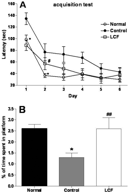
Fig. 1A shows mean group latencies to reach to hidden platform in the Morris water maze for all groups for 6 days. The escape latency differed among the groups when the results were averaged over all the session. The control group showed a worse performance than did the normal group (at the Day 1, 2, respectively). There were no significant main effects, but there was a slight trend for a significant interaction effect on the distance traveled to reach the platform.
Fig. 1.
(A) The latency to escape onto the hidden platform during the Morris water maze. The task was performed with 3 trials per day during 6 days for the acquisition test. The values are presented as means±S.E.M. *p<0.05 vs. normal group & #p<0.05 vs. control group, respectively. (B) Retention performance was tested on 7th day. The rats received a 1 min probe trial in which the platform was removed from the pool for retention testing. The values are presented as means±S.E.M. *p<0.05 vs. normal group & ##p<0.01 vs. control group, respectively.
To examine the spatial memory of rats, the time spent swimming to the platform was compared and analysis is illustrated in Fig. 1B. The times spent to the platform were significantly different among the groups [F2,24=7.6, p<0.05]; the normal group spent more time around the platform than the control group (p<0.05 for the normal group). However, treatment of LCF was significantly increased the time spent around the platform (p<0.01).
ChAT immunoreactive neurons of the hippocampus
The results of determining the ChAT immunoreative cells per section from different hippocampal formations are shown in Fig. 2. The number of ChAT neurons in the CA1 area was 19.8±4.6 in the normal group, 16.5.7±0.3 in the control group and 18.5±1.9 in the LCF group [F2,11=0.678]. Also, the ChAT immunoreactive cells in the CA3 area were 16.5±2.6 in the normal group, 16.5±1.6 in the control group and 17.5±1.6 in the LCF group [F2,11=0.907]. The number of ChAT positive neurons in the hippocampal CA1 was slightly increased in the LCF group compared to the control group. However, there were no statistically significant differences.
Fig. 2.
The number of choline acetyltransferase (ChAT) immunostained nuclei in different hippocampal CA1 and CA3 of the experimental groups. Each values represents the ±S.E.M.
cAMP immunoreactive neurons of the hippocampus
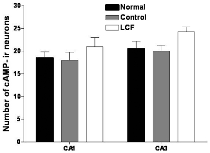
The results of determining the cAMP immunoreative cells per section from different hippocampal formations are shown in Fig. 3. The number of cAMP neurons in the CA1 area was 18.5±1.3 in the normal group, 18.0±1.8 in the control group and 21.0±1.95 in the LCF group [F2,11=0.446]. Also, the cAMP immunoreactive cells in the CA3 area were 20.5±1.7 in the normal group, 20±1.3 in the control group and 24.3±1.0 in the LCF group [F2,11=0.103]. The number of cAMP positive neurons was control group and 24.3±1.0 in the LCF group [F2,11=0.103]. The number of cAMP positive neurons was increased to 121.2% of the control in the LCF group (p<0.05). The number of cAMP positive neurons in the hippocampus was increased to 116~120% of the control in the LCF group. However, there were no statistically significant differences.
Fig. 3.
The number of cyclic adenosine monophosphate (cAMP) immunostained nuclei in different hippocampal CA1 and CA3 of the experimental groups. Each values represents the ±S.E.M.
DISCUSSION
The present study demonstrated that TMT injections produced severe deficits in rat performance in a Morris water maze along with signs of neurodegeration, including decreased ChAT and cAMP activity in the hippocampus. Treatment with LCF attenuated TMT-induced learning and memory deficits in the maze and had a protective effect against TMT-induced decrease in ChAT and cAMP positive neurons.
Intozication with trimethyltin (TMT) leads to profound behavioural and cognitive deficits in both humans (Fortemps et al., 1978) and experimental animals (Dyer et al., 1982a, b; Ishida et al., 1997). In rats, TMT induced the degeneration of pyramidal neurons in the hippocampus and cortical areas (pyriform cortex, entorhinal cortex, subiculum) connected to the hippocampus, but there is also neuronal loss in the association areas (Brown et al., 1979; Chang and Dyer, 1983a, b; Chang et al., 1983a, b; Chang and Dyer, 1985; Balaban et al., 1988; Koczyk et al., 1996a, b).
Futhermore, behavioural studies have shown increased disruption in memory and learning deficits in TMT-intoxicated rats (Swartzwelder et al., 1982; Andersson et al., 1995). TMT intoxication impairs the acquisition of water maze performance (Hagan et al., 1988; Segal, 1988; Earley et al., 1992). The Morris water maze is well-established paradigm for evaluating deficits in hippocampal-dependent memory and the MWM spatial learning task has been used in the validation of rodent models for neurocognitive disorders and for the evaluation of possible neurocognitive treatments (D'Hooge and De Deyn, 2001; Luine et al., 2003). The impairment in spatial learning produced by TMT in the current studies is consistent with previous reports of spatial learning impairments (Walsh et al., 1982a, b; Hagan et al., 1988; Earley et al., 1992; Alessandri et al., 1994). However, this study proved that spatial memory continued to improve in LCF group during the training days compared to the control group. Also, the data of spatial probe trial demonstrated that LCF protects against the TMT-induced decrease of the spatial retention, especially long-term memory. It has been previously reported that LCF has profound curative effects on improving the memory and cognitive function of AD-like animal model (Sun et al., 2003). Also, Deng et al showed that the treatment of Lucium barbarum polysaccharide, a compound of LCF, enhanced the learning and memory abilities in the rats (Deng et al., 2003). These results proved that LCF ameliorated disordered learning of TMT induced rat.
The neuroprotective effects of these herbal drugs on the central acetylcholine system were also examined by histochemistry of hippocampal neurons. The degeneration of the cholinergic innervation from the basal forebrain to the hippocampal formation in the temporal lobe is thought to be one of the factors determining the progression of memory decay, both during normal aging and AD (Wu et al., 1999). The best available marker for cholinergic neurons in the basal forebrain is ChAT activity. ChAT synthesizes the neurotransmitter acetylcholine, basal forebrain and the cortex, hippocampus, and amygdala. A significant reduction in ChAT activity in the postmortem brains of demented patients has been reported. In addition, there is a 20-50% decrease in ChAT activity in the hippocampus of the TMT-induced rats. However, LCF modulates the function of these neurons and plays a role in their maintenance by preventing the TMT-induced decrease in ChAT activity (Rabbani et al., 1997; Chen et al., 2000). The present results show that LCF exerts beneficial effects on cholinergic neurotransmission in the brain by increasing the hippocampus ChAT activities.
In the brain, activation of cAMP signaling occurs after stimulation of adenylyl cylases by stimulatory G-proteins after binding of an extracellular ligand to a G-protein-coupled receptor (GPCR) and by Ca2+ through the Ca2+ -binding protein calmodulin (Wang and Strom, 2003). Genetic and pharmacological studies provide strong evidence that the cAMP signaling pathway is crucial for learning and memory across species (Kandal, 2004; Skoulakis and Grammenoudi, 2006). However, critical questions remain about the potential of this pathway as a target for memory enhancement because of contradictory results from pharmacological and genetic studies. Our results showed that the levels of cAMP in the hippocampus had no significant difference among the groups. It has been previously reported that short-term memory may be dependent on cAMP signaling (Ahi et al., 2004) and that it may be enhanced after pharmacological immediate posttraining increases of hippocampal cAMP (Vianna et al., 2000). In contrast, by using our conditional system, there was no close relation between enhancement of memory and the change of cAMP expression in the hippocampus on TMT-induced rats.
In summary, treatment with LCF attenuated TMT-induced learning and memory deficits in the Morris water maze and had a protective effect against TMT-induced decreased in cholinergic neurons. LCF is thus a good candidate for further investigations that may ultimately result in its clinical use. Further work examining the effects of LCF activation on additional behavioral tasks will help to elucidate whether increasing cAMP signaling may also facilitate other types of memory.
References
- 1.Ahi J, Radulovic J, Spiess J. The role of hippocampal signaling cascades in consolidation of fear memory. Behav Brain Res. 2004;149:17–31. doi: 10.1016/s0166-4328(03)00207-9. [DOI] [PubMed] [Google Scholar]
- 2.Alessandri B, FitzGerald RE, Schaeppi U, Krinke GJ, Classen W. The use of an unbaited tunnel maze in neurotoxicology: I. Trimethyltin-induced brain lesions. Neurotoxicology. 1994;15:349–357. [PubMed] [Google Scholar]
- 3.Andersson H, Luthman J, Lindqvist E, Olson L. Time-course of trimethyltin effects on the monoamiergic systems of the rat brain. Neurotoxicology. 1995;16:201–210. [PubMed] [Google Scholar]
- 4.Balaban CD, O'Callaghan JP, Billingsley ML. Trimethyltin-induced neuronal damage in the rat brain: comparative studies using silver degeneration stains, immunocytochemistry and immuneassay for neuronotypic and gliotypic proteins. Neuroscience. 1988;26:337–361. doi: 10.1016/0306-4522(88)90150-9. [DOI] [PubMed] [Google Scholar]
- 5.Brabeck C, Michetti F, Geloso MC, Corvino V, Goezalan F, Meyermann R, Schluesener HJ. Expression of EMAP-II by activated monocytes/microglial cells in different regions of the rat hippocampus after trimethyltin-induced brain damage. Exp Neurol. 2002;177:341–346. doi: 10.1006/exnr.2002.7985. [DOI] [PubMed] [Google Scholar]
- 6.Brown AW, Aldridge WN, Street BW, Verschoyle RD. The behavioral and neuropathologic sequelae of intoxication by trimethyltin compounds in the rat. Am J Pathol. 1979;97:59–82. [PMC free article] [PubMed] [Google Scholar]
- 7.Bushnell PJ, Angell KE. Effect s of trimethyltin on repeated acquisition (learning) in the radial arm maze. Neurotoxicology. 1992;13:429–441. [PubMed] [Google Scholar]
- 8.Cannon RL, Hoover DB, Baisden RH, Woodruff ML. Effects of trimethyltin (TMT) on choline acetyltransferase activity in the rat hippocampus. Influence of dose and time following exposure. Mol Chem Neuropathol. 1994a;23:27–45. doi: 10.1007/BF02858505. [DOI] [PubMed] [Google Scholar]
- 9.Cannon RL, Hoover DB, Baisden RH, Woodruff ML. The effect of time following exposure to trimethyltin (TMT) on cholinergic muscarinic receptor binding in rat hippocampus. Mol Chem Neuropathol. 1994b;23:47–62. doi: 10.1007/BF02858506. [DOI] [PubMed] [Google Scholar]
- 10.Cannon RL, Hoover DB, Woodruff ML. Trimetyltin increases choline acethyltransferase in rat hippocampus. Neurotoxicol Teratol. 1991;13:241–244. doi: 10.1016/0892-0362(91)90017-q. [DOI] [PubMed] [Google Scholar]
- 11.Chang LW, Dyer RS. A time-course study of trimethyltin induced neuropathology in rats. Neurobehav Toxicol Teratol. 1983a;5:443–459. [PubMed] [Google Scholar]
- 12.Chang LW, Dyer RS. Trimethyltin induced pathology in sensory neurons. Neurobehav Toxicol Teratol. 1983b;5:673–696. [PubMed] [Google Scholar]
- 13.Chang LW, Dyer RS. Septotemporal gradients of trimethyltin-induced hippocampal lesions. Neurobehav Toxicol Teratol. 1985;7:43–49. [PubMed] [Google Scholar]
- 14.Chang LW, Tiemeyer TM, Wenger GR, McMillan DE. Neuropathology of trimethyltin intoxication. III. Changes in the brain stem neurons. Environ Res. 1983a;30:399–411. doi: 10.1016/0013-9351(83)90226-8. [DOI] [PubMed] [Google Scholar]
- 15.Chang LW, Wenger GR, McMillan DE, Dyer RS. Species and strain comparison of acute neurotoxic effects of trimethyltin in mice and rats. Neurobehav Toxicol Teratol. 1983b;5:337–350. [PubMed] [Google Scholar]
- 16.Chen G, Chen KS, Knox J, Inglis J, Bernard A, Martin SJ, Justice A, McConlogue L, Games D, Freedman SB, Morris RG. A learning deficit related to age and β-amyloid plaques in a mouse model of Alzheimer's disease. Nature. 2000;408:975–979. doi: 10.1038/35050103. [DOI] [PubMed] [Google Scholar]
- 17.Cohen CA, Messing RB, Sparber SB. Selective learning impairment of delayed reinforcement autoshaped behavior caused by low doses of trimethyltin. Psychopharmacology (Berl) 1987;93:301–307. doi: 10.1007/BF00187247. [DOI] [PubMed] [Google Scholar]
- 18.Deng HB, Cui DP, Jiang JM, Feng YC, Cai NS, Li DD. Inhibiting effects of Achyranthes bidentata polysaccharide and Lycium barbarum polysaccharide on nonenzyme glycation in D-galactose induced mouse aging model. Biomed Environ Sci. 2003;16:267–275. [PubMed] [Google Scholar]
- 19.Dyer RS. Physiological methods for assessment of Trimethyltin exposure. Neurobehav Toxicol Teratol. 1982a;4:659–664. [PubMed] [Google Scholar]
- 20.Dyer RS, Deshields TL, Wonderlin WF. Trimethyltin-induced changes in gross morphology of the hippocampus. Neurobehav Toxicol Teratol. 1982b;4:141–147. [PubMed] [Google Scholar]
- 21.Earley B, Burke M, Leonard BE. Behavioural, biochemical and histological effects of trimethyltin (TMT) induced brain damage in the rat. Neurochem Int. 1992;21:351–366. doi: 10.1016/0197-0186(92)90186-u. [DOI] [PubMed] [Google Scholar]
- 22.Fortemps E, Amand G, Bomboir A, Lauwerys R, Laterre EC. Trimethyltin poisoning: report of two cases. Int Arch Occup Environ Health. 1978;41:1–6. doi: 10.1007/BF00377794. [DOI] [PubMed] [Google Scholar]
- 23.Geloso MC, Corvino V, Cavallo V, Toesca A, Guadagni E, Passalacqua R, Michetti F. Expression of astrocytic nestin in the rat hippocampus during trimethyltin-induced neurodegeneration. Neurosci Lett. 2004;357:103–106. doi: 10.1016/j.neulet.2003.11.076. [DOI] [PubMed] [Google Scholar]
- 24.Geloso MC, Vercelli A, Corvino V, Repici M, Boca M, Haglid K, Zelano G, Michetti F. Cyclooxygenase-2 and caspase 3 expression in trimethyltin-induced apoptosis in the mouse hippocampus. Exp Neurol. 2002;175:152–160. doi: 10.1006/exnr.2002.7866. [DOI] [PubMed] [Google Scholar]
- 25.Hagan JJ, Jansen JH, Broekkamp CL. Selective behavioural impairment after acute intoxication with trimethyltin (TMT) in rats. Neurotoxicology. 1988;9:53–74. [PubMed] [Google Scholar]
- 26.D'Hooge R, De Deyn PP. Applications of the Morris water maze in the study of learning and memory. Brain Res Brain Res Rev. 2001;36:60–90. doi: 10.1016/s0165-0173(01)00067-4. [DOI] [PubMed] [Google Scholar]
- 27.Ishida N, Akaike M, Tsutsumi S, Kanai H, Masui A, Sadamatsu M, Kuroda Y, Watanabe Y, McEwen BS, Kato N. Trimethyltin syndrome as a hippocampal degeneration model: temporal changes and neurochemical features of seizure susceptibility and learning impairment. Neuroscience. 1997;81:1183–1191. doi: 10.1016/s0306-4522(97)00220-0. [DOI] [PubMed] [Google Scholar]
- 28.Kandel ER. The molecular biology of memory storage: a dialog between genes and synapses. Biosci Rep. 2004;24:475–522. doi: 10.1007/s10540-005-2742-7. [DOI] [PubMed] [Google Scholar]
- 29.Koczyk D. How does trimethyltin affect the brain: facts and hypotheses. Acta Neurobiol Exp (Wars) 1996a;56:587–596. doi: 10.55782/ane-1996-1164. [DOI] [PubMed] [Google Scholar]
- 30.Koczyk D, Skup M, Zaremba M, Oderfeld-Nowak B. Trimethyltin-induced plastic neuronal changes in rat hippocampus are accompanied by astrocytic trophic activity. Acta Neurobiol Exp (Wars) 1996b;56:237–241. doi: 10.55782/ane-1996-1126. [DOI] [PubMed] [Google Scholar]
- 31.Luine VN, Jacome LF, Maclusky NJ. Rapid enhancement of visual and place memory by estrogens in rats. Endocrinology. 2003;144:2836–2844. doi: 10.1210/en.2003-0004. [DOI] [PubMed] [Google Scholar]
- 32.Matthews HB, Lucier GW, Fisher KD. Medicinal herbs in the United States: research needs. Environ Health Perspect. 1999;107:773–778. doi: 10.1289/ehp.99107773. [DOI] [PMC free article] [PubMed] [Google Scholar]
- 33.McMillan DE, Wenger GR. Neurobehavioral toxicology of trialkyltins. Pharmacol Rev. 1985;37:365–379. [PubMed] [Google Scholar]
- 34.Messing RB, Devauges V, Sara SJ. Limbic forebrain toxin trimethyltin reduces behavioral suppression by clonidine. Pharmacol Biochem Behav. 1992;42:313–316. doi: 10.1016/0091-3057(92)90532-k. [DOI] [PubMed] [Google Scholar]
- 35.Miller DB, O'Callaghan JP. Biochemical, functional and morphological indicators of neurotoxicity: effects of acute administration of trimethyltin to the developing rat. J Pharmacol Exp Ther. 1984;231:744–751. [PubMed] [Google Scholar]
- 36.O'Connell A, Earley B, Leonard BE. Changes in muscarinic (M1 and M2 subtypes) and phencyclidine receptor density in the rat brain following trimethyltin intoxication. Neurochem Int. 1994a;25:243–252. doi: 10.1016/0197-0186(94)90068-x. [DOI] [PubMed] [Google Scholar]
- 37.O'Cconnell A, Earley B, Leonard BE. The neuroprotective effect of tacrine on trimethyltin induced memory and muscarinic receptor dysfunction in the rat. Neurochem Int. 1994b;25:555–566. doi: 10.1016/0197-0186(94)90154-6. [DOI] [PubMed] [Google Scholar]
- 38.O'Connell AW, Earley B, Leonard BE. The sigma ligand JO 1784 prevents trimethyltin-induced behavioural and sigma-receptor dysfunction in the rat. Pharmacol Toxicol. 1996;78:296–302. doi: 10.1111/j.1600-0773.1996.tb01378.x. [DOI] [PubMed] [Google Scholar]
- 39.Paxinos G, Watson C, Pennisi M, Topple A. Bregma, lambda and the interaural midpoint in stereotaxic surgery with rats of different sex, strain and weight. J Neurosci Methods. 1985;13:139–143. doi: 10.1016/0165-0270(85)90026-3. [DOI] [PubMed] [Google Scholar]
- 40.Rabbani O, Panickar KS, Rajakumar G, King MA, Bodor N, Meyer EM, Simpkins JW. 17β-Estradiol attenuates fimbrial lesion-induced decline of ChAT-immunoreactive neurons in the rat medial septum. Exp Neurol. 1997;146:179–186. doi: 10.1006/exnr.1997.6516. [DOI] [PubMed] [Google Scholar]
- 41.Segal M. Behavioral and physiological effects of trimethyltin in the rat hippocampus. Neurotoxicology. 1988;9:481–489. [PubMed] [Google Scholar]
- 42.Skoulakis EM, Grammenoudi S. Dunces and da Vincis: the genetics of learning and memory in Drosophila. Cell Mol Life Sci. 2006;63:975–988. doi: 10.1007/s00018-006-6023-9. [DOI] [PMC free article] [PubMed] [Google Scholar]
- 43.Sun H, Hu Y, Zhang JM, Li SY, He W. Effects of one Chinese herbs on improving cognitive function and memory of Alzheimer's disease mouse models. Zhongguo Zhong Yao Za Zhi. 2003;28:751–754. [PubMed] [Google Scholar]
- 44.Swartzwelder HS, Dyer RS, Holahan W, Myers RD. Activity changes in rats following acute trimethyltin exposure. Neurotoxicology. 1981;2:589–593. [PubMed] [Google Scholar]
- 45.Swartzwelder HS, Hepler J, Holahan W, King SE, Leverenz HA, Miller PA, Myers RD. Imparied maze performance in the rat caused by trimethyltin treatment: problem-solving deficits and perseveration. Neurobehav Toxicol Teratol. 1982;4:169–176. [PubMed] [Google Scholar]
- 46.Vianna MR, Izquierdo LA, Barros DM, Ardenghi P, Pereira P, Rodrigues C, Moletta B, Medina JH, Izquierdo I. Differential role of hippocampal cAMP-dependent protein kinase in short- and long-term memory. Neurochem Res. 2000;25:621–626. doi: 10.1023/a:1007502918282. [DOI] [PubMed] [Google Scholar]
- 47.Walsh TJ, Gallagher M, Bostock E, Dyer RS. Trimethyltin impairs retention of a passive avoidance task. Neurobehav Toxicol Teratol. 1982a;4:163–167. [PubMed] [Google Scholar]
- 48.Walsh TJ, Miller DB, Dyer RS. Trimethyltin, a selective limbic system neurotoxicant, impairs radial-arm maze performance. Neurobehav Toxicol Teratol. 1982b;4:177–183. [PubMed] [Google Scholar]
- 49.Wang H, Storm DR. Calmodulin-regulated adenylyl cyclases: cross-talk and plasticity in the central nervous system. Mol Pharmacol. 2003;63:463–468. doi: 10.1124/mol.63.3.463. [DOI] [PubMed] [Google Scholar]
- 50.Woodruff ML, Baisden RH, Cannon RL, Kalbfleisch J, Freeman JN., 3rd Effects of trimethyltin on acquisition and reversal of a light-dark discrimination by rats. Physiol Behav. 1994;55:1055–1061. doi: 10.1016/0031-9384(94)90387-5. [DOI] [PubMed] [Google Scholar]
- 51.Woodruff ML, Baisden RH, Nonneman AJ. Anatomical and behavioral sequelae of fetal brain transplants in rats with trimethyltin-induced neurodegeneration. Neurotoxicology. 1991;12:427–444. [PubMed] [Google Scholar]
- 52.Wu X, Glinn MA, Ostrowski NL, Su Y, Ni B, Cole HW, Bryant HU, Paul SM. Raloxifene and estradiol benzoate both fully restore hippocampal choline acetyltransferase activity in ovariectomized rats. Brain Res. 1999;847:98–104. doi: 10.1016/s0006-8993(99)02062-4. [DOI] [PubMed] [Google Scholar]
- 53.Xiao Y, Harry GJ, Pennypacker KR. Expression of AP-1 transcription factors in rat hippocampus and cerebellum after trimethyltin neurotoxicity. Neurotoxicology. 1999;20:761–766. [PubMed] [Google Scholar]


