Abstract
Introduction: An entirely new type of staphyloma has been recently described as dome-shaped macula (DSM). It is characterized by an abnormal convex macular contour within the concavity of a posterior staphyloma. We found DSM associated with serous macular detachment (SMD) and tilted disc in two consecutive cases. Case Reports: Case 1: A 37-year-old female presented to our department because of sudden onset blurred vision in her right eye (OD). The best-corrected visual acuity (BCVA) was 0.5 in both eyes. Funduscopy evidenced bilateral tilted disc associated with posterior staphyloma. Optical coherence tomography (OCT) demonstrated a DSM with SMD in her OD. After 15 months of follow-up, BCVA of her OD remained stable with chronic SMD. Case 2: A 32-year-old female presented to our department because of blurred vision in her OD. The BCVA was 0.4 in the OD and 1.0 in the left eye (OS). Bilateral tilted disc and posterior staphyloma were evidenced in the funduscopy. OCT demonstrated a bilateral DSM with SMD in her OD. After 45 months of follow-up, two further episodes of transient SMD were observed in her OD and seven in her OS. The final BCVA was 0.63 in the OD and 0.8 in the OS. Discussion: SMD associated with tilted disc constitutes a potential cause of subretinal fluid accumulation in myopic patients. OCT is essential for the detection of both SMD and DSM.
Key Words: Dome-shaped macula, Tilted disc, Serous macular detachment, Posterior staphyloma
Introduction
Myopia affects 1,600 million people worldwide, representing more than a quarter of the world's population, and its prevalence among the Spanish population is between 20 to 30%.
When myopia is no longer just a refractive error and associates with other ophthalmic abnormalities then we consider it a pathologic myopia, accounting for 9% of all cases. It is defined by the presence of refractive errors exceeding 6 negative diopters and/or axial length exceeding 26 mm. One of the typical anomalies is the protrusion of the posterior wall of the eyeball, known as posterior staphyloma. This is often associated with different pathologic changes: chorioretinal atrophy, choroidal neovascularization, foveal retinoschisis, neurosensory detachment and macular holes, all responsible for visual loss. In 2008, Gaucher et al. [1] described an entirely new type of staphyloma, known as dome-shaped macula (DSM); this type of macular staphyloma is associated with retinal pigment epithelial (RPE) disturbances coincident with the posterior rim of the posterior staphyloma wall, typically with a tilted disc, which can cause chronic or recurrent neuroepithelial detachments [2, 3].
The pathogenesis of DSM is unknown; Gaucher et al. [1] hypothesize that it is the result of resistance to deformation of scleral staphyloma or a localized choroidal thickening. On the contrary, Mehdizadeh et al. [4] point to a mechanism of hypotonia or vitreomacular traction. Tilted disc, meanwhile, may be associated with astigmatism and visual field defects. Even in eyes with normal macular anatomy, dysfunction of the retinal outer layers has been described [5].
Herein, we present two cases of myopic patients with foveal neurosensory detachment in relation to DSM and tilted disc.
Case Reports
Case 1
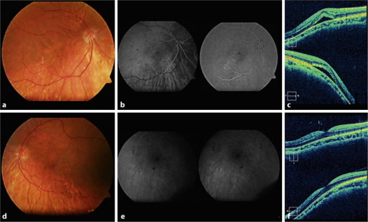
A 37-year-old female presented to our emergency department complaining of sudden onset vision loss in her right eye (OD). Best-corrected visual acuity (BCVA) was 0.5 in both eyes (OU). A posterior staphyloma and tilted disc in OU were evidenced in the funduscopy (fig. 1a–d), hinting serous macular detachment (SMD) in the OD was confirmed by optical coherence tomography (OCT) (fig. 1c). An abnormal convex profile of the macular region was noticed within the concavity of the posterior staphyloma (fig. 1c–f). The axial length measured 25.03 mm in the OD and 25.86 mm in the left eye (OS), with a spherical equivalent of −4.00 and −4.75 diopters, respectively.
Fig. 1.
a–d Color retinography of both eyes in which posterior staphyloma and tilted disc are evident. b–e Fluorescein angiography showing retinal epithelial disturbance within the inferior and temporal macular area of the right eye; the left eye shows no abnormality. c Optical coherence tomography evidences dome-shaped macula and serous macular detachment in the right eye. f Optical coherence tomography evidences the presence of dome-shaped macula without subretinal fluid in the left eye.
Fluorescein angiography (FA) ruled out the presence of a neovascular membrane and detected a leakage point corresponding to the disturbance of the retinal pigment epithelium by the posterior wall of the staphyloma (fig. 1b). After 3 months of watchful waiting, no change in the SMD was observed. Therefore, we decided to perform a focal argon laser treatment, but without any success. After 15 months of follow-up, the SMD was still present and the BCVA did not change from the initial 0.5.
Case 2
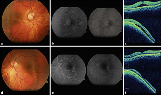
A 32-year-old female presented to our emergency department complaining of visual loss in her OD for several weeks. BCVA was 0.4 in her OD and 1 in her OS. A posterior staphyloma and tilted disc in OU were evidenced in the funduscopy (fig. 2a–d). OCT showed a DSM in OU with a SMD in her OD (fig. 2c–f). FA confirmed the presence of a subtle leakage in the OD and RPE disturbances in OU (fig. 2b–e). The axial length measured 26.39 mm in the OD and 25.24 mm in the OS, with a spherical equivalent of −7.25 and −5.50 diopters, respectively.
Fig. 2.
a–d Color retinography of both eyes in which posterior staphyloma and tilted disc are evident. b–e Fluorescein angiography showing juxtafoveal retinal pigment epithelial disturbance in the right eye, and extrafoveal retinal pigment epithelial disturbance in the left eye. c Optical coherence tomography evidences dome-shaped macula and serous macular detachment in the right eye. f Optical coherence tomography evidences the presence of dome-shaped macula without subretinal fluid in the left eye.
During the following 3 weeks, the SMD progressively disappeared. After 45 months of follow-up, two further episodes of transient SMD in her OD and 7 in her OS were observed. The final BCVA was 0.63 in the OD and 0.8 in the OS.
Discussion
The evaluation of cases with visual impairment in myopic patients has to be thorough. Typical alterations within the posterior pole (RPE disorders, chorioretinal atrophy, posterior staphyloma, lacked striae, etc.) should be added to a new OCT-based diagnosis of lesions responsible for vision loss, which is potentially treatable (neovascular membranes, macular holes, foveoschisis, etc.).
In our two cases, visual disturbance was proved to be a consequence of SMD in relation to tilted disc and DSM. OCT is useful in the detection of both SMD and DSM, which are nearly impossible to diagnose by means of a conventional funduscopy.
Currently, there is no effective treatment for SMD associated with tilted disc, although photocoagulation of accessible angiographic leaking areas has been assayed with variable outcomes. Intravitreal injection of the antiangiogenic drug bevacizumab has failed to show efficacy in a recent report [6]. Therefore, observation is recommended in these unusual cases of SMD associated with DSM and tilted disc.
Disclosure Statement
The authors have no financial/conflicting interests to disclose.
References
- 1.Gaucher D, Erginay A, Lecleire-Collet A, Haouchine B, Puech M, Cohen SY, Massin P, Gaudric A. Dome-shaped macula in eyes with myopic posterior staphyloma. Am J Ophthalmol. 2008;145:909–914. doi: 10.1016/j.ajo.2008.01.012. [DOI] [PubMed] [Google Scholar]
- 2.Leys AM, Cohen SY. Subretinal leakage in myopic eyes with a posterior staphyloma or tilted disk syndrome. Retina. 2002;22:659–665. doi: 10.1097/00006982-200210000-00025. [DOI] [PubMed] [Google Scholar]
- 3.Cohen SY, Quentel G, Guiberteau B, Delahaye-Mazza, A Gaudric. Macular serous retinal detachment caused by subretinal leakage in tilted disc syndrome. Ophthalmology. 1998;105:1831–1834. doi: 10.1016/S0161-6420(98)91024-7. [DOI] [PubMed] [Google Scholar]
- 4.Mehdizadeh M, Nowroozzadeh MH. Dome-shaped macula in eyes with myopic posterior staphyloma. Am J Ophthalmol. 2008;146:478. doi: 10.1016/j.ajo.2008.05.045. [DOI] [PubMed] [Google Scholar]
- 5.Feigl B, Zele AJ. Macular function in tilted disc syndrome. Doc Ophthalmol. 2010;120:201–203. doi: 10.1007/s10633-010-9215-4. [DOI] [PubMed] [Google Scholar]
- 6.Milani P, Pece A, Pierro K, Sidenari P, Radice P, Scialdone A. Bevacizumab for macular serous neuroretinal detachment in tilted disk syndrome. J Ophthalmol. 2010;2010:970580. doi: 10.1155/2010/970580. [DOI] [PMC free article] [PubMed] [Google Scholar]


