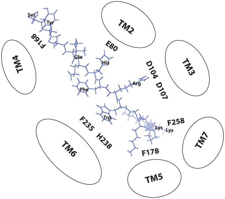
Figure 8.
Two-dimensional representation of a proposed three dimensional model illustrating ACTH1-16 docked inside the hMC2R. Based on our results, three main receptor binding pockets are proposed. The first is a predominantly ionic pocket formed by E80, D104 and D107. The second binding pocket is formed by aromatic residues F235 and H238 in TM6. The third binding pocket is formed by F178 in TM5 and F258 in TM7.

