Fig. 3.
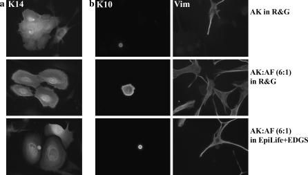
Indirect immunofluorescence of keratinocytes in different culture conditions. a Cytokeratin 14 (K14) staining showed the majority of AK were of a highly proliferative, basal phenotype. b Cytokeratin 10 (K10) staining identified a small population of differentiating AK surrounded by fibroblasts, stained with vimentin (Vim). The small areas labelled positive for the differentiation marker cytokeratin 10 (K10) show a small proportion cells that are differentiatiated. These cells were stratified in culture and more round in appearance, thus it might appear that they are smaller. The larger area stained in AK:AF is a more flattened but differentiated cell

