Abstract
Male occupants in frontal motor vehicle collisions have reduced tolerance for hip fractures than females in similar crashes. We studied 92 adult pelvic CT scans and found significant gender differences in bony pelvic geometry, including acetabular socket depth and femoral head width. Significant differences were also noted in the presentation angle of the acetabular socket to frontal loading. The observed differences provide biomechanical insight into why hip injury tolerance may differ with gender. These findings have implications for the future design of vehicle countermeasures as well as finite element models capable of more accurately predicting body tolerances to injury.
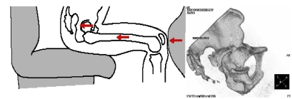
Examination of real life frontal vehicle crashes show consistent evidence of occupant contact with the knee bolster or lower instrument panel. During a frontal crash, contact of the occupant’s knee with the knee bolster or lower instrument panel results in loading to the knee, thigh, hip (KTH) complex along the long axis of the femur (Figure 1). Consistent with this manner of loading, fractures to the posterior wall of the acetabulum or hip socket are frequently observed in occupants involved in frontal vehicle crashes.
FIGURE 1.
The left panel shows the direction of loading to the knee-thigh-hip complex during a frontal crash. The right panel shows a 3-dimensional CT reconstruction of the bony pelvis from a frontal crash occupant with a typical fracture to the posterior wall of the acetabulum.
Recent analysis of real life frontal crash data in the NASS database showed that approximately 30,000 occupants sustain AIS2+ injuries to the knee thigh hip (KTH) complex in frontal crashes and that approximately 47% of these are to the hip (Kuppa). In general, injuries to the hip, particularly the acetabulum, are clinically more severe and difficult to treat than injuries to the femur or the knee. We have examined the real life crash data collected at the University of Michigan as part of the Crash Injury Research Engineering Network (CIREN) funded by the National Highway Traffic Safety Administration. We earlier reported that hip injuries occurred in significant numbers to young, middle aged, and older occupants, but the percentage of CIREN occupants with hip injuries increases with age (Sochor). We also found that hip injuries were observed more often in males than females of similar stature.
The bony geometry of the pelvis is complex and it is known that gender differences exist. While multiple studies have cataloged these differences, most of the measurements are not directly relevant to the issue of acetabular loading of a seated occupant in a frontal crash, in which the posterior wall of the acetabulum is the key reactive structure in the pelvis that must handle the crash forces being transmitted along the femur. Based on our observation that there were gender differences in tolerance to hip injuries, particularly acetabular fractures, we hypothesized that gender differences existed in the physical interaction of the femoral head with the posterior wall of the acetabulum when the pelvis is placed in a seated orientation. We further hypothesized that these differences might contribute to a lower threshold for injury tolerance in males compared to females.
METHODS
Subjects were recruited at the University of Michigan as part of the Crash Injury Research Engineering Network study. CIREN was established by the US Department of Transportation’s National Highway and Safety Administration (NHTSA). The University of Michigan is one of ten such centers around the US. Ninety percent of all eligible subjects at our institution consented to inclusion in this study. Study subjects are eligible for inclusion in CIREN if they are occupants of late model year passenger vehicles (less than 8 years old at the time of the crash) involved in frontal or side impact Motor Vehicle Crashes (MVCs). To qualify, subjects must have sustained significant injuries (Abreviated Inury Scale {AIS} ≥ 3 or two AIS ≥ 2). For frontal impacts, the subject must have been using some form of occupant restraint (e.g. deployed airbag, belt restraints, or a combination). The CIREN team is composed of physicians, engineers, and crash investigators (from the University of Michigan Transportation Research Institute). Each motor vehicle crash is researched in detail for determination of vehicle and occupant kinematics during the crash event. The NHTSA NASS CDS and CIREN databases operate on the same network and use the same basic crash data elements. CIREN expands on these basic elements to include a detailed medical dataset. The CIREN database provides valuable medical and engineering biomechanics data whereas the NASS database provides the nationally representative picture of crashes and injuries.
Ninety-two adult CIREN subjects, aged 18–65, who underwent helical CT scanning of the pelvis as part of their initial trauma evaluation were entered into this study. At the University of Michigan Trauma Center, abdominal-pelvic CT scans are performed as part of the standard initial evaluation of hemodynamically stable patients involved in high-energy trauma. Subjects from all crash configurations (not just frontal crashes) were analyzed for the current study.
In order to determine an appropriate orientation for the pelvis of an occupant involved in a motor vehicle collision, a preliminary study was first performed. Forty plain pelvic radiographs of subjects seated for defecography studies were analyzed and compared to scout radiographs from CT scans on supine subjects. Using bony landmarks, we and found that the pelvis in seated subjects is typically tipped up 66 degrees in females and 68 degrees in males from their orientation in the supine position.
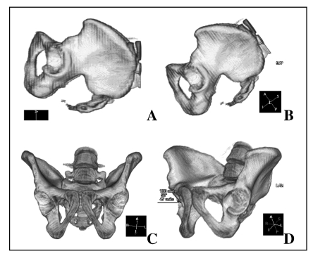
The bony pelvis of the supine study subject was reconstructed in three dimensions using Voxar 3D software. The femoral head was sculpted out of each hip joint. The pelvis was then rotated about its supine vertical axis as well as the subject body’s long axis until the iliac crests and pubic rami from both sides overlapped optimally. This was defined as the standard supine pelvis orientation (shown in Figure 2A). The long body axis of the pelvis was then tilted 68 degrees in males and 66 degrees in females above the horizontal to achieve the standard seated pelvic orientation as viewed from the side (Figure 2B). The pelvis was then rotated 90 degrees about its seated vertical axis to obtain the standard seated pelvic orientation as viewed from the front (Figure 2C). We are aware that the torso flexes around the hip joint and that the pelvis tilts during a frontal crash; however, there is no specific data available from cadaver tests regarding the average degree or range of tilting that occurs. We therefore used the average tilt obtained from our preliminary study of seated subject radiographs as the standard reference value.
FIGURE 2.
Technique of measuring pelvic angles. The bony pelvis of study subjects was reconstructed in 3-dimensional computed tomography. A) straight lateral view of the supine pelvis. B) lateral view of the pelvis tilted up to the reference seated position. C) frontal view of the seated pelvis. D) oblique view across the face of the right hip socket; this view was used to measure the slope angle.
Starting with the pelvis in standard seated position as viewed from the front (Figure 2C), we rotated the pelvis around its seated vertical axis until the front and back edges of the acetabular cup were aligned (Figure 2D). The amount of rotation necessary to achieve alignment of the front and back edge of the acetabular cup was defined as the Open Angle. With the pelvis in this latter position, a line was drawn between the superior and inferior edges of the acetabulum. The angle between this line and the horizontal was defined as the Slope Angle. Repeated measures of these angles were performed by multiple analysts and confirmed the precision and reproducibility of this protocol. Angle measurements taken on either side varied by no more than one or two degrees. Angles were measured only if the acetabulum was not fractured. An Open Angle of 0 degrees defined an acetabular cup that was directed laterally; an open angle of 90 degrees defined an acetabular cup that was directed straight anteriorly. A Slope Angle of 0 degrees defined an acetabular cup directed straight downward; a Slope Angle of 90 degrees defined an acetabular cup directed horizontally.
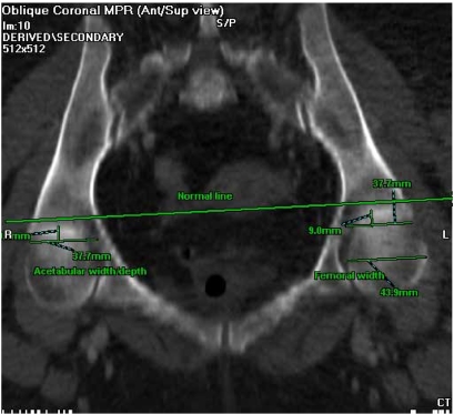
Following determination of the acetabular cup orientation, measurements of the posterior acetabular wall and femoral head dimensions were performed. Horizontal planar sections were then taken through the acetabulum to determine the acetabular cup and femoral head dimensions (Figure 3). All pelvic CTs were first tilted to the standard seated position (Figure 2C). Measurements were not performed if the acetabulum was fractured. We were unable to separately rotate the femoral head in the acetabulum to achieve normal seated configuration within the joint and the femoral head diameter was measured in this plane with the subject’s hip extended in supine position.
FIGURE 3.
A horizontal planar view of the seated pelvis, taken through the center of the hip socket, was used to measure acetabular width/depth and femoral head diameter.
RESULTS
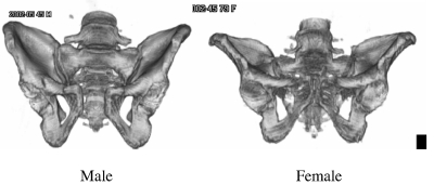
Overall comparison of the bony pelvis in the study population showed several gender differences. Figure 4 shows a representative male pelvis (Left) and representative female pelvis (Right) in the seated orientation as viewed from the front. The male pelvis (Left) is generally taller and narrower than the female pelvis (Right). The cup of the acetabulum is oriented more laterally in the male as opposed to the female.
FIGURE 4.
Representative male and female bony pelvis.
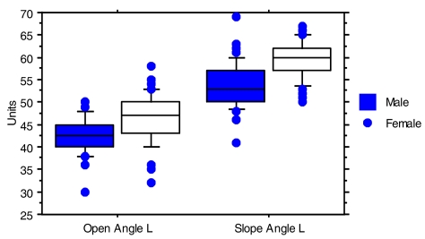
We found a significant gender difference in the Open Angle between males and females. Figure 5 shows the distribution of left acetabular Open Angle in male and female study subjects. Females have a significantly greater open angle (p<.0001). An Open Angle of 0 degrees means that in the horizontal plane of the seated pelvis, the acetabular cup faces directly laterally whereas an angle of 90 degrees means that the acetabular cup faces directly anterior. With a smaller average Open Angle, the pelvis of our male subjects therefore present less of their posterior acetabular cup as a reactive surface to take an anterior-to-posterior load directed along the long axis of the femur. The acetabular cup is generally thinner at its edges, therefore a more laterally-oriented cup (smaller Open Angle) would be more likely to fracture than one more anteriorly-oriented (larger Open Angle).
FIGURE 5.
Distribution of Open Angles and Slope Angles measured in the left acetabulum of male and female crash subjects. The box encompasses the 25th to 75th percentiles and is bisected by the median value. Observed values less than 10th percentile and greater than 90th percentile are shown as individual points.
Slope Angle was also noted to be significantly greater in females compared to males (p<.0001). The distribution of observed values shows that there is even less overlap between genders than was seen with Open Angle. An acetabular cup open directly downward has a Slope Angle of 0 degrees where as one directed horizontally has a Slope Angle of 90 degrees. This difference in Slope Angle suggests that females present a greater percentage of their available acetabular surface to take an anterior-to-posterior directed load than males.
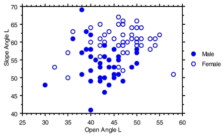
Figure 6 shows the Open Angle and Slope Angle of individual subjects in our study. There does not appear to be any strong association between the two angles that might synergize to affect the ability of an acetabulum to tolerate frontal loading.
FIGURE 6.
Bivariate plot of left acetabular Open Angle and Slope for all study subjects, divided by gender.
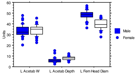
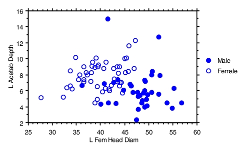
Acetabular width was noted to be quite similar between males and females (Figure 7.). However, females had significantly greater acetabular depth than males (p<.0001). In contrast, females had significantly smaller femoral head diameters than males (p<.0001). This latter finding regarding femoral head diameter differences between genders is consistent with previous reports. Purkait examined 280 femora of known age, sex and ethnicity and found gender differences in femoral head height and width, with male dimensions being significantly greater. Similar gender differences have also been earlier reported by Mall et. al. A greater acetabular depth and a smaller femoral head would make the hip joint more stable during anterior-to-posterior loading through the long axis of the femur and less likely to experience posterior hip dislocation.
FIGURE 7.
Distribution of Acetabular Width, Acetabular Depth and Femoral Head Diameter measurements in the left hip of male and female crash subjects (in millimeters). The box encompasses the 25th to 75th percentiles and is bisected by the median value. Observed values less than 10th percentile and greater than 90th percentile are shown as individual points.
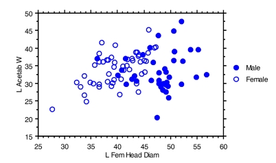
Figure 8 shows that male study subjects had a greater femoral head diameter but similar acetabular width compared to female study subjects. The presence of a larger femoral head diameter in males might result in more eccentric loading of the acetabulum, decreasing the posterior wall’s ability to tolerate crash forces before fracture or dislocation.
FIGURE 8.
Bivariate plot of left acetabular width and femoral head diameter (in millimeters) for all study subjects, divided by gender.
Figure 9 also shows clear separation by gender. The combination of deeper acetabular depth with smaller femoral head diameter would potentially make the female hip more stable to loading through the femur during a frontal crash. The presence of a more stable hip joint may not only decrease the likelihood of hip injury, but also increase the possibility of a knee or thigh injury during KTH loading during a crash.
FIGURE 9.
Bivariate plot of acetabular depth versus femoral head diameter for all study subjects, divided by gender.
DISCUSSION
Motor vehicle morbidity and mortality has had a steady decline with the advancement of occupant restraint systems (seatbelts, air bags). However, injuries are still occurring and many of these injuries involve the lower extremities. Thomas reported that nearly half of seriously injured (Average of 1.2 AIS 3+ injuries to any body region) survivors of MVCs sustained an AIS 3+ lower limb injury. Knee contact with the vehicle interior (particularly the knee restraint) is a consistent finding in investigations of real-life frontal crashes and approximately 30,000 fractures and dislocations of the knee-thigh-hip (KTH) complex occur in such crashes each year in the United States (Kuppa).
We have recently reported that the proportion of hip injuries compared to knee and femur injuries resulting from frontal crashes is increasing with newer vehicles (Sochor). This trend toward hip injuries is of significant concern since hip injuries are clinically more severe and more difficult to treat than injuries to either the knee or thigh. Pelvic fractures, particularly those involving disruption of the joint (acetabulum), often result in significant rehabilitation costs and long term disability. Hip injuries often result in lifelong impaired gait (Nerubay) and account for the majority of life years lost due to KTH injuries in motor-vehicle crashes (Kuppa).
Previous analysis of hip/pelvis injuries in frontal crashes in our center’s CIREN data indicated a high incidence of acetabular fractures relative to other AIS2+ hip/pelvis injuries (Sochor). As expected based on the orientation of the pelvis relative to the direction of force applied through the femur, most acetabular fractures in the UM CIREN database were to the posterior acetabulum. The rate of hip fracture occurrence increased somewhat with age, but KTH injuries occur in all adult age groups. Unexpectedly, we also found these hip injuries to be much more frequent among tall male drivers between 150 and 200 pounds of all ages (Sochor). Both men and women have increased percentages of hip injury for increased weight, but the percent of hip injuries sustained by men increased more at the lower weight groups than for women. For weight groups between 151–200 pounds, men had approximately twice the percentage of hip injuries. In weight groups above 200 pounds, men and women both had high percentages of hip injury. This data indicated that the higher percentage of hip injuries observed for heavier and taller occupants is principally related to their size but is also significantly related to gender (Sochor).
Our current study therefore sought to determine how hip anatomy differs between males and females under conditions applicable to anterior-to-posterior loading of the acetabulum in a seated pelvis through the femoral long axis. We determined the angle of presentation of the acetabulum to frontal loading and also characterized the size and shape of the posterior acetabular wall that would take the loading.
If the acetabulum or hip socket were oriented directly forward and in line with the force being transmitted along the femur, a maximal surface would be available to take the load. In this situation, a fracture of the knee or femur might occur before a fracture of the acetabulum. If however, the acetabulum were facing directly lateral, only the posterior edge of the socket would be able to absorb the load from the femur. In the latter situation, acetabular fracture and posterior dislocation would be more likely to occur. We found the orientation of the acetabular cup in the seated male pelvis to be directed more horizontally and laterally than in the female pelvis, providing less of a reactive surface to take femoral loading directed along the long axis of the femur.
We also found that females have a greater average acetabular depth than males while their femoral heads tended to be smaller. This combination of factors would potentially make the female hip more stable to loading through the femur during a frontal crash. The presence of a more stable hip joint may not only decrease the likelihood of hip injury, but also increase the possibility of a knee or thigh injury during KTH loading during a crash.
It is well known that significant differences exist in pelvic bony anatomy between males and females. Rissech et al examined 327 bony pelvis specimens ranging form birth to 97 years of age from 4 documented west European collections and found significant differences in acetabular anatomy between males and females. These gender differences began emerging between the ages of 15 and 19 and were significant by the age of 20, persisting thereafter. Maruyama determined the acetabular and femoral anteversion angles and femoral head offset in 50 male and 50 female human skeletons. He found the acetabular anteversion angle measured was significantly larger in females compared to males. These previous studies and others (Murphy) were conducted to determine how gender differences might contribute to the differential risk of hip fracture from falls between males and females or differential clinical outcome following surgical repair. To date, little has been done to determine if and how gender differences in hip anatomy might affect risk of injury from femur loading in frontal motor vehicle crashes.
The extent of adduction, internal rotation and flexion at the hip joint are thought to significantly affect the risk of acetabular injury by changing the contact area between femoral head and acetabulum (Letournel and Judet 1993). There are differences in preferred seating position and driving posture between males and females that may affect these three factors during a frontal crash. How these factors interact with the anatomic differences we found to affect hip loading in a frontal crash remain to be determined.
Early biomechanical research produced mostly knee or distal femur injuries. Early crash investigation data also documented more frequent knee and femur injuries compared to hip injuries. Therefore, the femur has traditionally been viewed as the weakest part of the KTH complex. We (Rupp) recently investigated the frontal-impact fracture tolerance of the hip in nineteen tests performed on the KTH complexes from sixteen unembalmed human cadavers and showed that, under the applied loading conditions, the hip is weaker than the knee or thigh, and that the weakest portion of the hip is the posterior acetabulum. The real-world data and clinical implications of hip injuries suggest a need for additional biomechanical research to better understand the response and tolerance of the KTH complex for knee loading conditions
CONCLUSION
Males and females differ significantly in the anatomy of their hips. Based on our current understanding of the biomechanics of hip loading during frontal crashes, these differences are consistent with the observed gender difference in hip injury tolerance from real-life crash investigations. Our findings have implications for the design, development and testing of knee restraints in the future. Our findings also suggest that consideration be given to developing anthropomorphic testing devices and computerized models that have greater anatomic fidelity to the gender they are meant to represent in crash testing.
ACKNOWLEDGMENT
This work was supported by grant # DTNH22-00-H-07444 from the US Department of Transportation - National Highway Traffic Safety Administration as well as educational grants from Johnson Controls, Breed Technologies and General Motors.
REFERENCES
- Kuppa S, Wang JU, Haffner M, Eppinger R. Lower extremity injuries and associated injury criteria. Proceedings of the 17th International Technical Conference on the Enhanced Safety of Vehicles. Paper No. 457; National Highway Traffic Safety Administration, Washington, DC. 2001. [Google Scholar]
- Letournel E, Judet R. Fractures of the acetabulum. Springer-Verlag; New York: 1993. [Google Scholar]
- Lewis PR, Molz FJ, Schmidke SZ, Bidez MW. Society of Automotive Engineers, Inc; 1996. A NASS-Based Investigation of Pelvic Injury within the Motor Vehicle Crash Environment. [Google Scholar]
- Mall G, Graw M, Gehring K, Hubig M. Determination of sex from femora. Forensic Science International. 2000;113:315–21. doi: 10.1016/s0379-0738(00)00240-1. [DOI] [PubMed] [Google Scholar]
- Maruyama M, Feinberg JR, Capello WN, D’Antonio JA. The Frank Stinchfield Award: Morphologic features of the acetabulum and femor: anteversion angle and implant positioning. Clinical Orthopaedics. 2001;393:52–65. [PubMed] [Google Scholar]
- Murphy D, Kaliszer M, Rice J, McElwain JP. Outcome after acetabular fracture, Prognostic factors and their inter-relationships. Injury. 2003;34(7):512–7. doi: 10.1016/s0020-1383(02)00349-2. [DOI] [PubMed] [Google Scholar]
- Nerubay J, Glancz G, Katznelson A. Fractures of the acetabulum. Journal of Trauma. 1973;13:1050–1062. doi: 10.1097/00005373-197312000-00003. [DOI] [PubMed] [Google Scholar]
- Purkait R. Sex determination from femoral head measurements: a new approach. Legal Medicine (Toyko) 2003;5 (Suppl 1):S347–50. doi: 10.1016/s1344-6223(02)00169-4. [DOI] [PubMed] [Google Scholar]
- Rissech C, Garcia M, Malgosa A. Sex and age diagnosis by ischium morphometric analysis. Forensic Science Int’l. 2003;135:188–96. doi: 10.1016/s0379-0738(03)00215-9. [DOI] [PubMed] [Google Scholar]
- Rupp JD, Reed MP, Van Ee CA, Kuppa S, Wang SC, Schneider LW. The tolerance of the human hip to dynamic knee loading. Stapp Car Crash Journal. 2002;46:211–228. doi: 10.4271/2002-22-0011. [DOI] [PubMed] [Google Scholar]
- Sochor MR, Faust DP, Schneider LW, Wang SC. Knee, Thigh and Hip Injury Patterns For Drivers and Right Front Passengers In Frontal Impacts. Society of Automotive Engineers paper # 2003-01-0164; 2003. [Google Scholar]
- Thomas P. Priorities in frontal crash protection. Proceedings of the 26th International Symposium on Automotive Technology and Automation – Dedicated conference on road and Vehicle Safety; Aachen. 1993. [Google Scholar]


