Abstract
Objective
To quantitatively evaluate the asymmetry of the multifidus and psoas muscles in unilateral sciatica caused by lumbar disc herniation using magnetic resonance imaging (MRI).
Methods
Seventy-six patients who underwent open microdiscectomy for unilateral L5 radiculopathy caused by disc herniation at the L4-5 level were enrolled, of which 39 patients (51.3%) had a symptom duration of 1 month or less (group A), and 37 (48.7%) had a symptom duration of 3 months or more (group B). The cross-sectional areas (CSAs) of the multifidus and psoas muscles were measured at the mid-portion of the L4-5 disc level on axial MRI, and compared between the diseased and normal sides in each group.
Results
The mean symptom duration was 0.6±0.4 months and 5.4±2.7 months for groups A and B, respectively (p<0.001). There were no differences in the demographics between the 2 groups. There was a significant difference in the CSA of the multifidus muscle between the diseased and normal sides (p<0.01) in group B. In contrast, no significant multifidus muscle asymmetry was found in group A. The CSA of the psoas muscle was not affected by disc herniation in either group.
Conclusion
The CSA of the multifidus muscle was reduced by lumbar disc herniation when symptom duration was 3 months or more.
Keywords: Multifidus, Psoas, Cross-sectional area, Lumbar disc herniation
INTRODUCTION
Sciatica is characterized by radiating pain in an area of the leg typically served by one nerve root in the lumbar or sacral spine. The most common cause of sciatica is lumbar disc herniation. The clinical course of sciatica is considered favorable, with resolution of leg pain via conservative treatment in a majority of the patients15). However, atrophy of the ipsilateral multifidus or psoas muscles has been reported in cases of sciatica caused by lumbar disc herniation2,3,10). Dangaria and Naesh3) noted a significant reduction in the cross sectional area (CSA) of the ipsilateral psoas in patients with unilateral lumbar disc herniation. Hyun et al.10) reported a significant decrease in the CSA of the ipsilateral multifidus in patients with unilateral lumbosacral radiculopathy.
The aim of the current study was to investigate whether asymmetry of the multifidus and psoas muscle occurred and whether it was related to the duration of the unilateral sciatica caused by lumbar disc herniation. For this purpose, we assessed the CSAs of these muscles using magnetic resonance imaging (MRI). Based on the results, we suggest possible remedies to hasten the recovery after lumbar disc surgery.
MATERIALS AND METHODS
Patient population
We retrospectively analyzed data obtained from 76 consecutive patients who underwent conventional open microdiscectomy for severe leg pain caused by lumbar disc herniation in 2005, of which 39 patients (51.3%) had a symptom duration of 1 month or less (group A), and 37 (48.7%) had a symptom duration of 3 months or more (group B). The inclusion criteria of group A were as follows : 1) single-level lumbar disc herniation at L4-5 level on computed tomography and/or MRI; 2) severe leg pain that was consistent with the radiologic findings; 3) leg pain that did not respond to conservative treatment; and 4) interval from symptom onset to surgery of 1 month or less. Patients with chronic low back pain, motor weakness, and/or previous history of lumbosacral spinal surgery were excluded. Group A comprised 27 men and 12 women with a mean age of 42.2±7.9 years (range, 25-58 years). The mean symptom duration in group A was 0.6±0.4 months (range, 0.1-1 months). Group B had the same inclusion and exclusion criteria except that the interval from symptom onset to surgery was 3 months or more. Group B comprised 22 men and 15 women with a mean age of 44.6±9.1 years (range, 22-58 years). Their mean symptom duration was 5.4±2.7 months (range, 3-12 months). There was no significant difference in patient demographics between groups A and B (Table 1).
Table 1.
The demographic characteristics of the group A and B
*Chi-square test. †Independent t-test
Radiological evaluation
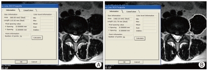
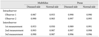
All patients underwent MRI examination of the lumbar spine before the operation. The patients were placed supine with a pillow positioned underneath their knees, ensuring that they were lying symmetrically with their weight evenly distributed across both sides. MRI scans was performed using Magnetom Symphony Ultragradient 1.5-Tesla MRI system (Siemens, Erlangen, Germany) with a slice thickness of 4 mm. Axial T2-weighted images were obtained under the following scan setting : repetition time, 3,700 msec and echo time, 120 msec. Axial scans were aligned parallel to the vertebral end-plate. The MRI scan measurements were taken using PiView program (Infinitt Co. Ltd., Seoul, Korea). All measurements were performed 'blind' by two neurosurgeons who did not know the purpose of this study. Each observer measured radiological parameters 3 times. The axial T2-weighted image of the lumbar spine at the mid-portion of the L4-5 disc level was selected. The image was enlarged, and the area of the multifidus and psoas muscles was identified. The software then calculated the area identified (Fig. 1). The parameters for quantitative analysis included the CSAs of the multifidus and psoas muscles, CSA of the diseased and normal side, and ratio of the CSA of the diseased side to that of the normal side. The means of the two observers' first tmeasurements were used for data analysis. Data are presented as mean±standard deviation. Intraobserver and interobserver reliability tests on the measurements of the CSA of the multifidus and psoas muscles showed high reliability (Table 2).
Fig. 1.
Cross-sectional area measurement of the multifidus (A) and psoas (B) muscles using a magnetic resonance imaging at the L4-5 intervertebral disc level.
Table 2.
The intra- and interobserver reliability using intra- and interclass correlation coefficient for measurements of multifidus and psoas muscles
Statistical analysis
Differences between the 2 groups were analyzed using an independent t-test and chi-square test. The CSAs of the multifidus muscle between the diseased and normal sides were compared using a paired t-test, and the CSAs of the psoas muscle between the diseased and normal sides were compared using the Wilcox signed-rank test. Associations between the ratio of the CSA of the diseased side to that of the normal side as well as the clinical variables, such as age, body mass index, and the straight leg raising test, were tested using Spearman's test. Differences in the CSAs between the diseased and normal sides according to gender and smoking were tested using an independent t-test. The inter- and intraclass correlation coefficient test was performed to determine the inter- and intraobserver reliability of the three sets of measurements obtained by the two different observers. Significance was set at p<0.05.
RESULTS
In group A, the mean CSA of the multifidus muscle was not significantly different between the diseased side (680.1±171.9 mm2) and the normal side (664.7±156.6 mm2) (p=0.085). The CSA of the psoas muscle was not significantly different between the diseased side (1,408.3±415.7 mm2) and the normal side (1,411.9±414.5 mm2) (p=0.967). In group B, the mean CSA of the multifidus muscle on the diseased side was 632.9±123.0 mm2, which was significantly smaller than that on the normal side (675.3±133.9 mm2) (p<0.01). There was no significant difference in the mean CSA of the psoas muscle between the diseased side (1,341.0±436.0 mm2) and the normal side (1,346.6±439.0 mm2) (p=0.829) (Table 3).
Table 3.
The cross-sectional area of the multifidus and psoas muscles in the group A and B
*Paired t-test. †Wilcox signed rank test
In group A, there was no significant associations between the ratios of the CSAs of the same muscles and clinical variables; age [r=0.232; p=0.155 (multifidus) and r=0.059; p=0.723 (psoas)], gender [p=0.064 (multifidus) and p=0.667 (psoas)], body mass index [r=0.198; p=0.228 (multifidus) and r=-0.261; p=0.108 (psoas)], smoking [p=0.068 (multifidus) and p=0.241 (psoas)], and the straight leg raising test [r=-0.006; p=0.972 (multifidus) and r=0.077; p=0.642 (psoas)]. In group B, there was no significant correlation between the ratios of either the multifidus or the psoas CSAs and clinical variables such as age [r=0.273; p=0.101 (multifidus) and r=0.011; p=0.948 (psoas)], gender [p=0.891 (multifidus) and p=0.984 (psoas)], body mass index [r=0.282; p=0.091 (multifidus) and r=0.030; p=0.858 (psoas)], smoking [p=0.878 (multifidus) and p=0.471 (psoas)], and the straight leg raising test [r=-0.072; p=0.670 (multifidus) and r=-0.137; p=0.418 (psoas)].
DISCUSSION
The multifidus muscle is the largest and the most medial of the lumbar paraspinal muscles9). It is innervated by the medial branch of the dorsal ramus of the segmental nerve12), and the innervations are always unisegmental6). The psoas muscle is the largest muscle at the cross section of the lower levels of the lumbar spine13). The multifidus and psoas muscles both act to provide stability to the spine and are sensitive to pathologic changes1). Atrophy of the multifidus and psoas muscles has been linked to low back pain4,14). Parkkola et al.14) reported that the psoas and the back muscles (erector spinae and multifidus) of the patients with chronic low back pain were smaller than those of healthy volunteers. Danneels et al.4) reported a significant decrease in the CSA of the multifidus muscle, but not the psoas muscle of patients with chronic low back pain as compared to healthy controls. In the present study, patients with chronic low back pain were excluded from the analysis.
The changes in the multifidus and psoas muscles were also found in patients with lumbar disc herniation. Hyun et al.10) conducted a prospective clinical study examining the CSA of the multifidus muscle in patients with unilateral lumbosacral radiculopathy, lumbosacral disc herniation, and in healthy volunteers. They demonstrated that the CSA of the multifidus muscle was changed in both patient groups as compared with the controls and that asymmetric multifidus muscle atrophy occurred more frequently in patients with radiculopathy than in those with disc herniation only. Zhao et al.18) noted differences in the characteristics of the multifidus muscle between the diseased and normal sides in patients with lumbar disc herniation. The results of the present study correspond well with those previously reported. The CSA of the multifidus muscle was influenced by lumbar disc herniation in cases in which the symptom duration was 3 months or more, as shown by the significant difference in the CSAs of the multifidus muscles between the diseased and normal sides in group B subjects. Dangaria and Naesh3) conducted a prospective clinical cohort study examining the CSA of the psoas major in healthy volunteers and subjects with unilateral sciatica caused by disc herniation. They demonstrated that most patients with lumbar disc herniation showed a significant reduction in the CSA of the psoas muscle on the affected side only, most prominently at the level of the disc herniation. They suggested that a correlation exists between the reduction in the CSA of the psoas muscle of the affected side and the duration of conscious sciatica. In contrast to Dangaria's study, the CSA of the psoas muscle in our subjects was not influenced by unilateral sciatica caused by disc herniation. The cause of this difference remains to be determined.
The changes in the multifidus and psoas muscles have been reported to be influenced by the duration of the symptoms1,3,6,8,9). The atrophy of these muscles tends to occur more frequently in patients with longer symptom duration. In the present study, atrophy of the multifidus muscle ipsilateral to the lumbar disc herniation was observed in the patients who suffered from sciatica for 3 months or more. In contrast, there was no significant difference in the CSA of the multifidus muscle in the patients who suffered from sciatica for 1 month or less.
There is evidence for a strong relationship between multifidus muscle dysfunction and poor functional outcome as well as recurrence of low back pain after disc surgery9,16,17). Furthermore, the atrophy and dysfunction of the multifidus muscle did not recover spontaneously on remission of the painful syndrome9). Therefore, rehabilitation exercises have been recommended for patients with paraspinal muscle dysfunction7,11). An exercise program conducted following lumbar discectomy reportedly improved outcomes with respect to pain, disability, and functional recovery3,5). Choi et al.2) proposed the positive effects of the postoperative early lumbar extension muscle-strengthening program on pain, duration of recuperation, and strength of back muscles in patients after operation for a herniated lumbar disc. The results of the present study also strongly support the necessity of a lumbar extension muscle-strengthening program, especially in patients suffering from sciatica for 3 months or more.
There are several limitations to the present study. First, the medical treatment prior to surgery and daily activity were not fully assessed. Pain relief through medical treatment may affect the daily activity of these patients, which may also affect the structure and function of the multifidus and psoas muscles. Secondly, the measurements of the muscle CSA were confined to the L4-5 level and may not individually reflect the status of the psoas and multifidus muscles. Consequently, there might have been underestimation or overestimation of muscle atrophy. Lastly, muscle degeneration may be detected by decreased muscle CSA and by increased amounts of fatty deposit within the muscle. The measurement technique we used only gave a gross measurement of muscle CSA and did not allow for any computation of replacement of muscle by fatty or connective tissues.
CONCLUSION
The CSA of the multifidus muscle was reduced by lumbar disc herniation when the symptom duration was 3 months or more.
Acknowledgements
This study was supported by a grant from the Wooridul Spine Foundation.
References
- 1.Barker KL, Shamley DR, Jackson D. Changes in the cross-sectional area of multifidus and psoas in patients with unilateral back pain : the relationship to pain and disability. Spine (Phila Pa 1976) 2004;29:E515–E519. doi: 10.1097/01.brs.0000144405.11661.eb. [DOI] [PubMed] [Google Scholar]
- 2.Choi G, Raiturker PP, Kim MJ, Chung DJ, Chae YS, Lee SH. The effect of early isolated lumbar extension exercise program for patients with herniated disc undergoing lumbar discectomy. Neurosurgery. 2005;57:764–772. doi: 10.1093/neurosurgery/57.4.764. discussion 764-772. [DOI] [PubMed] [Google Scholar]
- 3.Dangaria TR, Naesh O. Changes in cross-sectional area of psoas major muscle in unilateral sciatica caused by disc herniation. Spine (Phila Pa 1976) 1998;23:928–931. doi: 10.1097/00007632-199804150-00016. [DOI] [PubMed] [Google Scholar]
- 4.Danneels LA, Vanderstraeten GG, Cambier DC, Witvrouw EE, De Cuyper HJ. CT imaging of trunk muscles in chronic low back pain patients and healthy control subjects. Eur Spine J. 2000;9:266–272. doi: 10.1007/s005860000190. [DOI] [PMC free article] [PubMed] [Google Scholar]
- 5.Dolan P, Greenfield K, Nelson RJ, Nelson IW. Can exercise therapy improve the outcome of microdiscectomy? Spine (Phila Pa 1976) 2000;25:1523–1532. doi: 10.1097/00007632-200006150-00011. [DOI] [PubMed] [Google Scholar]
- 6.Franke J, Hesse T, Tournier C, Schuberth W, Mawrin C, LeHuec JC, et al. Morphological changes of the multifidus muscle in patients with symptomatic lumbar disc herniation. J Neurosurg Spine. 2009;11:710–714. doi: 10.3171/2009.7.SPINE08448. [DOI] [PubMed] [Google Scholar]
- 7.Häkkinen A, Ylinen J, Kautiainen H, Airaksinen O, Herno A, Tarvainen U, et al. Pain, trunk muscle strength, spine mobility and disability following lumbar disc surgery. J Rehabil Med. 2003;35:236–240. doi: 10.1080/16501970306096. [DOI] [PubMed] [Google Scholar]
- 8.Hides JA, Richardson CA, Jull GA. Multifidus muscle recovery is not automatic after resolution of acute, first-episode low back pain. Spine (Phila Pa 1976) 1996;21:2763–2769. doi: 10.1097/00007632-199612010-00011. [DOI] [PubMed] [Google Scholar]
- 9.Hides JA, Stokes MJ, Saide M, Jull GA, Cooper DH. Evidence of lumbar multifidus muscle wasting ipsilateral to symptoms in patients with acute/subacute low back pain. Spine (Phila Pa 1976) 1994;19:165–172. doi: 10.1097/00007632-199401001-00009. [DOI] [PubMed] [Google Scholar]
- 10.Hyun JK, Lee JY, Lee SJ, Jeon JY. Asymmetric atrophy of multifidus muscle in patients with unilateral lumbosacral radiculopathy. Spine (Phila Pa 1976) 2007;32:E598–E602. doi: 10.1097/BRS.0b013e318155837b. [DOI] [PubMed] [Google Scholar]
- 11.Leinonen V, Kankaanpää M, Luukkonen M, Kansanen M, Hänninen O, Airaksinen O, et al. Lumbar paraspinal muscle function, perception of lumbar position, and postural control in disc herniation-related back pain. Spine (Phila Pa 1976) 2003;28:842–848. [PubMed] [Google Scholar]
- 12.Macintosh JE, Bogduk N. 1987 Volvo award in basic science. The morphology of the lumbar erector spinae. Spine (Phila Pa 1976) 1987;12:658–668. doi: 10.1097/00007632-198709000-00004. [DOI] [PubMed] [Google Scholar]
- 13.McGill SM, Patt N, Norman RW. Measurement of the trunk musculature of active males using CT scan radiology : implications for force and moment generating capacity about the L4/L5 joint. J Biomech. 1988;21:329–341. doi: 10.1016/0021-9290(88)90262-x. [DOI] [PubMed] [Google Scholar]
- 14.Parkkola R, Rytökoski U, Kormano M. Magnetic resonance imaging of the discs and trunk muscles in patients with chronic low back pain and healthy control subjects. Spine (Phila Pa 1976) 1993;18:830–836. doi: 10.1097/00007632-199306000-00004. [DOI] [PubMed] [Google Scholar]
- 15.Peul WC, van Houwelingen HC, van den Hout WB, Brand R, Eekhof JA, Tans JT. Surgery versus prolonged conservative treatment for sciatica. N Engl J Med. 2007;356:2245–2256. doi: 10.1056/NEJMoa064039. [DOI] [PubMed] [Google Scholar]
- 16.Rantanen J, Hurme M, Falck B, Alaranta H, Nykvist F, Lehto M, et al. The lumbar multifidus muscle five years after surgery for a lumbar intervertebral disc herniation. Spine (Phila Pa 1976) 1993;18:568–574. doi: 10.1097/00007632-199304000-00008. [DOI] [PubMed] [Google Scholar]
- 17.Sihvonen T, Herno A, Paljärvi L, Airaksinen O, Partanen J, Tapaninaho A. Local denervation atrophy of paraspinal muscles in postoperative failed back syndrome. Spine (Phila Pa 1976) 1993;18:575–581. doi: 10.1097/00007632-199304000-00009. [DOI] [PubMed] [Google Scholar]
- 18.Zhao WP, Kawaguchi Y, Matsui H, Kanamori M, Kimura T. Histochemistry and morphology of the multifidus muscle in lumbar disc herniation : comparative study between diseased and normal sides. Spine (Phila Pa 1976) 2000;25:2191–2199. doi: 10.1097/00007632-200009010-00009. [DOI] [PubMed] [Google Scholar]


