Abstract
The infection of the root canal system is considered to be a polymicrobial infection, consisting of both aerobic and anaerobic bacteria. Because of the complexity of the root canal infection, it is unlikely that any single antibiotic could result in effective sterilization of the canal. A combination of antibiotic drugs (metronidazole, ciprofloxacin, and minocycline) is used to eliminate target bacteria, which are possible sources of endodontic lesions. Three case reports describe the nonsurgical endodontic treatment of teeth with large periradicular lesions. A triple antibiotic paste was used for 3 months. After 3 months, teeth were asymptomatic and were obturated. The follow-up radiograph of all the three cases showed progressive healing of periradicular lesions. The results of these cases show that when most commonly used medicaments fail in eliminating the symptoms then a triple antibiotic paste can be used clinically in the treatment of teeth with large periradicular lesions.
Keywords: Ciprofloxacin, metronidazole, minocycline, nonsurgical root canal treatment, periradicular lesion, triple antibiotic paste
Around 33.8 million people worldwide are living with HIV-AIDS (WHO 2008 report) of which around 3.8 million are in the Indian subcontinent. The first case of AIDS was reported in the year 1981. Since then the disease has gone through various stages of changes with respect to its epidemiology as well as its manifestations.
HIV disease has an effect over the entire body. It is not practical in the present scenario for any health personnel dealing with diagnosis and treatment in humans to not encounter this dreaded disease and its manifestations. Thus it becomes imperative to be aware of the various forms of HIV manifestations.
Oral health is an important component of the overall health status in HIV infection. Awareness of the variety of oral disorders which can develop throughout the course of HIV infection and coordination of health care services between a physician and a dentist may improve the overall health of the patient. The spectrum of oral manifestations is very vast in HIV-AIDS.
Oral manifestations of HIV infection occur in 30–80% of the affected patient population.[1,2] The overall prevalence of oral manifestations in HIV disease has changed since the advent of HAART.
The various oral manifestations can be categorized into
Infections: bacterial, fungal, viral
Neoplasms: Kaposi's sarcoma, non-Hodgkin's lymphoma
Immune mediated: major aphthous, necrotizing stomatitis
Others: parotid diseases, nutritional, xerostomia
Oral manifestations as adverse effects of antiretroviral therapy.
There is no particular oral lesion which is associated only with HIV-AIDS but there are certain manifestations like oral candidiasis, oral hairy leukoplakia (OHL) which are associated very frequently and are considered AIDS-defining diseases and have also been included in the clinical classification of HIV by CDC in category B.[3]
Fungal Infections
Candidiasis: Oral or pharyngeal candidiasis are the commonest fungal infections observed as the initial manifestation of symptomatic HIV infection.[4–7] Many patients can have esophageal candidiasis as well. It is usually observed at CD4 counts of less than 300/μl. The commonest species of candida involved are Candida albicans although nonalbicans species have also been reported. There are four frequently observed forms of oral candidiasis: erythematous candidiasis, pseudomembranous candidiasis, angular cheilitis, and hyperplastic or chronic candidiasis:[8]
Erythematous candidiasis presents as a red, flat, subtle lesion either on the dorsal surface of the tongue and/or the hard/soft palates. Patients complain of a burning sensation in the mouth more so while eating salty and spicy food. Diagnosis is made on the basis of clinical examination, a potassium hydroxide preparation which demonstrates the fungal hyphae can be used for confirmation.
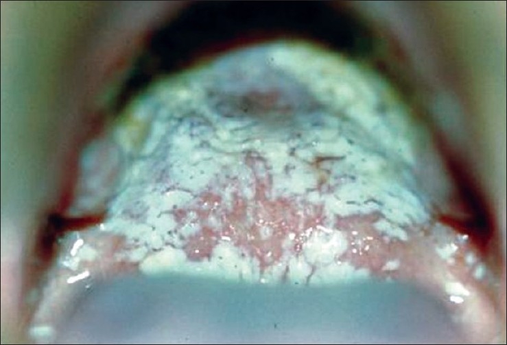
Pseudomembranous candidiasis appears as creamy white curd-like plaques on the buccal mucosa, tongue, and other oral mucosal surfaces that can be wiped away, leaving a red or bleeding surface [Figure 1].
Angular cheilitis is erythema and/or fissuring and cracks of the corners of the mouth. Angular cheilitis can occur with or without the presence of erythematous candidiasis or pseudomembranous candidiasis.
Hyperplastic or chronic candidiasis presents as white nonremovable plaques over the mucosal surface; hence they cannot be scraped off.
Figure 1.

Hyperplastic candidiasis
Oral candidiasis can extend to involve the pharynx, larynx, and esophagus as well. Treatment of oral candidiasis depends on the clinical type, distribution, and severity of infection. Topical treatment is effective for limited and accessible lesions. Clotrimazole troches, nystatin pastilles, and nystatin oral suspension are effective for mild-to-moderate erythematous and pseudomembranous candidiasis. However, prolonged use of these agents can result in significant dental caries due to the fermentable carbohydrate substrates present in the formulations. Increased risk of caries can be avoided by using a nystatin oral suspension (100,000 units/5 ml, rinsing mouth, and expectorating 3 times/day). Chlorhexidine 0.12% oral rinses do not contain a cariogenic substrate and may be similarly effective.
Topical amphotericin B can also be used in the treatment for resistant candidiasis and can be prepared by dissolving 50 mg in 500 ml of sterile saline (0.1 mg/ml). Clotrimazole 1% cream, miconazole or ketoconazole 2% cream, and nystatin ointment are useful medications for angular cheilitis and for application to a removable denture base when there is candidal infection involving the underlying mucosa.
Systemic treatment for oral candidiasis involves the use of imidazole (ketoconazole) and triazole (fluconazole and itraconazole) antifungal medications. Fluconazole is given in the dose of 100–200 mg/day. The duration of treatment with oral imidazoles usually is around 7–10 days but in cases of suspicion of esophageal involvement, the duration can be extended to 21 days. As per the recent guidelines there is no role of prophylaxis for candidiasis in HIV patients.
Histoplasmosis: Histoplasmosis is a granulomatous fungal disease caused by Histoplasma capsulatum. The clinical presentation ranges from an asymptomatic or mild lung infection to an acute or chronic disseminated form. Oral histoplasmosis appears as chronic ulcerated areas located on the dorsum of the tongue, palate, floor of the mouth, and vestibular mucosa. Focal or multiple sites can be involved. In AIDS patients, histoplasmosis is rarely curable, but it can be controlled with long-term suppressive therapy consisting of the administration of amphotericin B and ketoconazole.
Cryptococcosis: Oral manifestations are quite unusual and only two cases have been reported in the literature.[9,10] The lesions consist of ulcerations of the oral mucosa, but the clinical diagnosis of oral cryptococcus may be difficult since other microbial infections and trauma may show similar appearances. Tissue biopsy may be required for the diagnosis and treatment involves use of amphotericin B.
Viral Infections
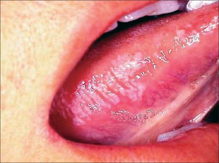
Oral hairy leukoplakia: These lesions are usually seen on the lateral surface of the tongue, but may extend to the dorsal and ventral surfaces [Figure 2]. Lesions may be variably sized and may appear as vertical white striations, corrugations, or as flat plaques, or raised, shaggy plaques with hair-like keratin projections. In most cases, OHL is bilateral and asymptomatic. When it leads to discomfort it is usually associated with superimposed candidal infection. OHL has been shown to be associated with a localized Epstein–Barr virus (EBV) infection and occurs most commonly in individuals whose CD4 lymphocyte count is less than 200/μl. Histological investigations reveal typical epithelial hyperplasia suggestive of EBV infection. This condition usually does not require treatment but use of oral acyclovir, topical podophyllum resin, retinoids, and surgical removal have all been reported as successful treatments.[11]
Figure 2.

Pseudomembranous candidiasis
Herpes simplex and varicella zoster virus infections: Herpes simplex virus (HSV) is responsible for both primary and recurrent infections of the oral mucosa. These infections are acquired in childhood and after initial pustular lesions. The virus remains dormant, but in later stages of immunosuppresion the virus can be activated and can lead to various manifestations. Oral manifestations, represented by diffuse mucosal ulcerations, are accompanied by fever, malaise, and cervical lymphadenopathy. Ulcerations that follow the rupture of vesicles are painful and may persist for several weeks. Recurrent HSV usually appears in keratinizing oral mucosa (i.e., palate, dorsum of tongue, and gingiva) as ulcerations but in most HIV-seropositive patients, this rule is not followed. In these patients, the lesions may show unusual clinical aspects and persist for many weeks. Contact with the varicella zoster virus (VZV) may result in varicella (chicken pox) as a primary infection and herpes zoster (shingles) as a reactivated infection. In HIV infection, herpes zoster frequently presents with early cranial nerve involvement and carries a poor prognostic significance. There may be involvement of multiple dermatomes and these lesions might get secondarily infected as well. The lesions are usually associated with severe postherpetic neuralgias.
Cytomegalovirus: CMV-related oral ulcerations, although infrequent, are a recognized complication of HIV infection. The diagnosis of oral CMV is based upon the presence of large intranuclear and smaller cytoplasmic CMV inclusions in the endothelial cells at the base of the ulcerations. These infections usually manifest in stage IV of the infection when there is advanced immunosupression with a CD4 count below 50. Presently, the drug of choice for CMV infection is intravenous gancyclovir.
Human papilloma virus: In some patients with HIV infection, human papilloma virus (HPV) causes a focal epithelial and connective tissue hyperplasia, forming an oral wart. In HIV-infected patients, oral HPV-related lesions have a papillomatous appearance, either pedunculated or sessile, and are mainly located on the palate, buccal mucosa, and labial commissure. The most common genotypes found in the mouth of patients with HIV infection are 2, 6, 11, 13, 16, and 32. Surgical removal, with or without intraoperative irrigation with podophyllum resin, is the treatment of choice.[12]
Molluscum contagiosum: Molluscum contagiosum is caused by an unclassified DNA virus of the poxvirus family. Lesions appear as single or multiple papules on the skin of the buttocks, back, face, and arms. Molluscum contagiosum usually affects children and young adults and is spread by direct and indirect contact. The typical lesion is an umbilicated papule that may itch, leading to autoinoculation. Lesions may persist for years and eventually regress spontaneously. The occurrence of disseminated molluscum contagiosum has been reported in HIV-infected patients. These lesions usually subside with immune reconstitution when patients are started on HAART.
Bacterial Infections
The most common oral lesions associated with bacterial infection are linear erythematous gingivitis, necrotizing ulcerative periodontitis, and, much less commonly, bacillary epithelioid angiomatosis and syphilis. In the case of the periodontal infections, the bacterial flora is no different from that of a healthy individual with periodontal disease. Thus, the clinical lesion is a manifestation of the altered immune response to the pathogens.
Linear erythematous gingivitis: This entity appears as a band of marginal gingival erythema, often with petechiae. It is typically associated with no symptoms or only mild gingival bleeding and mild pain. Histological examination fails to reveal any significant inflammatory response, suggesting that the lesions represent an incomplete inflammatory response, principally with only hyperemia present. Oral rinsing with chlorhexidine gluconate often reduces or eliminates the erythema and may require prophylactic use to avoid recurrence.
Necrotizing ulcerative periodontitis (NUP): This periodontal lesion is characterized by generalized deep osseous pain, significant erythema that is often associated with spontaneous bleeding, and rapidly progressive destruction of the periodontal attachment and bone. The destruction is progressive and can result in loss of the entire alveolar process in the involved area. It is a very painful lesion and can adversely affect the oral intake of food, resulting in significant and rapid weight loss. Patients also have severe halitosis. Because the periodontal microflora is no different from that seen in healthy patients, the lesion probably results from the altered immune response in HIV infection. More than 95% of patients with NUP have a CD4 lymphocyte count of less than 200/mm3. Treatment consists of rinsing twice daily with chlorhexidine gluconate 0.12%, metronidazole (250 mg orally four times daily for 10 days), and periodontal debridement, which is done after antibiotic therapy has been initiated.[13]
Bacillary Epithelioid Angiomatosis (BEA) This lesion appears to be unique to HIV infection and is often clinically indistinguishable from oral Kaposi's sarcoma (KS). Since both may present as an erythematous, soft mass which may bleed upon gentle manipulation, biopsy and histological examination are required to distinguish bacillary epithelioid angiomatosis (BEA) from KS. The presumed etiological pathogen, Rochalimaea henselae, can be identified using Warthin–Starry staining. Both KS and BEA are histologically characterized by atypical vascular channels, extravasated red blood cells, and inflammatory cells. However, prominent spindle cells and mitotic figures occur only in KS. Erythromycin is the treatment of choice for BEA.
Syphilis: While the prevalence of syphilis infection has risen significantly over the past decade, it is an uncommon cause of intraoral ulceration, even in HIV infection. Its appearance is no different from that observed in healthy individuals; it is a chronic, nonhealing, deep, solitary ulceration; often clinically indistinguishable from that due to tuberculosis, deep fungal infection, or malignancy. Dark field examination may demonstrate treponema. Positive reactive plasma reagin (RPR) and histological demonstration of Treponema pallidum is diagnostic. Combination treatment with penicillin, erythromycin, and tetracycline is the treatment of choice, the dosage and duration of the treatment depending on the presence or absence of neurosyphilis.
Neoplasms
Kaposi's sarcoma: It is the most common intraoral malignancy associated with HIV infection. The lesion may appear as a red-purple macule, an ulcer, or as a nodule or mass. Intraoral KS occurs on the heavily keratinized mucosa, the palate being the site in more than 90% of reported cases. KS is especially common among homosexual and bisexual males and is rarely found in HIV-infected women. Human herpes virus (HHV8) has been demonstrated to be an important cofactor in the development of KS. A histological examination is warranted for the definitive diagnosis of KS. There is no cure for KS. Therapy for intraoral KS should be instituted at the earliest sign of the lesion, the goal being local control of the size and number of lesions. When only a few lesions exist and the lesions are small (< 1 cm), intralesional chemotherapy with vinblastine sulfate or sclerotherapy with 3% sodium tetradecyl sulfate is usually effective. Radiation therapy (800–2,000 cGy) is required for larger or multiple lesions; stomatitis and glossitis are common side effects of radiation. Although this entity has been reported in the western literature but its incidence in Indian patients is quite low with only nine cases been reported till date.[14]
Non-Hodgkin's lymphoma: NHL is the most common lymphoma associated with HIV infection and is usually seen in late stages with CD4 lymphocyte counts of less than 100/mm3. It appears as a rapidly enlarging mass, less commonly as an ulcer or plaque, and most commonly on the palate or gingivae. A histological examination is essential for diagnosis and staging. Prognosis is poor, with mean survival time of less than 1 year, despite treatment with multidrug chemotherapy.
Immune-Mediated Oral Lesions
In HIV there is immune suppression of cell-mediated immunity as the disease progresses but at the same time there is abnormal activation of B-cell immunity. These disorders of the immune system also lead to various oral manifestations.
Aphthous ulcers: They are the most common immune-mediated HIV-related oral disorder, with a prevalence of approximately 2–3%. These ulcers are either large solitary or multiple, chronic, deep, and painful often lasting much longer in the seronegative population and are less responsive to therapy. Treatment requires the use of a potent topical steroid such as clobetesol when the lesions are accessible or dexamethasone oral rinse when not accessible. Systemic glucocorticosteroid therapy may be required (prednisone 1 mg/kg) in the case of large multiple ulcers and those not responding to topical preparations. Alternative therapies such as dapsone 50–100 mg daily and thalidomide 200 mg daily for 4 weeks should be considered in recalcitrant cases. When immunosuppressant drugs are used in order to prevent superadded fungal or bacterial infection, concurrent antifungal medications such as fluconazole, itraconazole, and antibacterial medications such as chlorhexidine gluconate oral rinse may be required.
Necrotizing stomatitis: It is an acute, painful ulceration which often exposes the underlying bone and leads to considerable tissue destruction. This lesion may be a variant of major aphthous ulceration, but occurs in areas overlying the bone and is associated with severe immune deterioration. These lesions can also occur in edentulous areas. Like in major aphthous ulceration, systemic corticosteroid medication or topical steroid rinse is the treatment of choice.
Xerostomia: Xerostomia is common in HIV disease, most often as a side effect of antiviral medications or other medications commonly prescribed for patients with HIV infection, like angiolytics, antifungals, etc. The oral dryness is a significant risk factor for caries and can lead to rapid dental deterioration. Xerostomia also can lead to oral candidiasis, mucosal injury, and dysphagia, and is often associated with pain and reduced oral intake of food. Patient who has residual salivary gland function which is determined by gustatory challenge, oral pilocarpine often provides improved salivary flow and consistency. Oral hygiene should be scrupulously maintained along with the use of dental floss.
Parotid Gland Disease
HIV infection is associated with parotid gland disease. There can be gland enlargement and diminished flow of secretions. Histologically, there may be lymphoepithelial infiltration and benign cyst formation. The enlargement typically involves the tail of the parotid gland or, less commonly, the submandibular gland, and it may present uni- or bilaterally with periods of increased or decreased size. The enlargement can be mistaken for a malignancy but in such cases a needle aspiration with yellow secretions in aspiration would help in making the diagnosis and in such cases further biopsy is unnecessary. Occasional swelling can be managed simply by repeated aspiration and rarely is radical removal of the gland necessary. The pathophysiological mechanism is not known, though cytomegalovirus has been suggested to play a role.
Oral Manifestations As Adverse Effects of Antiretroviral Therapy
With the widespread availability and usage of antiretroviral therapy for the management of HIV, the clinical picture now has shown a paradigm shift. The manifestations due to adverse effects of the HAART are also observed along with the above-mentioned features of immunosuppresion. Thus one should be aware about them as well. Oral hyperpigmentation can be observed if a patient is on zidovudine.
Erythema multiformes is a known side effect of NNRTIs. Xerostomia is also observed in patients on lamivudine, didanosine, indinavir and ritonavir. Lipodystrophy with loss of subcutaneous fat has been reported extensively in patients on stavudine. Other oral effects like paresthesias, lip edema, chelitis, and taste disturbances have been observed in patients on protease inhibitors.[15]
The above-mentioned list is not the complete panorama of manifestations which can be observed in an HIV patient but only an illustration of important lesions. It is thus essential that oral healthcare professionals recognize the hallmarks of the illness and provide timely management for better survival of these patients.
Footnotes
Source of Support: Nil
Conflict of Interest: None declared.
References
- 1.Grando LJ, Yurgel LS, Machado DC, Nachman S, Ferguson F, Berentsen B, et al. The association between oral manifestations and the socioeconomic and cultural characteristics of HIV-infected children in Brazil and in the United States of America. Rev Panam Salud Publica. 2003;14:112–8. doi: 10.1590/s1020-49892003000700006. [DOI] [PubMed] [Google Scholar]
- 2.Ceballos-Salobreña A, Gaitán-Cepeda LA, Ceballos-Garcia L, Lezama-Del Valle D. Oral lesions in HIV/AIDS patients undergoing highly active antiretroviral treatment including protease inhibitors: A new face of oral AIDS? AIDS Patient Care STDS. 2000;14:627–35. doi: 10.1089/10872910050206540. [DOI] [PubMed] [Google Scholar]
- 3.Interim WHO clinical staging of HIV/AIDS and HIV/AIDS case definitions for surveillance: WHO-HIV. 2005:2. [Google Scholar]
- 4.Klein RS, Harris CA, Small CB, Moll B, Lesser M, Friedland GH. Oral candidiasis in high risk patients as the initial manifestation of the acquired immunodeficiency syndrome. N Engl J Med. 1984;311:354–8. doi: 10.1056/NEJM198408093110602. [DOI] [PubMed] [Google Scholar]
- 5.Silverman S, Jr, Migliorati CA, Lozada-Nur F, Greenspan D, Conant MA. Oral findings in people with or at risk for AIDS: A study of 375 homosexual males. J Am Dent Assoc. 1986;112:187–92. doi: 10.14219/jada.archive.1986.0321. [DOI] [PubMed] [Google Scholar]
- 6.Schiodt M, Pindborg JJ. AIDS and the oral cavity: Epidemiology and clinical oral manifestations of human immune deficiency virus infection: A review. Int J Oral Maxillofac Surg. 1987;16:1–14. doi: 10.1016/s0901-5027(87)80025-5. [DOI] [PubMed] [Google Scholar]
- 7.Barone R, Ficarra G, Gaglioti D, Orsi A, Mazzotta F. Prevalence of oral lesions among HIV-infected intravenous drug abusers and other risk groups. Oral Surg Oral Med Oral Pathol. 1990;69:169–73. doi: 10.1016/0030-4220(90)90322-j. [DOI] [PubMed] [Google Scholar]
- 8.Sirois DA. Oral manifestations of HIV disease. Mt Sinai J Med. 1998;65:322–32. [PubMed] [Google Scholar]
- 9.Glick M, Cohen SG, Cheney RT, Crooks GW, Greenberg MS. Oral manifestation of disseminated Cryptococcus neoformans in a patient with acquired immune deficiency syndrome. Oral Surg Oral Med Oral Pathol. 1987;64:454–9. doi: 10.1016/0030-4220(87)90152-6. [DOI] [PubMed] [Google Scholar]
- 10.Lynch DP, Naftolin LZ. Oral Cryptococcus neoformans infection in AIDS. Oral Surg Oral Med Oral Pathol. 1987;64:449–53. doi: 10.1016/0030-4220(87)90151-4. [DOI] [PubMed] [Google Scholar]
- 11.Syrjanen S, Laine P, Niemela M, Happonen RP. Oral hairy leukoplakia is not a specific sign of HIV infection but related to immunosuppression in general. J Oral Pathol Med. 1989;18:28–31. doi: 10.1111/j.1600-0714.1989.tb00728.x. [DOI] [PubMed] [Google Scholar]
- 12.Ficarra G, Shillitoe EJ. HIV-related infections of the oral cavity. Crit Rev Oral Biol Med. 1992;3:207–31. doi: 10.1177/10454411920030030301. [DOI] [PubMed] [Google Scholar]
- 13.Moore LV, Moore WE, Riley C, Brooks CN, Burmeister JA, Smibert RM. Periodontal microflora of HIV positive subjects with gingivitis or adult periodontitis. J Periodontol. 1993;64:48–56. doi: 10.1902/jop.1993.64.1.48. [DOI] [PubMed] [Google Scholar]
- 14.Kura MM, Khemani UN, Lanjewar DN, Raghuwanshi SR, Chitale AR, Joshi SR. Kaposi's sarcoma in a patient with AIDS. J Assoc Physicians India. 2008;56:262–4. [PubMed] [Google Scholar]
- 15.Cherry-Peppers G, Daniels CO, Meeks V, Sanders CF, Reznik D. Oral manifestations in the era of HAART. J Natl Med Assoc. 2003;95:21S–32S. [PMC free article] [PubMed] [Google Scholar]


