Abstract
Esthetics is a primary concern amongst young patients and represents a challenge to the dentist. Discolored teeth are frequently seen in the general population. Several techniques have been devised to minimize or completely eliminate tooth discoloration. This case report highlights the successful management of enamel discoloration via modified microabrasion technique followed by in office bleaching.
Keywords: Discoloration, fluorosis, hydrochloric acid, microabrasion
Introduction
A beautiful smile contributes a lot to esthetics. Many attractive smiles are marred by some discoloration or stain, either on an individual tooth or on all teeth. Isolated yellow, brown, or white hypoplastic areas on an otherwise normal enamel surface are common. Such defects usually occur due to dental fluorosis amongst patients residing in areas with high fluoride content in drinking water. Dental fluorosis has been categorized under various grades as follows: Grade 0: Normal, translucent, smooth, and glossy teeth; Grade I: White opacities, faint yellow line; Grade II: Changes as in Grade I and brown stains; Grade III: Brown line, pitting, and chipped off edges; Grade IV: Brown, black and/or loss of teeth.[1] Various methods have been suggested to remove or mask discoloration such as vital or nonvital bleaching, microabrasion, macroabrasion, and direct or indirect veneering procedure. Isolated brown or white defects of less than few tenths of a millimeter depth can be successfully treated with microabrasion.[2] It is impossible to correct deeper enamel defects via microabrasion alone. However, a combination of various techniques such as microabrasion/macroabrasion along with bleaching or full or partial veneering are available to effectively mask deeper defects.
Originally, the use of 18% hydrochloric acid was recommended to remove superficial fluorosis stains.[3] Later on, the procedure was modified to incorporate the use of pumice and hydrochloric acid paste and was termed microabrasion.[4] Croll further modified the technique, reducing the concentration of acid and increasing the abrasiveness of the paste using silicon carbide particles instead of pumice.[5]
Currently, microabrasion is performed by applying abrasive slurry of silicon carbide and hydrochloric acid using a manual or handpiecedriven rubbing action. This slow removal of enamel is easy to control. The depth of the discoloration cannot be known until attempts are made to remove it. If the discoloration is too deep, a restorative solution should be considered.[6]
Abrasive disks are used for gross contouring, finishing and polishing of ceramic and resin materials. Super Snap™ (Shofu Inc., Japan) is one such system that is used at low speed (10000–15000 rpm). This system provides high gloss finish when used sequentially from coarse to ultra fine grit.[7] These abrasive discs along with hydrochloric acid may be utilized during microabrasion to reduce the chair side time and to improve efficacy.
The mechanism of color improvement appears to be the removal of discolored surface enamel and the creation of a highly reflective enamel surface that may mask any remaining discoloration.[8]
The combined benefits of enamel microabrasion followed by home tooth whitening using carbamide eroxide have been reported in case studies.[9–11] In vitro studies favor the application of neutral sodium fluoride after microabrasion treatment to create enamel that is significantly more resistant to demineralization than untreated enamel.[12] This report presents a modified microabrasion technique followed by in office bleaching that resulted in a significant reduction of the discoloration.
Case Report
A 21-year-old female patient reported to the Department of Conservative Dentistry and Endodontics with the complaint of discoloration in upper front teeth [Figure 1]. History of the patient revealed the presence of yellowish brown and white patches on teeth since childhood. Clinical examination showed the presence of dark yellow patches in maxillary central incisors and white patches on the facial surface of all teeth. Careful exploration of teeth revealed intact and smooth enamel surfaces. Several treatment options suggested to the patient were microabrasion, microabrasion along with bleaching, and direct or indirect veneers. Patient declined any procedure that included preparation of her intact tooth surfaces. After discussion, it was agreed to use enamel microabrasion followed by bleaching to improve the appearance of her teeth. The maxillary canines and incisors to be treated were first cleaned with pumice and water slurry. Petroleum jelly was applied around the cervical portion of the teeth to prevent leakage of the hydrochloric acid or damage to the gingiva. The teeth were then isolated using rubber dam. Freshly prepared solution of 11% hydrochloric acid was applied using cotton pellet on the discolored area followed by application of coarse composite contouring discs (Super Snap, Shofu Inc., Japan) at low speed for 10 seconds per tooth [Figure 2]. Same procedure was repeated 3 times at the same visit, which resulted in lightening of the dark areas to a great extent [Figure 3]. White spots were still evident on the labial surfaces of teeth. Thereafter, the remaining acid was rinsed from the teeth by use of gentle air water pressure and a high-volume aspirator. All teeth were finished using fine and extra fine grit abrasive discs (Super Snap). After finishing, the bleaching agent (XX-White™, DENT’N Co., France) was applied over the labial surfaces of isolated teeth, followed by light activation using a bleaching light (Flexi Light whitening system, DENT’N Co., France) for 4 minutes [Figure 4]. Bleaching procedure was repeated one more time for 8 minutes. The bleaching agent was removed by means of moist cotton followed by rinsing with air water spray. Lightening of the teeth shade was noted and rubber dam was removed. All the treated teeth were polished using fine polisher (Compo Site, Shofu Inc, Japan) to obtain high luster. High gloss finish obtained after fine polishing, camouflaged white spots as well as remaining dark areas on the enamel surface [Figure 5]. The patient was informed regarding the likelihood of developing dentinal hypersensitivity, which would subside in a few days. The patient was prescribed the calcium sodium phosphosilicate-containing desensitizing tooth paste (Vantej™, Dr. Reddy's Laboratories Ltd., India) for 3 weeks and then discharged. At 1 month follow-up, the results were stable and the patient did not complain of any symptoms related to dentinal hypersensitivity [Figure 6].
Figure 1.
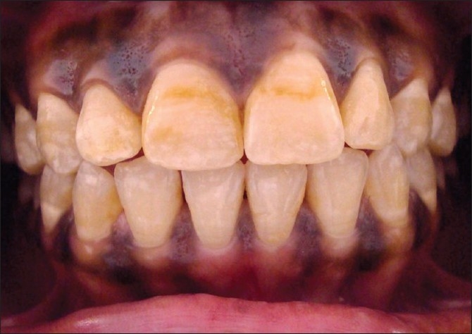
Preoperative view showing yellowish discoloration on labial aspect of both central incisors and chalky labial surface of all teeth
Figure 2.
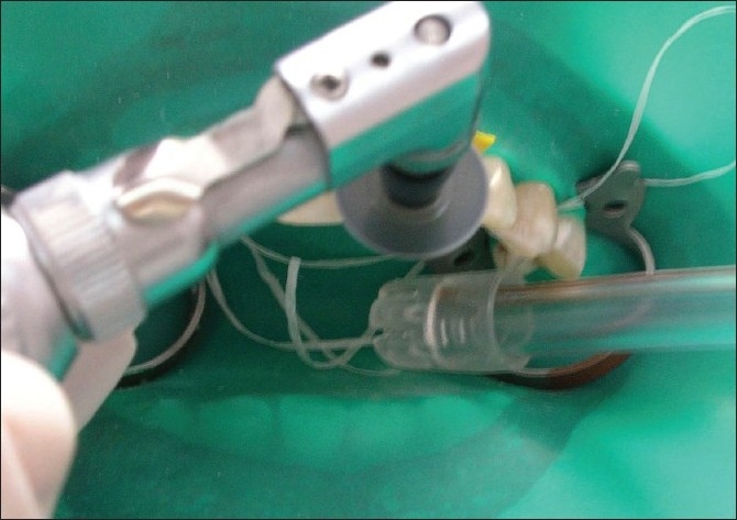
Application of acid via cotton pellet followed by use of coarse grit abrasive discs for 10 seconds at low speed under rubber dam isolation
Figure 3.
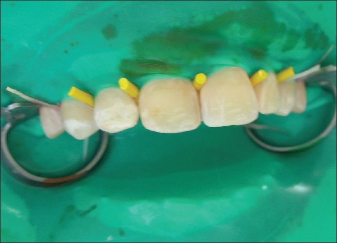
Note the color change immediately after completion of microabrasion
Figure 4.
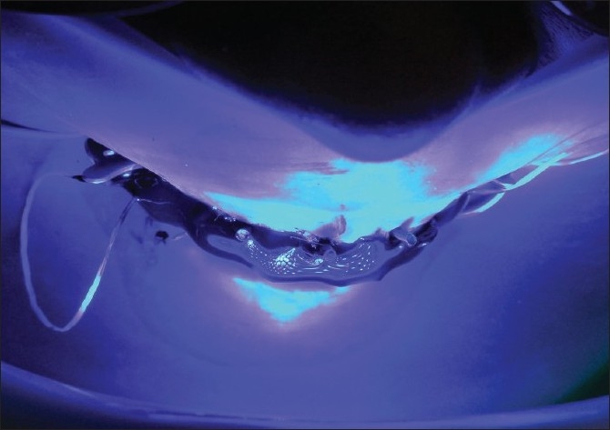
In office power bleaching procedure carried immediately after microabrasion
Figure 5.
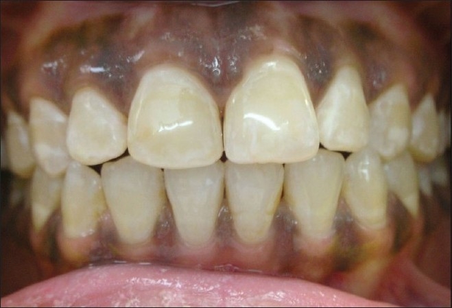
Completion of finishing and polishing procedure. Note the high luster obtained producing camouflage effect and masking the white chalky patches
Figure 6.
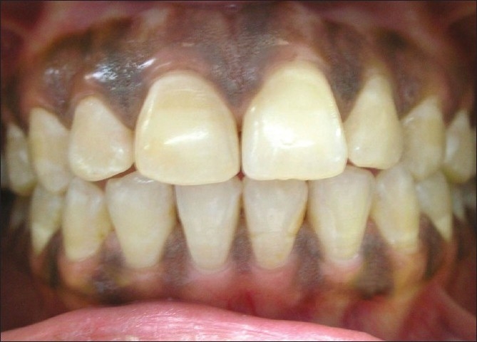
One month follow-up visit showing constancy of esthetics achieved after treatment
Discussion
Microabrasion is a noninvasive, conservative, and economical approach to partially or completely remove superficial enamel discoloration. Lower cost of this technique makes it a viable treatment alternative in rural/semi urban dental practice. Microabrasion technique has been modified several times to enhance the esthetic results as well as simplify the treatment procedure.
Discolored teeth without loss of surface integrity of enamel are ideally suited for microabrasion treatment. However, if discoloration is accompanied with pitting of the surface as in grade III or grade IV fluorosis, clinicians can utilize a combination of microabrasion/bleaching followed by direct or indirect veneering procedures.
It is prudent to carry out microabrasion and in office bleaching under strict isolation using rubber dam as the acid and 35% hydrogen peroxide used in the procedures may damage the adjacent soft tissues. Application of petroleum jelly over the gingival tissue prior to placement of rubber dam provides added protection from the acid. Also, the use of protective eye wear during the procedure by the patient as well as operator is mandatory.
Long term effectiveness of microabrasion is clinically proven in several studies and case reports with no or minimal intra-operative and post-operative discomfort such as dentinal hypersensitivity.[13–15] Not only does microabrasion mask and remove stained tooth structure, thus improving tooth color, but the surface layer created during treatment is a highly polished, densely compacted, mineralized structure.[16] The technique is believed to modify the optical properties of enamel. Abrasion of enamel prisms combined with acid erosion results in the development of a densely compacted prism-free layer on the enamel surface.[17] In addition, subsurface stains may be camouflaged by the optical properties of the newly microabraded surface.[18]
The original microabrasion technique utilized the slurry of 11% hydrochloric acid and silicon carbide using a manual or handpiece-driven rubbing action. In the present case, technique was modified in an attempt to reduce chair side time and enhance effectiveness as well as convenience. Instead of slurry of abrasive and acid, the acid was applied directly on the tooth surface and the abrasive effect was achieved by the use of coarse composite contouring discs (Super Snap™). Thus, the abrasive action was limited only to the area of contact of the disc, thereby, avoiding unnecessary loss of enamel surface.
The combined regimen (microabrasion and bleaching) is most effective in patients who have prominent white areas on teeth that are yellowish or that have darkened with age. In office bleaching was preferred in this case over home bleaching regimen because it is quick and the dentist is in total control of the treatment.[19] Several bleaching agents are available in the market such as hydrogen peroxide and carbamide peroxide in various concentrations. In general, the efficacy of hydrogen peroxide containing products are approximately the same when compared with carbamide peroxide containing products with equivalent or similar hydrogen peroxide content and delivered using similar format and formulations.[20] Peroxide and light treatment significantly lighten the color of teeth to a greater extent than does peroxide or light alone, with a low and transient incidence of tooth sensitivity.[21]
Use of casein phosphopeptide-amorphous calcium phosphate containing tooth paste after microabrasion have been found to significantly reduce the enamel surface roughness thus, minimizing the risk of developing smooth surface caries.[22,23] Bioactive glasses like calcium sodium phosphosilicate when in contact with saliva, rapidly release sodium, calcium, and phosphorous ions into the saliva, which are then available for the remineralization of the tooth surface and result in the formation of hydroxyl carbonate apatite.[23]
To conclude, the case presented supports a feasible and conservative technique to manage enamel discoloration.
Footnotes
Source of Support: Nil
Conflict of Interest: None declared.
References
- 1.Rajyalakshmi K, Rao NV, Krishna N. Studies in Environmental Science. Vol. 27. India: Publishers BV; 1985. Investigations on the relevance of defluoridated water and nutritional supplements in fluorosis endemic areas in Andhra Pradesh. Fluoride Research 1985; p. 358. [Google Scholar]
- 2.Croll TP. Enamel microabrasion: Observations after 10 years. J Am Dent Assoc. 1997;128:45S–50. doi: 10.14219/jada.archive.1997.0424. [DOI] [PubMed] [Google Scholar]
- 3.McCloskey RJ. A Technique for removal of fluorosis stains. J Am Dent Assoc. 1984;109:63–4. doi: 10.14219/jada.archive.1984.0297. [DOI] [PubMed] [Google Scholar]
- 4.Croll TP, Cavanaugh RR. Enamel color modification by controlled hydrochloric acid-pumice abrasion: Part 1. Technique and examples. Quintessence Int. 1986;17:81–7. [PubMed] [Google Scholar]
- 5.Croll TP. Enamel microabrasion for removal of superficial dysmineralization and decalcification defects. J Am Dent Assoc. 1990;120:411–5. doi: 10.14219/jada.archive.1990.0127. [DOI] [PubMed] [Google Scholar]
- 6.Sarrett DC. Tooth whitening today. J Am Dent Assoc. 2002;133:1535–8. doi: 10.14219/jada.archive.2002.0085. [DOI] [PubMed] [Google Scholar]
- 7.Srinivasan M. Finishing composite veneer restorations: The rainbow technique. J Prosthodont Soc. 2007;7:95–101. [Google Scholar]
- 8.Donly KJ, O’Neill MO, Croll TP. Enamel microabrasion: A microscopic evaluation of the “abrasion effect”. Quintessence Int. 1992;23:175–9. [PubMed] [Google Scholar]
- 9.Cvitko E, Swift EJ, Denehy GE. Improved esthetics with a combined bleaching technique: A case report. Quintessence Int. 1992;23:91–3. [PubMed] [Google Scholar]
- 10.Killian CM. Conservative color improvement for teeth with fluorosis- type stain. J Am Dent Assoc. 1993;124:72–4. doi: 10.14219/jada.archive.1993.0111. [DOI] [PubMed] [Google Scholar]
- 11.Croll TP. Esthetic correction for teeth with fluorosis and fluorosislike enamel dysmineralization. J Esthet Dent. 1998;10:21–9. doi: 10.1111/j.1708-8240.1998.tb00332.x. [DOI] [PubMed] [Google Scholar]
- 12.Segura A, Dunly KJ, Wefel JS. The effects of microabrasion on demineralization inhibition of enamel surfaces. Quintessence Int. 1997;28:463–6. [PubMed] [Google Scholar]
- 13.Price RB, Loney RW, Doyle MG, Moulding An evaluation of a technique to remove stains from teeth using microabrasion. J Am Dent Assoc. 2003;134:1066–71. doi: 10.14219/jada.archive.2003.0320. [DOI] [PubMed] [Google Scholar]
- 14.Welbury RR, Shaw L. A simple technique for removal of mottling, opacities and pigmentation from enamel. Dent Update. 1990;17:161–3. [PubMed] [Google Scholar]
- 15.Kilpatrick NM, Welbury RR. Hydrochloric acid/pumice microabrasion technique for the removal of enamel pigmentation. Dent Update. 1993;20:105–7. [PubMed] [Google Scholar]
- 16.Croll TP. Enamel microabrasion. Chicago: Quintessence Publishing; 1991. pp. 27–60. [Google Scholar]
- 17.Olin PS, Lehner CR, Hilton JA. Enamel surface modification in vitro usinghydrochloric acid pumice: An SEM investigation. Quintessence Int. 1988;19:733–6. [PubMed] [Google Scholar]
- 18.Croll TP, Killian CM, Miller AS. Effect of enamel microabrasion compound on human gingiva: Report of a case. Quintessence Int. 1990;21:959–63. [PubMed] [Google Scholar]
- 19.Goldstein CE, Goldstein RE, Feinman RA, Garber DA. Bleaching vital teeth: State of the art. Quintessence Int. 1989;20:729–37. [PubMed] [Google Scholar]
- 20.Joiner A, Thakker G. In vitro evaluation of a novel 6% hydrogen peroxide tooth whitening product. J Dent. 2004;32:19–25. doi: 10.1016/j.jdent.2003.10.007. [DOI] [PubMed] [Google Scholar]
- 21.Tavares M, Stultz J, Newman M, Smith V, Kent R, Carpino E. Light augments tooth whitening with peroxide. J Am Dent Assoc. 2003;134:167–75. doi: 10.14219/jada.archive.2003.0130. [DOI] [PubMed] [Google Scholar]
- 22.Mathias J, Kavitha S, Mahalaxmi S. A comparison of surface roughness after microabrasion of enamel with and without using CPP-ACP: An in vitro study. J Conserv Dent. 2009;12:22–5. doi: 10.4103/0972-0707.53337. [DOI] [PMC free article] [PubMed] [Google Scholar]
- 23.Chalmers JM. Minimal Intervention Dentistry: Part 1. Strategies for addressing the new caries challenge in older patients. J Can Dent Assoc. 2006;72:427–33. [PubMed] [Google Scholar]


