

Abstract
A patient presented with an acute abdomen at the Emergency Department. The patient, a 69-year-old man, was admitted and underwent surgery with a provisional diagnosis of acute appendicitis. During surgery, omental torsion was diagnosed and the involved omentum was removed. The patient had no previous surgical history. Omental torsion is a rare cause of acute abdomen in children and adults who may present with various signs and symptoms; a preoperative diagnosis may therefore be difficult and can usually only be established during surgery.
Keywords: Omentum, Torsion, Abdominal pain
INTRODUCTION
Omental torsion is a rare cause of acute abdomen. When the greater omentum is twisted around its axis, perfusion defects and vascular impairment of the organ are possible. As a result, different pathological modifications are possible, from simple edema to ischemia and gangrene of the omentum[1]. Omental torsion can be either primary (idiopathic) or secondary, depending on the predisposing factors that cause it. Primary torsion of the omentum was first described in 1899[2]. However, very few cases have been reported in adults[3] and children[4-8]. Omental torsion is responsible for 0.1% of laparotomies performed for acute appendicitis in children[7]. This report describes one case of a male adult who presented with acute abdomen and in whom omental torsion was the definitive surgical diagnosis.
CASE REPORT
A 69-year-old man went to the Emergency Department complaining of abdominal pain. The pain, which started 2 d earlier, was constant and mainly located over the upper abdomen; the symptoms had increased in severity in the last few hours. The patient also presented with nausea, vomiting and anorexia. His medical history revealed untreated irritable bowel syndrome but no previous surgical history. On physical examination, tenderness over the upper abdominal area was recognized with rebound and guarding. A biochemical analysis revealed leukocytosis (19 000 leukocytes/mm3). All other laboratory tests were normal. Abdominal computed tomography (CT) was performed which revealed an inflammatory mass in the upper right side of the abdomen (Figure 1). With an incorrect diagnosis of appendicitis, the patient underwent a laparotomy via a midline incision. At laparotomy, the surgeons observed torsion of the right part of the omentum that was twisted several times around its long axis in a counter clockwise manner (Figure 2A). The omentum was removed (Figure 2B), the postoperative recovery of the patient was uneventful and he was discharged 5 d later. The pathologist confirmed the diagnosis of omental torsion.
Figure 1.
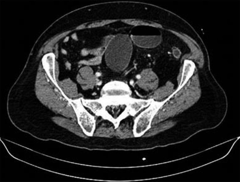
Computed tomography scan images of the 69-year-old man showing a large inflammatory mass of the upper right side of the abdomen.
Figure 2.

Torsion of the right part of the omentum that was twisted several times around its long axis (A) and omentum removed, showing ischemia and necrosis (B).
DISCUSSION
Omental torsion is a rare condition and difficult to diagnose preoperatively. It can mimic various other causes of acute abdomen; surgeons should always consider it in the differential diagnosis of acute abdominal pain. Unfortunately, the symptoms and clinical findings do not present in any characteristic pattern that suggests the diagnosis. The differential diagnosis includes acute appendicitis, acute cholecystitis, cecal diverticulitis and other diseases[1,9]. Omental torsion has an incidence of 0.0016%-0.37% when compared with appendicitis (ratio of less than 4 cases per 1000 cases of appendicitis)[8,10,11].
The correct etiology is not clear in idiopathic omental torsion. No pathological findings can be found in the abdomen of the patient; sometimes surgeons observe a large and mobile omentum which has been rotated one or more times around a fixed spot, usually the right epiploic artery[1,9].
Infarction of the right side of the omentum is more frequent because of its greater length and mobility[12]. Other authors explain this as being due to a different embryological origin of the right side of omentum with congenitally anomalous fragile blood vessels[13]. “Bifid omentum” is an accessory omentum originating from a narrow route and excessive adipose tissue accumulation on the omentum.
Obesity has been identified as a predisposing factor. One study documents that almost 70% of patients with omental torsion are obese[14,15]. In children, obesity is considered an important factor in omental torsion, especially when the body mass index is above the 95th percentile[9,16]. Precipitating factors leading to an increased risk for omental torsion include trauma, coughing, a sudden change of body position, hyperperistalsis after a copious meal, or compression between the liver and the abdominal wall[1,17]. In secondary omental torsion, some associated abdominal pathology has been frequently observed such as cysts, tumors, inflammation, prior surgery or hernias. These conditions increase abdominal pressure as in the case of heavy exercise, sneezing or coughing and the occupational use of vibrating tools. Primary omental torsion is difficult to diagnose preoperatively and an accurate preoperative diagnosis is reported in only 0.6%-4.8% of all cases[17]. Clinical presentations vary; they include a sudden increase of pain on the right side enhanced with abdominal movements, with signs of peritoneal irritation in the right upper quadrant. If the omentum involved is a large part, a mass might be palpable. Other symptoms may be present, such as nausea and vomiting, fever and leukocytosis. Many authors have stressed the importance of imaging in the diagnosis of omental torsion. Abdominal ultrasound is important to exclude acute cholecystitis and shows an ovoid or cake-like hyperechoic mass adherent to the peritoneum located in the umbilical region or anterolaterally to the right half of the colon[12,18]. Doppler sonography sometimes shows vessels within the mass and peripheral hyperaemia[19]. CT scans play a important role in the diagnosis of torsion of the greater omentum[17,20,21]. Omental torsion can be easily differentiated from acute cholecystitis, appendicitis and cecum diverticulitis which have different characteristics. In the case of omental torsion, the CT-scan shows an infarcted omentum as an area of high-attenuated fat containing hyperattenuated streaks just beneath the parietal peritoneum with thickening of the overlying anterior abdominal wall[20]. Another finding can be a whirling pattern of the mesentery or fluid accumulation within the abdomen. Unfortunately, all these findings can be observed in various other conditions, such as in lipoma, liposarcoma, angiomyolipoma, teratoma, mesenteric lipodystrophy, pseudomyxoma peritonei, epiploic appendagitis, segmental infarction of the omentum and intestinal volvulus[20]. To make the correct diagnosis, some authors recommend laparoscopy as the diagnostic and therapeutic method of choice in cases of omental torsion[10,22-24]. In many reports of individual cases as well as larger series of patients with omental torsion, the diagnosis was mainly based on CT findings and the treatment was frequently conservative. Miguel Perelló et al[25] reviewed six patients who were diagnosed with primary omental torsion based on CT scans and thereafter underwent conservative treatment. In addition, Abadir et al[26] reported that 12 of 15 patients who had primary omental torsion were diagnosed using a CT scan and were managed without surgery. In our case, no predisposing factors could be identified. Acute appendicitis was the initial clinical possibility. The CT findings were not diagnostic and the diagnosis was finally established intraoperatively. Traditionally, the standard treatment for omental torsion is a resection of the involved segment of omentum[4]. However, with the success of imaging tools there are many reported cases of omental torsion that have been successfully managed by conservative treatment, especially in patients with no associated complications[10,21,24-27].
In conclusion, primary omental torsion appears with a wide variety of clinical manifestations. It can mimic various other causes of acute abdomen; surgeons should always consider it in the differential diagnosis of acute abdominal pain. A preoperative diagnosis in most cases is difficult. For an early preoperative diagnosis, a high index of suspicion is required as well as abdominal CT scans. In the majority of cases, the surgical removal of the diseased omentum remains the treatment of choice. Patients with uncomplicated omental torsion can be safely managed with conservative treatment.
Footnotes
Peer reviewer: Yoshihiro Moriwaki, MD, PhD, Department of Critical Care and Emergency Center, Yokohama City University Medical Center, 4-57, Urafune-cho, Minami-ku, Yokohama 232-0024, Japan
S- Editor Wang JL L- Editor Roemmele A E- Editor Zheng XM
References
- 1.Adams JT. Torsion of the omentum. Abdominal wall, omentum, mesentery and retroperitoneum. In: Schwartz SI, Shires GT, Spencer FC, editors. Principles of surgery. 5th ed. New York, NY: McGraw-Hill; 1989. pp. 1495–1496. [Google Scholar]
- 2.Eitel GG. Rare omental tumor. Med Rec. 1899;55:715–716. [Google Scholar]
- 3.Basson SE, Jones PA. Primary torsion of the omentum. Ann R Coll Surg Engl. 1981;63:132–134. [PMC free article] [PubMed] [Google Scholar]
- 4.Karayiannakis AJ, Polychronidis A, Chatzigianni E, Simopoulos C. Primary torsion of the greater omentum: report of a case. Surg Today. 2002;32:913–915. doi: 10.1007/s005950200180. [DOI] [PubMed] [Google Scholar]
- 5.Ozbey H, Salman T, Celik A. Primary torsion of the omentum in a 6-year-old boy: report of a case. Surg Today. 1999;29:568–569. doi: 10.1007/BF02482356. [DOI] [PubMed] [Google Scholar]
- 6.Saraç AM, Yeğen C, Aktan AO, Yalin R. Primary torsion of the omentum mimicking acute appendicitis: report of a case. Surg Today. 1997;27:251–253. doi: 10.1007/BF00941655. [DOI] [PubMed] [Google Scholar]
- 7.Sweeney MJ, Blestel GA, Ancalmo N. Primary torsion of the greater omentum. A rare cause of abdominal pain in children. JAMA. 1983;249:3073. [PubMed] [Google Scholar]
- 8.Kimber CP, Westmore P, Hutson JM, Kelly JH. Primary omental torsion in children. J Paediatr Child Health. 1996;32:22–24. doi: 10.1111/j.1440-1754.1996.tb01535.x. [DOI] [PubMed] [Google Scholar]
- 9.Theriot JA, Sayat J, Franco S, Buchino JJ. Childhood obesity: a risk factor for omental torsion. Pediatrics. 2003;112:e460. doi: 10.1542/peds.112.6.e460. [DOI] [PubMed] [Google Scholar]
- 10.Itenberg E, Mariadason J, Khersonsky J, Wallack M. Modern management of omental torsion and omental infarction: a surgeon's perspective. J Surg Educ. 2010;67:44–47. doi: 10.1016/j.jsurg.2010.01.003. [DOI] [PubMed] [Google Scholar]
- 11.Pinedo-Onofre JA, Guevara-Torres L. [Omental torsion. An acute abdomen etiology] Gac Med Mex. 2007;143:17–20. [PubMed] [Google Scholar]
- 12.Puylaert JB. Right-sided segmental infarction of the omentum: clinical, US, and CT findings. Radiology. 1992;185:169–172. doi: 10.1148/radiology.185.1.1523302. [DOI] [PubMed] [Google Scholar]
- 13.Epstein LI, Lempke RE. Primary idiopathic segmental infarction of the greater omentum: case report and collective review of the literature. Ann Surg. 1968;167:437–443. doi: 10.1097/00000658-196803000-00020. [DOI] [PMC free article] [PubMed] [Google Scholar]
- 14.van Breda Vriesman AC, Lohle PN, Coerkamp EG, Puylaert JB. Infarction of omentum and epiploic appendage: diagnosis, epidemiology and natural history. Eur Radiol. 1999;9:1886–1892. doi: 10.1007/s003300050942. [DOI] [PubMed] [Google Scholar]
- 15.LEITNER MJ, JORDAN CG, SPINNER MH, REESE EC. Torsion, infarction and hemorrhage of the omentum as a cause of acute abdominal distress. Ann Surg. 1952;135:103–110. doi: 10.1097/00000658-195201000-00014. [DOI] [PMC free article] [PubMed] [Google Scholar]
- 16.Styne DM. Childhood and adolescent obesity. Prevalence and significance. Pediatr Clin North Am. 2001;48:823–54, vii. doi: 10.1016/s0031-3955(05)70344-8. [DOI] [PubMed] [Google Scholar]
- 17.Poujade O, Ghiles E, Senasli A. Primary torsion of the greater omentum: case report--review of literature: diagnosis cannot always be performed before surgery. Surg Laparosc Endosc Percutan Tech. 2007;17:54–55. doi: 10.1097/01.sle.0000213763.40429.f6. [DOI] [PubMed] [Google Scholar]
- 18.Schlesinger AE, Dorfman SR, Braverman RM. Sonographic appearance of omental infarction in children. Pediatr Radiol. 1999;29:598–601. doi: 10.1007/s002470050657. [DOI] [PubMed] [Google Scholar]
- 19.Baldisserotto M, Maffazzoni DR, Dora MD. Omental infarction in children: color Doppler sonography correlated with surgery and pathology findings. AJR Am J Roentgenol. 2005;184:156–162. doi: 10.2214/ajr.184.1.01840156. [DOI] [PubMed] [Google Scholar]
- 20.Kim J, Kim Y, Cho OK, Rhim H, Koh BH, Kim YS, Han DS, Baek HK. Omental torsion: CT features. Abdom Imaging. 2003;29:502–504. doi: 10.1007/s00261-003-0155-2. [DOI] [PubMed] [Google Scholar]
- 21.Rimon A, Daneman A, Gerstle JT, Ratnapalan S. Omental infarction in children. J Pediatr. 2009;155:427–431.e1. doi: 10.1016/j.jpeds.2009.03.039. [DOI] [PubMed] [Google Scholar]
- 22.Mallick MS, Al-Bassam AA. Primary omental torsion in children. The pre-disposing factors and role of laparoscopy in diagnosis and treatment. Saudi Med J. 2006;27:194–197. [PubMed] [Google Scholar]
- 23.Chan KW, Chow CS, Tam YH, Lee KH. Laparoscopy: an excellent tool in the management of primary omental torsion in children. J Laparoendosc Adv Surg Tech A. 2007;17:821–824. doi: 10.1089/lap.2007.0034. [DOI] [PubMed] [Google Scholar]
- 24.Kavalakat AJ, Varghese CJ. Laparoscopic management of an uncommon cause for right lower quadrant pain: A case report. Cases J. 2008;1:164. doi: 10.1186/1757-1626-1-164. [DOI] [PMC free article] [PubMed] [Google Scholar]
- 25.Miguel Perelló J, Aguayo Albasini JL, Soria Aledo V, Aguilar Jiménez J, Flores Pastor B, Candel Arenas MF, Girela Baena E. [Omental torsion: imaging techniques can prevent unnecessary surgical interventions] Gastroenterol Hepatol. 2002;25:493–496. [PubMed] [Google Scholar]
- 26.Abadir JS, Cohen AJ, Wilson SE. Accurate diagnosis of infarction of omentum and appendices epiploicae by computed tomography. Am Surg. 2004;70:854–857. [PubMed] [Google Scholar]
- 27.Soobrah R, Badran M, Smith SG. Conservative management of segmental infarction of the greater omentum: a case report and review of literature. Case Report Med. 2010;2010 doi: 10.1155/2010/765389. [DOI] [PMC free article] [PubMed] [Google Scholar]