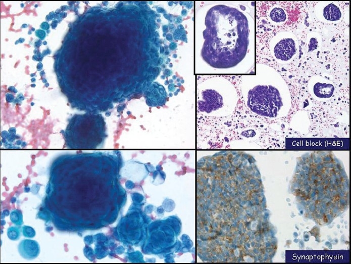
Figure 3.

Small-cell carcinoma with a predominant single-cell pattern mimicking lymphoma in pleural fluid. The nuclei show a finely distributed granular chromatin texture and small nucleoli, left and middle (Pap stain, left ×200 and middle ×400). Chains of small cells with nuclear molding (middle, center of the image) and karyorrhexis (left) are seen. In cell block sections (right, H and E stain, ×400) tumor cells display a discohesive pattern
