Abstract
Purpose
The efficacy of cisplatin, irinotecan, and bevacizumab was evaluated in patients with extensive-stage small-cell lung cancer (ES-SCLC).
Patients and Methods
Patients with ES-SCLC received cisplatin 30 mg/m2 and irinotecan 65 mg/m2 on days 1 and 8 plus bevacizumab 15 mg/kg on day 1 every 21 days for six cycles on this phase II study. The primary end point was to differentiate between 50% and 65% 12-month survival rates.
Results
Seventy-two patients were enrolled between March 2005 and April 2006; four patients canceled, and four were ineligible. Grade 3 or 4 toxicities included neutropenia (25%), all electrolyte (23%), diarrhea (16%), thrombocytopenia (10%), fatigue (10%), nausea (10%), hypertension (9%), anemia (9%), infection (7%), vascular access thrombosis (2%), stroke (2%), and bowel perforation (1%). Three deaths (5%) occurred on therapy as a result of pneumonitis (n = 1), stroke (n =1), and heart failure (n = 1). Complete response, partial response, and stable disease occurred in three (5%), 45 (70%), and 11 patients (17%), respectively. Progressive disease occurred in one patient (2%). Overall response rate was 75%. Median progression-free survival (PFS) was 7.0 months (95% CI, 6.4 to 8.4 months). Median overall survival (OS) was 11.6 months (95% CI, 10.5 to 15.1 months). Hypertension ≥ grade 1 was associated with improved OS after adjusting for performance status (PS) and age (hazard ratio [HR], 0.55; 95% CI, 0.31 to 0.97; P = .04). Lower vascular endothelial growth factor levels correlated with worse PFS after adjusting for age and PS (HR, 0.90; 95% CI, 0.83 to 0.99; P = .03).
Conclusion
PFS and OS times were higher compared with US trials in ES-SCLC with the same chemotherapy. However, the primary end point of the trial was not met. Hypertension was associated with improved survival after adjusting for age and PS.
INTRODUCTION
Most of the 30,000 new patients with small-cell lung cancer (SCLC) in the United States each year have extensive stage (ES) at presentation.1 Platinum-based chemotherapy can achieve response rates of up to 80% and improve survival from approximately 3 months to 10 months.2–4 The Japanese Clinical Oncology Group (JCOG) 9511 trial compared etoposide-based therapy with irinotecan-based therapy for untreated ES-SCLC and reported improved survival of 12.8 months with irinotecan.5 Phase III trials conducted primarily in the United States comparing etoposide- and irinotecan-based regimens showed that the regimens had similar efficacy.6,7
Angiogenesis is a validated cancer therapy target.8 Vascular endothelial growth factor (VEGF) levels are elevated in a number of malignancies, including SCLC.9–13 Bevacizumab is a monoclonal antibody against VEGF. Toxicities related to bevacizumab have included hypertension, hemoptysis, thrombosis, proteinuria, leukopenia, GI perforation, and defective wound healing.14–16 A phase III trial comparing chemotherapy with or without bevacizumab for nonsquamous, non–small-cell lung cancer (NSCLC) showed a survival advantage with bevacizumab.16 A phase III trial in advanced colon cancer showed a survival advantage for the addition of bevacizumab to irinotecan-based chemotherapy.17 Promising phase II activity was seen when bevacizumab was added to cisplatin and irinotecan in advanced gastric cancer and to irinotecan in recurrent glioblastoma multiforme.18,19 The current trial evaluated cisplatin, irinotecan, and bevacizumab as therapy for untreated ES-SCLC.
PATIENTS AND METHODS
Eligible patients had histologic documentation of ES-SCLC. ES was defined as extrathoracic metastatic disease, malignant pleural effusion, bilateral or contralateral supraclavicular adenopathy, or contralateral hilar adenopathy. Eligibility criteria included Eastern Cooperative Oncology Group (ECOG) performance status (PS) of 0 to 2 and standard initial laboratory tests. Patients with CNS metastases were eligible if they had recovered from toxicity and it was a minimum of 1 week after completion of radiotherapy. Patients were not eligible if they had recent major surgery, significant hemoptysis, or open wounds or were receiving full-dose anticoagulation. Each participant signed an institutional review board–approved, protocol-specific informed consent in accordance with federal and institutional guidelines. Registration and data collection were managed by the Cancer and Leukemia Group B (CALGB) Statistical Center. Data quality was ensured by careful review by CALGB Statistical Center staff and the study chairperson following CALGB policies. Data analysis was performed by CALGB statisticians.
Chemotherapy
Patients received cisplatin 30 mg/m2 and irinotecan 65 mg/m2 on days 1 and 8 and bevacizumab 15 mg/kg on day 1 at 21-day intervals. Bevacizumab was not continued after chemotherapy. After every two cycles, imaging studies were repeated to assess tumor response. Patients with stable disease, partial response, or complete response received a maximum of six cycles of therapy. The initial 10 evaluable patients were monitored for safety with biweekly conference calls. Accrual was held until initial cohort safety evaluation was complete.
Chemotherapy dose modifications were based on treatment day counts. If platelets were less than 100,000/μL or granulocytes were less than 1,500/μL, then day 1 therapy was held. Therapy was discontinued if the granulocyte count did not return to ≥ 1,500/μL and the platelet count did not return to ≥ 100,000/μL after a treatment delay of 3 weeks.
Biomarker Analysis
Whole blood was collected and centrifuged, and plasma was then stored at −80°C. VEGF 165 and platelet-derived growth factor (PDGF) AB were determined by enzyme-linked immunosorbent assay kits according to the manufacturers' protocol (VEGF; R&D Systems, Minneapolis, MN; PDGF, Insight Genomics, Falls Church, VA) and expressed in picograms per milliliter. Assays were performed in triplicate for each sample, and results were presented as mean and SE.
At the time that CALGB 30306 was designed, hypertension was not known to be associated with outcome on bevacizumab therapy, so there was no preplanned analysis of hypertension and outcome. Hypertension was known to be an adverse effect associated with bevacizumab therapy, and collection of hypertension data was preplanned on the study-specific case report form.
Statistical Analysis
Overall survival (OS) time was measured from the day of registration until date of death; living patients were censored at the date of last follow-up. Progression-free survival (PFS) was measured from the day of registration until disease progression or death, whichever occurred first; living patients who did not experience progression were censored at the date of last follow-up. Progression was defined as at least a 20% increase in the sum of the longest diameter of target lesions, taking as references the smallest sum of the longest diameter recorded since the treatment started, or the appearance of one or more new lesions. Kaplan-Meier estimates were used to illustrate PFS and OS curves.
The log-rank test and likelihood ratio test were used to investigate survival difference for patients with or without hypertension adverse events. Nonparametric Kruskal-Wallis test was used to compare between the two batches of biomarkers. Cox proportional hazard models were used to examine the correlation between survival time and covariates, such as age, PS, and the biomarkers VEGF and PDGF. Pearson χ2 statistic was used to test the relationship between survival time and all the covariates in the model.
The primary end point of the study was based on the proportion of patients who were alive 12 months after initiation of protocol therapy. For efficacy end points, the evaluable population consisted of patients enrolled, excluding patients who canceled, never received any treatment, or were deemed ineligible based on eligibility criteria specified in the protocol. For tabulation of safety data, all patients who had grade 3 or greater adverse events are included.
The study was prospectively designed to differentiate between a 50% and 65% 12-month survival rate. A one-stage phase II design was used. A sample size of 72 patients would provide approximately 90% power to differentiate 12-month survival rates of 50% or less and 65% or greater, with a one-sided type I error of 0.097. If a patient remained alive 12 months after the initial administration of the experimental treatment regimen, treatment was considered a success; otherwise, it was considered a failure. If less than 57% of patients were successful, it was concluded that the treatment regimen is not worthy of additional investigation. Secondary objectives were to assess the response rates, median OS and PFS times, and toxicity. Analyses of associations between hypertension, VEGF and PDGF levels (treated as continuous variables), and PFS or OS were performed using univariate and multivariate Cox regression hazard models. Members of the Audit Committee visit all participating institutions at least once every 3 years to review source documents.
RESULTS
Study activation occurred in December 2004, and closure occurred in April 2006. Seventy-two patients were registered, and four patients were canceled, receiving no treatment. Of the 68 remaining patients, four patients were ineligible as a result of elevated baseline AST, no baseline brain imaging, NSCLC, and being deemed ineligible by treating institution after cycle 1, day 1. All results presented exclude ineligible patients except for those presented in Table 1, which lists adverse events. Baseline characteristics are listed in Appendix Table A1 (online only). The median number of treatment cycles was six cycles, and 41 patients finished six cycles.
Table 1.
Grade 3 or 4 Toxicities Occurring in ≥ 5% of Patients and All Grade 5 Toxicities (n = 68)
| Adverse Event | Grade 3 |
Grade 4 |
Grade 5 |
|||
|---|---|---|---|---|---|---|
| No. of Patients | % | No. of Patients | % | No. of Patients | % | |
| Hematologic | ||||||
| Hemoglobin | 6 | 9 | 0 | 0 | 0 | 0 |
| Leukocytes (total WBC) | 5 | 7 | 3 | 4 | 0 | 0 |
| Neutrophils (ANC/AGC) | 13 | 19 | 4 | 6 | 0 | 0 |
| Platelets | 4 | 6 | 3 | 4 | 0 | 0 |
| Maximum hematologic | 18 | 26 | 7 | 10 | 0 | 0 |
| Nonhematologic | ||||||
| Hypertension | 6 | 9 | 0 | 0 | 0 | 0 |
| Fatigue | 7 | 10 | 0 | 0 | 0 | 0 |
| Anorexia | 4 | 6 | 0 | 0 | 0 | 0 |
| Dehydration | 8 | 12 | 0 | 0 | 0 | 0 |
| Diarrhea | 9 | 13 | 2 | 3 | 0 | 0 |
| Nausea/vomiting | 7 | 10 | 0 | 0 | 0 | 0 |
| Febrile neutropenia | 4 | 6 | 0 | 0 | 0 | 0 |
| Infection | 5 | 7 | 0 | 0 | 0 | 0 |
| Hypokalemia | 6 | 9 | 0 | 0 | 0 | 0 |
| Hyponatremia | 4 | 6 | 2 | 3 | 0 | 0 |
| Congestive heart failure | 0 | 0 | 0 | 0 | 1 | 1 |
| Hemorrhage CNS | 0 | 0 | 0 | 0 | 1 | 1 |
| Pneumonitis | 0 | 0 | 0 | 0 | 1 | 1 |
| Maximum nonhematologic | 24 | 35 | 5 | 7 | 3 | 4 |
| Maximum overall adverse events | 30 | 44 | 11 | 16 | 3 | 4 |
NOTE. Three deaths occurred on therapy (congestive heart failure, n = 1; pneumonia, n = 1; and embolic/thrombotic stroke that later became hemorrhagic, n = 1).
Abbreviations: AGC, absolute granulocyte count; ANC, absolute neutrophil count.
Adverse event data were collected on 68 patients, and treatment-related grade ≥ 3 events are listed in Table 1. Adverse events affecting 10% or more of patients were neutropenia (25%), all electrolyte (23%), diarrhea (16%), dehydration (12%), leukocytes (11%), thrombocytopenia (10%), fatigue (10%), and nausea (10%). There were three grade 5 adverse events (congestive heart failure, pneumonitis/pulmonary infiltrates, and CNS hemorrhage/bleeding). The patient who died of CNS bleed presented with a stroke without hemorrhage on the initial brain computed tomography scan performed to assess an acute change in neurologic status. There was no greater than grade 2 hemoptysis or fistula between an airway and an adjacent structure reported. Additional adverse events included one bowel perforation, one typhlitis, two thromboses related to vascular access devices, and two incidents of cerebrovascular ischemia, including one in the patient who ultimately died of CNS bleed.
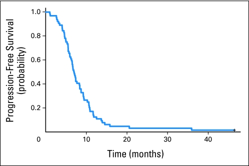
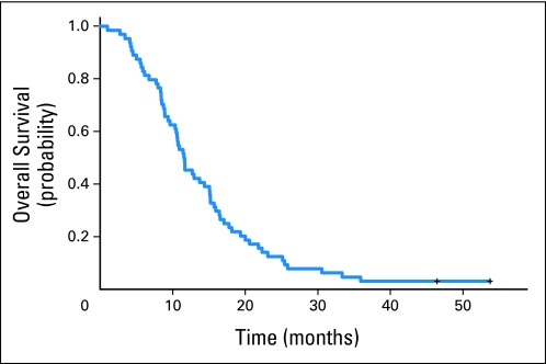
Of the 64 patients assessable for efficacy, three patients (5%) achieved complete response and 45 patients (70%) achieved partial response, for an overall response rate of 75% (Appendix Table A2, online only). Median PFS was 7.0 months (95% CI, 6.4 to 8.4 months; Fig 1). Median OS was 11.6 months (95% CI, 10.5 to 15.1 month; Fig 2). Twelve-month survival was 43.8% (95% CI, 33.1% to 57.8%), with 28 patients who survived more than 12 months (Table 2). This percentage is less than the prespecified 57% needed to declare that this study is worthy of further investigation.
Fig 1.
Kaplan-Meier curve for progression-free survival.
Fig 2.
Kaplan-Meier curve for overall survival.
Table 2.
Median and 12-Month Progression-Free Survival and Overall Survival
| End Point | No. of Patients Who Experienced Disease Progression or Death | 12-Month Survival (%) |
Survival (months) |
|||
|---|---|---|---|---|---|---|
| Rate | 95% CI | 90% CI | Median | 95% CI | ||
| Overall survival | 62 | 43.8 | 33.1 to 57.8 | 34.7 to 55.2 | 11.6 | 10.5 to 15.1 |
| Progression-free survival | 63 | 10.9 | 5.4 to 22.0 | 6.1 to 19.7 | 7.0 | 6.4 to 8.4 |
NOTE. The 12-month survival rates were computed using the Kaplan-Meier estimator (n = 64).
Seventy-two patients were registered for correlative studies. Four patients were cancelled and did not receive therapy, and four patients were deemed ineligible. Blood samples from 59 patients were collected and sent for analysis in two separate batches (48 samples in batch 1 and 11 samples in batch 2) several months apart. Of the 64 patients, five patients either had unusable samples or did not consent for use of their blood sample. There was no difference in age and PS between patients who provided samples and those who did not. There were significant differences in both VEGF and PDGF levels between batches (tested using nonparametric Kruskal-Wallis test; P = .0027 and P < .001 for VEGF and PDGF, respectively). All P values were two-sided tests at the P = .05 level. For the 48 samples in batch 1, the median VEGF level was 78 pg/mL (range, 0.05 to 1,812 pg/mL), whereas the median PDGF level was 26 pg/mL (range, 3.95 to 113 pg/mL). There was no correlation between tumor response and VEGF or PDGF level. Lower VEGF level, but not PDGF level, before starting therapy was associated with worse PFS (hazard ratio [HR], 0.904; 95% CI, 0.825 to 0.991; P = .0309) when analyzed with a multivariate Cox proportional hazards model after adjusting for age greater than 65 years and ECOG PS of 0, 1, or 2. Lower VEGF level, but not PDGF level, showed a trend of association with worse OS (HR, 0.924; 95% CI, 0.848 to 1.008) after adjusting for age and PS, but the trend was not significant (P = .0745; Appendix Table A3, online only). However, univariate VEGF levels were not significantly associated with PFS (HR, 0.626; 95% CI, 0.288 to 1.360; P = .24) or OS (HR, 0.626; 95% CI, 0.29 to 1.35; P = .23). We have also investigated the inclusion of the second batch of 11 patients with adjustments for batch effect. Both the PFS and OS models for VEGF have P < .10 (Appendix Tables A4 and A5, online only).
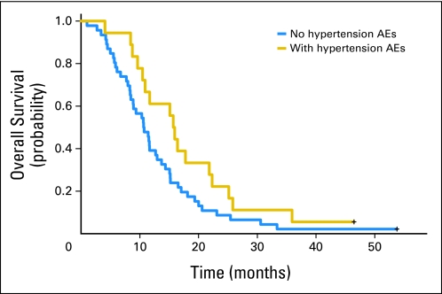
Of 64 patients, 18 patients had grade 1 or greater hypertension, whereas 46 patients did not have grade 1 or greater hypertension. Two patients who were still alive at final data analysis were censored, one with and one without hypertension. Patients developing grade 1 or greater hypertension had a trend toward improved OS, with an HR of 0.591 (95% CI, 0.336 to 1.039; P = .0676) relative to patients not developing grade 1 hypertension when analyzed with a Cox proportional hazards model (Table 3). Hypertension was associated with significantly improved OS after adjusting for age and PS (HR, 0.55; 95% CI, 0.31 to 0.97; P = .04; Fig 3). The proportional hazards assumption was checked for both models, and the assumption holds. The median OS time for patients experiencing ≥ grade 1 hypertension was 15.8 months (95% CI, 10.5 to 21.8 months), and the median OS time for patients not experiencing hypertension was 10.7 months (95% CI, 8.4 to 12.9 months). To investigate whether the outcomes for patients who experienced hypertension differed from the outcomes for patients who did not experience hypertension, Cox models with age and PS were fitted in one model, and an indicator for experiencing hypertension was added in addition to age and PS in the second model. The likelihood ratio test comparing these two models for OS was statistically significant (P = .03), indicating that patients on cisplatin, irinotecan, and bevacizumab who experienced hypertension had better outcomes than patients who did not experience hypertension.
Table 3.
Log-Rank Test and Cox Model for Association Between Grade ≥ 1 Hypertension and Overall Survival
| Measure | Result |
|---|---|
| Grade ≥ 1 hypertension | |
| No. of deaths | 17 |
| No. of patients censored | 1 |
| No grade ≥ 1 hypertension | |
| No. of deaths | 45 |
| No. of patients censored | 1 |
| Log-rank P | .0645 |
| Cox model | |
| P | .0676 |
| Hazard ratio | 0.591 |
| 95% CI | 0.336 to 1.039 |
Fig 3.
Kaplan-Meier curve for overall survival for patients with or without hypertensive adverse events (AEs), either grade ≥ 1 hypertension or no hypertension.
DISCUSSION
The median PFS of 7.0 months (95% CI, 6.4 to 8.4 months) and OS of 11.6 months (95% CI, 10.5 to 15.1 months) seen in this phase II trial of cisplatin, irinotecan, and bevacizumab were modestly higher compared with the results with irinotecan-based regimens in ES-SCLC of two recent phase III trials for ES-SCLC conducted in the United States and recent trials with etoposide-based regimens.4,6,7,20 However, this trial did not meet the primary objective, which was to improve on the 12.8-month survival reported by the JCOG 9511 trial with the cisplatin and irinotecan combination.5
The JCOG 9511 trial showed superior survival for irinotecan compared with etoposide, but large randomized trials conducted in North America have found survival to be similar between etoposide- and irinotecan-based regimens.6,7 In the Hoosier Oncology Group (HOG) trial, the same cisplatin and irinotecan chemotherapy regimen was studied as was used in our trial, and PFS was 4.1 months and OS was 9.3 months.7 In a Southwest Oncology Group (SWOG) trial, the cisplatin and irinotecan combination was the same as was used in JCOG 9511, and PFS was 5.8 months and OS was 9.9 months.6 Therefore, the cisplatin and irinotecan arms for the two large phase III trials conducted primarily in the United States had survival times substantially lower than JCOG 9511.6,7 A comparative analysis of response rates and toxicities between the cisplatin and irinotecan arms of JCOG 9511 and SWOG 0124 suggests that there may be pharmacogenomic factors that account for differences in the efficacy and toxicities seen on the two trials.21 In the irinotecan arms, the incidence of grade 3 or higher neutropenia was 65% on the JCOG 9511 trial and 34% on the SWOG 0124 trial (P < .001). It is now apparent from the results of the HOG and SWOG trials that it might have been better to base the primary survival end point for CALGB 30306 on the control arm of a phase III US trial such as the 9.9-month survival time reported in CALGB 9732.4
An ECOG phase II trial that studied cisplatin, etoposide, and bevacizumab in ES-SCLC reported a median PFS of 4.7 months and a median OS of 10.9 months.22 The Study of Bevacizumab in Previously Untreated Extensive-Stage Small Cell Lung Cancer (SALUTE) trial, a randomized phase II trial of cisplatin or carboplatin and etoposide with or without bevacizumab in untreated ES-SCLC, showed improved PFS but no improvement in OS for the bevacizumab arm.23 Similarly, the PFS of 7.0 months observed in CALGB 30306 is favorable compared with the PFS of 5.8 months and 4.1 months reported for irinotecan-based chemotherapy alone in previously untreated ES-SCLC. 6,7 Bevacizumab was continued after chemotherapy until disease progression in the ECOG and SALUTE trials. In CALGB 30306, bevacizumab was not continued after chemotherapy because there were no data comparing bevacizumab concurrent with chemotherapy with or without maintenance bevacizumab showing improved survival with maintenance therapy. The OS seen in phase II trials of standard chemotherapy plus bevacizumab in ES-SCLC does not justify further study of such regimens in unselected populations.
Life-threatening hemoptysis has been reported with bevacizumab in advanced NSCLC, especially with squamous cell histology.15,17 It has been speculated that central tumors may be a risk for hemoptysis with bevacizumab therapy. Histology may be the most important factor for hemoptysis risk because CALGB 30306 and a similar ECOG trial did not observe significant hemoptysis with bevacizumab in SCLC, which is often a centrally located tumor.22 Tracheoesophageal fistulas were seen in studies of limited-stage SCLC and NSCLC with the combination of chemoradiotherapy and bevacizumab, but no fistulas were reported for the patients treated in our trial.24 The nonhemorrhagic stroke that later became hemorrhagic and fatal could possibly be bevacizumab related. Hypertension was seen as expected for bevacizumab-containing regimens. Two cases of thrombosis associated with catheters and one bowel perforation may be related to bevacizumab. On CALGB 30306, three (4.4%) of 68 patients experienced treatment-related death, whereas on the HOG trial, 22% of patients with PS of 2 and 3.6% of patients with PS of 0 to 1 experienced treatment-related death, and on the irinotecan arm of the SWOG trial, 11 (3.5%) of 317 patients experienced treatment-related death.6,7 In the other two trials in which bevacizumab was added to a standard etoposide chemotherapy combination in untreated ES-SCLC, similar toxicities were reported.22,23 One patient on the SALUTE trial had grade 5 hemoptysis during cycle 1 of chemotherapy plus bevacizumab.23
Hypertension is a common adverse effect of bevacizumab therapy and may reflect effective inhibition of the VEGF pathway.25,26 There has been an association between the development of hypertension with bevacizumab and improved outcome in some clinical trials of breast cancer, colorectal cancer, and NSCLC.27–29 In this trial, there was a significant association between the development of hypertension and improved survival after adjusting for age and PS. An analysis of hypertension and outcome was not presented for the ECOG trial or SALUTE trial that studied etoposide-based chemotherapy and bevacizumab in ES-SCLC.22,23 Our results are in agreement with randomized trials in other cancers that show that the development of hypertension may be a biomarker for bevacizumab benefit and suggest that a subset of patients with ES-SCLC might benefit from the addition of bevacizumab to chemotherapy.
VEGF levels increase with progression of SCLC, and a recent meta analysis confirmed that VEGF overexpression indicates poor prognosis in patients with SCLC.30,31 High pretreatment serum VEGF level was reported to be associated with poor response and survival in patients with SCLC treated with combination therapy.32,33 In our study, lower pretreatment levels of VEGF were associated with worse PFS. This is in contrast to a similar trial in which only baseline vascular cell adhesion molecule levels predicted survival.22 Overall, there is no predictor biomarker in blood that has been identified for bevacizumab therapy in ES-SCLC.
In conclusion, the combination of cisplatin, irinotecan, and bevacizumab for untreated ES-SCLC had an acceptable toxicity profile without clinically significant bleeding or fistula formation. The efficacy was modestly promising compared with standard chemotherapy alone in the same setting for trials performed in the United States. The results do not justify a phase III trial in an unselected population. The development of hypertension was associated with improved survival after adjusting for age and PS. It would be appropriate to study this combination further if a biomarker were identified to select the patients who benefit from the addition of bevacizumab to chemotherapy.
Appendix
The following institutions participated in this study: Dana-Farber Cancer Institute, Boston, MA–Harold J. Burstein, MD, PhD, supported by Grant No. CA32291; Duke University Medical Center, Durham, NC–Jeffrey Crawford, MD, supported by Grant No. CA47577; Hematology-Oncology Associates of Central New York Community Clinical Oncology Program (CCOP), Syracuse, NY–Jeffrey Kirshner, MD, supported by Grant No. CA45389; Illinois Oncology Research Association, Peoria, IL–John W. Kugler, MD, supported by Grant No. CA35113; Kansas City CCOP, Kansas City, MO–Rakesh Gaur, MD; Mount Sinai Medical Center, Miami, FL–Rogerio C. Lilenbaum, MD, supported by Grant No. CA45564; New Hampshire Oncology-Hematology PA, Concord, NH–Douglas J. Weckstein; Rhode Island Hospital, Providence, RI–William Sikov, MD, supported by Grant No. CA08025; Roswell Park Cancer Institute, Buffalo, NY–Ellis Levine, MD, supported by Grant No. CA59518; Southeast Cancer Control Consortium Inc, CCOP, Goldsboro, NC–James N. Atkins, MD, supported by Grant No. CA45808; State University of New York Upstate Medical University, Syracuse, NY–Stephen L. Graziano, MD, supported by Grant No. CA21060; The Ohio State University Medical Center, Columbus, OH–Clara D. Bloomfield, MD, supported by Grant No. CA77658; University of California at San Diego, San Diego, CA–Barbara A. Parker, MD, supported by Grant No. CA11789; University of Nebraska Medical Center, Omaha, NE–Anne Kessinger, MD, supported by Grant No. CA77298 University of Illinois MBCCOP, Chicago, IL–David J. Peace, MD, supported by Grant No. CA74811; University of Iowa, Iowa City, IA–Daniel A. Vaena, MD, supported by Grant No. CA47642; University of Maryland Greenebaum Cancer Center, Baltimore, MD–Martin Edelman, MD, supported by Grant No. CA31983; University of Minnesota, Minneapolis, MN–Bruce A. Peterson, MD, supported by Grant No. CA16450; University of Missouri/Ellis Fischel Cancer Center, Columbia, MO–Michael C. Perry, MD, supported by Grant No. CA12046; University of North Carolina at Chapel Hill, Chapel Hill, NC–Thomas C. Shea, MD, supported by Grant No. CA47559; University of Vermont, Burlington, VT–Steven M. Grunberg, MD, supported by Grant No. CA77406; Wake Forest University School of Medicine, Winston-Salem, NC–David D. Hurd, MD, supported by Grant No. CA03927; Washington University School of Medicine, St Louis, MO–Nancy Bartlett, MD, supported by Grant No. CA77440.
Table A1.
Baseline Patient Demographics and Clinical Characteristics
| Demographic or Clinical Characteristic | No. of Patients (n = 64) | % |
|---|---|---|
| Sex | ||
| Male | 31 | 48 |
| Female | 33 | 52 |
| Age, years | ||
| < 50 | 5 | 8 |
| 50-59 | 21 | 33 |
| 60-69 | 30 | 47 |
| 70+ | 8 | 13 |
| Median | 62 | |
| Range | 38-81 | |
| Race | ||
| White | 56 | 88 |
| Black | 7 | 11 |
| Unknown | 1 | 2 |
| Performance status | ||
| 0 | 13 | 20 |
| 1 | 44 | 69 |
| 2 | 7 | 11 |
| Pleural effusion | ||
| No | 39 | 61 |
| Yes | 25 | 39 |
Table A2.
Best Overall Response to Protocol Therapy
| Response | No. of Patients (N = 64) | % |
|---|---|---|
| Complete response | 3 | 5 |
| Partial response | 45 | 70 |
| Stable disease | 11 | 17 |
| Progressive disease | 1 | 2 |
| Unevaluable* | 4 | 6 |
| Overall response rate | 75 | |
| 95% CI, % | 63 to 85 | |
Patients did not receive tumor assessment before death.
Table A3.
Multivariate Cox Proportional Hazards Model With Covariates of Age and Performance Status for PFS and OS
| Model | P* | HR | 95% CI |
|---|---|---|---|
| Progression-free survival, n = 48 | |||
| VEGF model† | .0309 | 0.904‡ | 0.825 to 0.991 |
| Age | .0368 | 0.438 | 0.202 to 0.951 |
| Performance status§ | .0155 | 2.477 | 1.188 to 5.166 |
| PDGF model | .1306 | 0.992 | 0.981 to 1.002 |
| Age | .0918 | 0.516 | 0.239 to 1.113 |
| Performance status§ | .0514 | 2.035 | 0.996 to 4.158 |
| Overall survival, n = 48 | |||
| VEGF model† | .0745 | 0.924‡ | 0.848 to 1.008 |
| Age | .1172 | 0.561 | 0.273 to 1.156 |
| Performance status§ | .1591 | 1.627 | 0.826 to 3.202 |
| PDGF model | .5853 | 0.997 | 0.987 to 1.007 |
| Age | .2846 | 0.682 | 0.339 to 1.374 |
| Performance status§ | .3506 | 1.372 | 0.706 to 2.668 |
Abbreviations: HR, hazard ratio; PDGF, platelet-derived growth factor; VEGF, vascular endothelial growth factor.
All P values are two-sided.
There were eight VEGF measurements below the detectable range; therefore, we imputed those values by the lowest value of the VEGF samples measured.
VEGF raw values were divided by 100 to give a more interpretable HR.
Performance status indicates Eastern Cooperative Oncology Group performance status of 0 v 1 or 2.
Table A4.
Multivariate Cox Proportional Hazards Model for PFS and OS After Adjusting for Batch Effect
| Model | P* | HR† | 95% CI |
|---|---|---|---|
| Progression-free survival, n = 59 | |||
| VEGF | .0755 | 0.566 | 0.302 to 1.060 |
| Age | .0463 | 0.517 | 0.270 to 0.989 |
| Performance status‡ | .0120 | 2.329 | 1.204 to 4.504 |
| Batch | .6948 | 0.873 | 0.444 to 1.717 |
| PDGF model | .0959 | 0.991 | 0.981 to 1.002 |
| Age | .0410 | 0.503 | 0.260 to 0.972 |
| Performance status‡ | .0204 | 2.318 | 1.125 to 4.064 |
| Batch | .1884 | 0.612 | 0.294 to 1.272 |
| Overall survival, n = 59 | |||
| VEGF | .0937 | 0.616 | 0.349 to 1.086 |
| Age | .1672 | 0.649 | 0.273 to 1.156 |
| Performance status‡ | .0283 | 2.078 | 1.081 to 3.994 |
| Batch | .9955 | 0.998 | 0.489 to 2.039 |
| PDGF | .5517 | 0.997 | 0.987 to 1.007 |
| Age | .2238 | 0.681 | 0.367 to 1.265 |
| Performance status‡ | .0830 | 1.738 | 0.930 to 3.245 |
| Batch | .4499 | 0.752 | 0.359 to 1.576 |
Abbreviations: HR, hazard ratio; PDGF, platelet-derived growth factor; VEGF, vascular endothelial growth factor.
All P values are two sided.
VEGF raw values were divided by 100 to give a more interpretable HR.
Performance status indicates Eastern Cooperative Oncology Group performance status of 0 v 1 or 2.
Table A5.
Patients at Risk at Different Time Points
| End Point | No. of Patients at Risk |
|||||
|---|---|---|---|---|---|---|
| At Registration | 6 Months | 12 Months | 18 Months | 24 Months | 54 Months | |
| Overall survival | 64 | 52 | 28 | 14 | 7 | 0 |
Footnotes
Written on behalf of the Cancer and Leukemia Group B (CALGB).
Supported, in part, by National Cancer Institute Grant No. CA31946 to CALGB (Monica M. Bertagnolli, MD, Chair), Grant No. CA33601 to the CALGB Statistical Center (Daniel J. Sargent, PhD, Statistician), and Grant No. CA41287 (E.E.V). See Appendix (online only) for additional grant information.
Presented in part at the 43rd Annual Meeting of the American Society of Clinical Oncology, June 1-5, 2007, Chicago, IL.
The content of this article is solely the responsibility of the authors and does not necessarily represent the official views of the National Cancer Institute.
Authors' disclosures of potential conflicts of interest and author contributions are found at the end of this article.
Clinical trial information can be found for the following: NCT00118235.
AUTHORS' DISCLOSURES OF POTENTIAL CONFLICTS OF INTEREST
Although all authors completed the disclosure declaration, the following author(s) indicated a financial or other interest that is relevant to the subject matter under consideration in this article. Certain relationships marked with a “U” are those for which no compensation was received; those relationships marked with a “C” were compensated. For a detailed description of the disclosure categories, or for more information about ASCO's conflict of interest policy, please refer to the Author Disclosure Declaration and the Disclosures of Potential Conflicts of Interest section in Information for Contributors.
Employment or Leadership Position: None Consultant or Advisory Role: Neal E. Ready, Genentech (C); Arkadiusz Z. Dudek, Genentech (C) Stock Ownership: None Honoraria: None Research Funding: None Expert Testimony: None Other Remuneration: None
AUTHOR CONTRIBUTIONS
Conception and design: Neal E. Ready, Arkadiusz Z. Dudek, Herbert H. Pang, Mark R. Green, Everett E. Vokes
Provision of study materials or patients: Neal E. Ready, Stephen L. Graziano, Everett E. Vokes
Collection and assembly of data: Arkadiusz Z. Dudek, Herbert H. Pang, Lydia D. Hodgson, Stephen L. Graziano
Data analysis and interpretation: Arkadiusz Z. Dudek, Herbert H. Pang, Lydia D. Hodgson, Everett E. Vokes
Manuscript writing: All authors
Final approval of manuscript: All authors
REFERENCES
- 1.Travis WD, Travis LB, Percy C, et al. Lung cancer incidence and survival by histologic type. Cancer. 1995;75:191–202. [Google Scholar]
- 2.Miller AA, Herndon J, Hollis DR, et al. Schedule dependency of 21-day oral versus 3-day intravenous etoposide in combination with intravenous cisplatin in extensive-stage small-cell lung cancer: A randomized phase III study of the Cancer and Leukemia Group B. J Clin Oncol. 1995;13:1871–1879. doi: 10.1200/JCO.1995.13.8.1871. [DOI] [PubMed] [Google Scholar]
- 3.Chute JP, Chen T, Feigal E, et al. Twenty years of phase III trials for patients with extensive-stage small-cell lung cancer: Perceptible progress. J Clin Oncol. 1999;17:1794–1801. doi: 10.1200/JCO.1999.17.6.1794. [DOI] [PubMed] [Google Scholar]
- 4.Niell HB, Herndon JE, 2nd, Miller AA, et al. Randomized phase III intergroup trial of etoposide and cisplatin with or without paclitaxel and granulocyte colony-stimulating factor in patients with extensive-stage small-cell lung cancer: Cancer and Leukemia Group B Trial 9732. J Clin Oncol. 2005;23:3752–3759. doi: 10.1200/JCO.2005.09.071. [DOI] [PubMed] [Google Scholar]
- 5.Noda K, Nishiwaki Y, Kawahara M, et al. Irinotecan plus cisplatin compared with etoposide plus cisplatin for extensive small-cell lung cancer. N Engl J Med. 2002;346:85–91. doi: 10.1056/NEJMoa003034. [DOI] [PubMed] [Google Scholar]
- 6.Lara PN, Jr, Natale R, Crowley J, et al. Phase III trial of irinotecan/cisplatin compared with etoposide/cisplatin in extensive-stage small-cell lung cancer: Clinical and pharmacogenomic results from SWOG S0124. J Clin Oncol. 2009;27:2530–2535. doi: 10.1200/JCO.2008.20.1061. [DOI] [PMC free article] [PubMed] [Google Scholar]
- 7.Hanna N, Bunn PA, Jr, Langer C, et al. Randomized phase III trial comparing irinotecan/cisplatin with etoposide/cisplatin in patients with previously untreated extensive-stage disease small-cell lung cancer. J Clin Oncol. 2006;24:2038–2043. doi: 10.1200/JCO.2005.04.8595. [DOI] [PubMed] [Google Scholar]
- 8.Folkman J. Role of angiogenesis in tumor growth and metastasis. Semin Oncol. 2002;29(suppl 16):15–18. doi: 10.1053/sonc.2002.37263. [DOI] [PubMed] [Google Scholar]
- 9.Ferrara N. Role of vascular endothelial growth factor in physiologic and pathologic angiogenesis: Therapeutic implications. Semin Oncol. 2002;29(suppl 16):10–14. doi: 10.1053/sonc.2002.37264. [DOI] [PubMed] [Google Scholar]
- 10.Waltenberger J, Claesson-Welsh L, Siegbahn A, et al. Different signal transduction properties of KDR and Flt 1, two receptors for vascular endothelial growth factor. J Biol Chem. 1994;269:26988–26995. [PubMed] [Google Scholar]
- 11.Dvorak HF, Brown LF, Detmar M, et al. Vascular permeability factor/vascular endothelial growth factor, microvascular hyperpermeability, and angiogenesis. Am J Pathol. 1995;146:1029–1039. [PMC free article] [PubMed] [Google Scholar]
- 12.Dvorak HF. Vascular permeability factor/vascular endothelial growth factor: A critical cytokine in tumor angiogenesis and a potential target for diagnosis and therapy. J Clin Oncol. 2002;20:4368–4380. doi: 10.1200/JCO.2002.10.088. [DOI] [PubMed] [Google Scholar]
- 13.Dowell J, Amirkhan RH, Lai WS, et al. Survival in small cell lung cancer (SCLC) is independent of vascular endothelial growth factor (VEGF) and cyclooxygenase-2 (COX-2) expression. Proc Am Soc Clin Oncol. 2003;22:632. abstr 2541. [Google Scholar]
- 14.Margolin K, Gordon MS, Holmgren E, et al. Phase Ib trial of intravenous recombinant humanized monoclonal antibody to vascular endothelial growth factor in combination with chemotherapy in patients with advanced cancer: Pharmacologic and long-term safety data. J Clin Oncol. 2001;19:851–856. doi: 10.1200/JCO.2001.19.3.851. [DOI] [PubMed] [Google Scholar]
- 15.Johnson DH, Fehrenbacher L, Novotny WF, et al. Randomized phase II trial comparing bevacizumab plus carboplatin and paclitaxel with carboplatin and paclitaxel alone in previously untreated locally advanced or metastatic non–small-cell lung cancer. J Clin Oncol. 2004;22:2184–2191. doi: 10.1200/JCO.2004.11.022. [DOI] [PubMed] [Google Scholar]
- 16.Hurwitz H, Fehrenbacher L, Novotny W, et al. Bevacizumab plus irinotecan, fluorouracil, and leucovorin for metastatic colorectal cancer. N Engl J Med. 2004;350:2335–2342. doi: 10.1056/NEJMoa032691. [DOI] [PubMed] [Google Scholar]
- 17.Sandler A, Gray R, Perry MC, et al. Paclitaxel-carboplatin alone or with bevacizumab for non–small-cell lung cancer. N Engl J Med. 2006;355:2542–2550. doi: 10.1056/NEJMoa061884. [DOI] [PubMed] [Google Scholar]
- 18.Shah M, Ramanathan R, Ilson D, et al. Multicenter phase II study of irinotecan, cisplatin, and bevacizumab in patients with metastatic gastric or gastroesophageal junction adenocarcinoma. J Clin Oncol. 2006;24:5201–5206. doi: 10.1200/JCO.2006.08.0887. [DOI] [PubMed] [Google Scholar]
- 19.Friedman HS, Prados MD, Wen PY, et al. Bevacizumab alone and in combination with irinotecan in recurrent glioblastoma. J Clin Oncol. 2009;27:4733–4740. doi: 10.1200/JCO.2008.19.8721. [DOI] [PubMed] [Google Scholar]
- 20.Rudin CM, Salgia R, Wang X, et al. Randomized phase II study of carboplatin and etoposide with or without the bcl-2 antisense oligonucleotide oblimersen for extensive-stage small-cell lung cancer: CALGB 30103. J Clin Oncol. 2008;26:870–876. doi: 10.1200/JCO.2007.14.3461. [DOI] [PMC free article] [PubMed] [Google Scholar]
- 21.Lara PN, Jr, Chansky K, Shibata T, et al. Common arm comparative outcomes analysis of phase 3 trials of cisplatin + irinotecan versus cisplatin + etoposide in extensive stage small cell lung cancer: Final patient-level results from Japan Clinical Oncology Group 9511 and Southwest Oncology Group 0124. Cancer. 2010;116:5710–5715. doi: 10.1002/cncr.25532. [DOI] [PMC free article] [PubMed] [Google Scholar]
- 22.Horn L, Dahlberg SE, Sandler AB, et al. Phase II study of cisplatin plus etoposide and bevacizumab for previously untreated, extensive-stage small-cell lung cancer: Eastern Cooperative Oncology Group Study E3501. J Clin Oncol. 2009;27:6006–6011. doi: 10.1200/JCO.2009.23.7545. [DOI] [PMC free article] [PubMed] [Google Scholar]
- 23.Spigel DR, Townley PM, Waterhouse DM, et al. Randomized phase II study of bevacizumab in combination with chemotherapy in previously untreated extensive-stage small-cell lung cancer: Results from the SALUTE trial. J Clin Oncol. 2011;29:2215–2222. doi: 10.1200/JCO.2010.29.3423. [DOI] [PubMed] [Google Scholar]
- 24.Spigel DR, Hainsworth JD, Yardley DA. Tracheoesophageal fistula formation in patients with lung cancer treated with chemoradiation and bevacizumab. J Clin Oncol. 2010;28:43–48. doi: 10.1200/JCO.2009.24.7353. [DOI] [PubMed] [Google Scholar]
- 25.Murukesh N, Dive C, Jayson GC. Biomarkers of angiogenesis and their role in the development of VEGF inhibitors. Br J Cancer. 2010;102:8–18. doi: 10.1038/sj.bjc.6605483. [DOI] [PMC free article] [PubMed] [Google Scholar]
- 26.Jubb AM, Harris AL. Biomarkers to predict the clinical efficacy of bevacizumab in cancer. Lancet Oncol. 2010;11:1172–1183. doi: 10.1016/S1470-2045(10)70232-1. [DOI] [PubMed] [Google Scholar]
- 27.Schneider BP, Wang M, Radovich M, et al. Association of vascular endothelial growth factor and vascular endothelial growth factor receptor-2 genetic polymorphisms with outcome in a trial of paclitaxel compared with paclitaxel plus bevacizumab in advanced breast cancer: ECOG 2100. J Clin Oncol. 2008;26:4672–4678. doi: 10.1200/JCO.2008.16.1612. [DOI] [PMC free article] [PubMed] [Google Scholar]
- 28.Scartozzi M, Galizia E, Chiorrini S, et al. Arterial hypertension correlates with clinical outcome in colorectal cancer patients treated with first-line bevacizumab. Ann Oncol. 2009;20:227–230. doi: 10.1093/annonc/mdn637. [DOI] [PubMed] [Google Scholar]
- 29.Dahlberg SE, Sandler AB, Brahmer JR, et al. Clinical course of advanced non–small-cell lung cancer patients experiencing hypertension during treatment with bevacizumab in combination with carboplatin and paclitaxel on ECOG 4599. J Clin Oncol. 2010;28:949–954. doi: 10.1200/JCO.2009.25.4482. [DOI] [PMC free article] [PubMed] [Google Scholar]
- 30.Mall JW, Schwenk W, Philipp AW, et al. Serum vascular endothelial growth factor levels correlate better with tumour stage in small cell lung cancer than albumin, neuron-specific enolase or lactate dehydrogenase. Respirology. 2002;7:99–102. doi: 10.1046/j.1440-1843.2002.00386.x. [DOI] [PubMed] [Google Scholar]
- 31.Zhan P, Wang J, Lv XJ, et al. Prognostic value of vascular endothelial growth factor expression in patients with lung cancer: A systematic review with meta-analysis. J Thorac Oncol. 2009;4:1094–1103. doi: 10.1097/JTO.0b013e3181a97e31. [DOI] [PubMed] [Google Scholar]
- 32.Salven P, Ruotsalainen T, Mattson K, et al. High pre-treatment serum level of vascular endothelial growth factor (VEGF) is associated with poor outcome in small-cell lung cancer. Int J Cancer. 1998;79:144–146. doi: 10.1002/(sici)1097-0215(19980417)79:2<144::aid-ijc8>3.0.co;2-t. [DOI] [PubMed] [Google Scholar]
- 33.Ustuner Z, Saip P, Yasasever V, et al. Prognostic and predictive value of vascular endothelial growth factor and its soluble receptors, VEGFR-1 and VEGFR-2 levels in the sera of small cell lung cancer patients. Med Oncol. 2008;25:394–399. doi: 10.1007/s12032-008-9052-4. [DOI] [PubMed] [Google Scholar]


