Abstract
Pathogenic bacteria interact not only with the host organism but most probably also with the resident microbial flora. In the knot disease of the olive tree (Olea europaea), the causative agent is the bacterium Pseudomonas savastanoi pv. savastanoi (Psv). Two bacterial species, namely Pantoea agglomerans and Erwinia toletana, which are not pathogenic and are olive plant epiphytes and endophytes, have been found very often to be associated with the olive knot. We identified the chemical signals that are produced by strains of the three species isolated from olive knot and found that they belong to the N-acyl-homoserine lactone family of QS signals. The luxI/R family genes responsible for the production and response to these signals in all three bacterial species have been identified and characterized. Genomic knockout mutagenesis and in planta experiments showed that virulence of Psv critically depends on QS; however, the lack of signal production can be complemented by wild-type E. toletana or P. agglomerans. It is also apparent that the disease caused by Psv is aggravated by the presence of the two other bacterial species. In this paper we discuss the potential role of QS in establishing a stable consortia leading to a poly-bacterial disease.
Keywords: quorum sensing, interspecies, signaling
Introduction
Bacterial diseases result from the ability of the pathogens to colonize and regulate gene expression in response to the host environment. Consequently, most studies thus far have been centered on the molecular interactions that take place between host and bacteria. Are there any interactions between the pathogen and other bacteria, which occupy the same niche (‘residents')? For instance Sibley et al. (2008) have shown recently that there are interspecies bacterial interactions between the human pathogen Pseudomonas aeruginosa and the indigenous non-pathogenic bacterial residents present in the host (Duan et al., 2003; Sibley et al., 2008). With this work we intend to initiate a systematic study into the role of interspecies signaling in plant bacterial pathogenesis by using as model the olive knot disease caused by the bacterial pathogen Pseudomonas savastanoi pv. savastanoi (Psv).
P. savastanoi is closely related to Pseudomonas syringae, which is a plant pathogen able to cause many different symptoms in various plants (Hirano and Upper, 2000; Hofte and De Vos, 2006). In recent years, considerable progress has been made in understanding the virulence mechanisms caused by P. syringae on herbaceous plants and several strains have also been sequenced (Feil et al., 2005; Joardar et al., 2005). By contrast, progress in the understanding of the pathogenicity of P. savastanoi on woody plants has been very slow. P. savastanoi strains can infect various woody host species such as olive, ash and oleander, often inducing an over-growth of the infected tissues, causing knots, cankers and wart-like excrescences (Hirano and Upper, 2000; Kennelly et al., 2007). Various pathovars are distinguished within this species, including pv. savastanoi, pv. fraxini and pv. nerii causing knots and galls on members of the Oleaceae family and oleander (Gardan et al., 1992; Vivian and Mansfield, 1993).
Psv is the causative agent of the olive knot disease in the olive plant (Olea europaea L.) (Gardan et al., 1992; Vivian and Mansfield, 1993; Hirano and Upper, 2000). Surprisingly, very few molecular studies on the Psv virulence determinants have thus far been performed, and these initial investigations have established that a type-III secretion system, and the phytohormones indole-3-acetic acid (IAA) and cytokinins are involved in knot development (Surico et al., 1985; Glass and Kosuge, 1988; Sisto et al., 2004; Rodriguez-Moreno et al., 2008).
Interestingly, apart from Psv, two other bacterial species have been found very often to be associated with the olive knot, namely Pantoea agglomerans (Gavini et al., 1989; Fernandes and Marcelo, 2002; Marchi et al., 2006) and Erwinia toletana (Rojas et al., 2004). To date little is known about the possible synergistic and community effects of these Enterobacteriaceae on knot development. P. agglomerans is widespread in many diverse natural and agricultural habitats; in particular, it is associated with many plants as a common epiphyte and endophyte (Lindow and Brandl, 2003). It can either depress the growth of Psv in olive plants probably through antibiotic production or in some cases it can also increase the knot size (Marchi et al., 2006). Their frequent presence and isolation from the same environment, where they coexist as common endophytic residents of olive knots, hints that interactions, community formation and synergisms might take place. The olive knot bacterial community therefore provides a special niche to study the role of interspecies communication and community interplay between the pathogen and resident bacteria in the development of disease.
A cell–cell signaling mechanism, which is known to have an important role in virulence in plant pathogenic bacteria, is the quorum sensing (QS) intercellular communication system (Von Bodman et al., 2003). QS regulates gene expression in response to cell density through the production and detection of signal molecules (for reviews, see references Bassler (2002) and Fuqua and Greenberg (2002)). In Gram-negative bacteria, the most common signal molecules are the N-acyl homoserine lactones (AHLs), which are produced by an acyl homoserine lactone synthase belonging in most cases to the LuxI-protein family. A transcriptional sensor/regulator belonging to the LuxR family then forms a complex with the cognate AHL at threshold (‘quorum') concentrations thereby affecting the transcription of target genes (Fuqua et al., 2001). Bacteria in nature mostly grow as poly-microbial consortia, which most likely involve interspecies signaling through the action of diffusible signal molecules (Ryan and Dow, 2008; Duan et al., 2009). Understanding the signaling taking place in poly-bacterial communities will be a challenge for future studies as most investigations on QS thus far have involved mono-culture set-ups.
The role of AHLs in interspecies signaling and community formation is not clear and, at least in our view, not studied sufficiently. This work is meant as the beginning of a systematic study of this issue using the olive knot community between Psv, P. agglomerans and E. toletana as a model system. Do all three species produce AHL signals and are they involved in inter-species signaling? AHL QS thus far is regarded as being species/strain-specific, however it is not known if AHL QS has a major role in niches involving stable cooperation between different bacterial members. Results have shown that all three species possess AHL QS systems producing similar AHLs and in two cases producing the same AHLs. AHL QS has been shown here for the first time to be pivotal for Psv pathogenicity as virulence AHL QS-knockout mutants is strongly reduced. Pairwise co-inoculations of the olive plant with wild-type residents and Psv AHL synthase mutants restore full Psv virulence. This shows that the different species can form stable microbial consortia where AHL QS has a major role. Co-inoculation studies in planta have also shown that synergisms among the different bacterial species take place, indicating that the knots of the olive tree can be caused by a poly-bacterial disease.
Materials and methods
Bacterial strains, plasmids and media
The Psv, P. agglomerans and E. toletana strains and plasmids used in this study are listed in Table 1. Bacterial strains were grown at 28 °C in M9 minimal medium supplemented with glucose (Sambrook et al., 1989), in King's B medium (King et al., 1954) or in Luria–Bertani. Six AHL bacterial biosensors were used for AHL detection: Chromobacterium violaceum strain CVO26 (McClean et al., 1997), Agrobacterium tumefaciens NTL4/pZLR4 (Shaw et al., 1997), Escherichia coli MT102/pJBA132 (Andersen et al., 2001), Pseudomonas putida F117/pASC8 and P. putida F117/pKRC12 (Riedel et al., 2001). The Chromobacterium, Agrobacterium and Pseudomonas AHL detector strains were grown at 30 °C as recommended, whereas the E. coli detector strains were grown at 37 °C. Antibiotics at the following final concentrations were added when required: ampicillin, 100 μg ml−1; streptomycin, 100 μg ml−1; tetracycline, 15 μg ml−1 (E. coli) or 40 μg ml−1 (Pseudomonas); gentamicin, 10 μg ml−1 (E. coli), 30 μg ml−1 (Agrobacterium) and 40 μg ml−1 (Pseudomonas); kanamycin, 50 μg ml−1 (E. coli and C. violaceum) or 100 μg ml−1 (Pseudomonas, Pantoea and Erwinia); nitrofurantoin, 50 μg ml−1.
Table 1. Bacterial strains isolated from olive or oleander knots caused by Pss, the plasmids and the primers used in this study.
| Strain, plasmid or primer | Relevant characteristics | Reference or source |
|---|---|---|
| P. savastanoi pv. savastanoi | ||
| LMG 2209T | Olive knots (Ex-Yugoslavia) | — |
| DAPP-PG 536 | Olive (Italy) | Moretti et al. (2008) |
| DAPP-PG 722 | Olive (Italy) | This study |
| DAPP-PG 723 | Oleander knots (Italy) | This study |
| DAPP-PG 724 | Oleander knots (Italy) | This study |
| DAPP-PG 725 | Olive knots (Italy) | This study |
| DAPP-PG 726 | Oleander knots (Malta) | This study |
| DAPP-PG 727 | Oleander knots (Italy) | This study |
| DAPP-PG 728 | Olive knots (Italy) | This study |
| DAPP-PG 685 | pssI: Km of Pss DAPP-PG 722 | This study |
| DAPP-PG 686 | pssR: Km of Pss DAPP-PG 722 | This study |
| Pantoea agglomerans | ||
| CFBP 3845T | — | — |
| DAPP-PG 729 | Olive knots (Italy) | This study |
| DAPP-PG 730 | Oleander knots (Italy) | This study |
| DAPP-PG 731 | Olive knots (Italy) | This study |
| DAPP-PG 732 | Oleander knots (Malta) | This study |
| DAPP-PG 733 | Oleander knots (Italy) | This study |
| DAPP-PG 734 | Olive knots (Italy) | This study |
| DAPP-PG 691 | pagI: Km of P. agglomerans DAPP-PG 734 | This study |
| DAPP-PG 692 | pagR: Km of P. agglomerans DAPP-PG 734 | This study |
| E. toletana | ||
| CFBP 6631T | Olive knots (Spain) | Rojas et al. (2004) |
| DAPP-PG 735 | Olive knots (Italy) | This study |
| DAPP-PG 736 | tolI: Km of E. toletana DAPP-PG 735 | This study |
| DAPP-PG 737 | tolR: Km of E. toletana DAPP-PG 735 | This study |
| Plasmid | ||
| pLAFR3 | Broad-host-range cloning vector, IncP1; TetR | Staskawicz et al. (1987) |
| pMOSBlue | E. coli cloning vector; AmpR | GE Healthcare, Milan, Italy |
| pJD100 | pLAFR3 containing P. savastanoi DNA | This study |
| pTH100 | pLAFR3 containing E. toletana DNA | This study |
| pBBR mcs-5 | Broad-host-range vector; GmR | Kovach et al. (1995) |
| pMOSPSSIR | pMOSBlue with a 5.8-kb fragment containing the QS genes of Psv; AmpR | This study |
| pBBRtolIR | pBBR mcs-5 with an 8-kb HindIII fragment containing the QS genes of E. toletana; GmR | This study |
| pKNOCK-Km | Conjugative suicide vector; KmR | Alexeyev (1999) |
| pKNOCK-pssI | Internal PCR EcoRV pssI fragment of P. savastanoi cloned in pKNOCK-Km | This study |
| pKNOCK-pssR | Internal PCR EcoRV pssR fragment of P. savastanoi cloned in pKNOCK-Km | This study |
| pKNOCK-pagI | Internal PCR EcoRV pagI fragment of P. agglomerans cloned in pKNOCK-Km | This study |
| pKNOCK-pagR | Internal PCR EcoRV pagR fragment of P. agglomerans cloned in pKNOCK-Km | This study |
| pKNOCK-tolI | Internal PCR EcoRV tolI fragment of E. toletana cloned in pKNOCK-Km | This study |
| pKNOCK-tolR | Internal PCR EcoRV tolR fragment of E. toletana cloned in pKNOCK-Km | This study |
| Primers | ||
| PssI For | 5′-CCTGTGATCCCTACATGC-3′ | This study |
| PssI Rev | 5′-AGACTTGCGCACGATCCG-3′ | This study |
| pssR For | 5′-AGCGATTTCGCTGTACGACT-3′ | This study |
| pssR Rev | 5′-ACCACATTGGCGATTTCACT-3′ | This study |
| PagI For | 5′-ACTGGCTTTGTCCCTGGTAA-3′ | This study |
| PagI Rev | 5′-AACGGGAAGTTGCTATGTGG-3′ | This study |
| pagR For | 5′-TGCCTACGCCATTATGAACA-3′ | This study |
| pagR Rev | 5′-GCATGTGTGGCGTTATTCAC-3′ | This study |
| TolI For | 5′-TTAAATGTACGGGTGAGATG-3′ | This study |
| TolI Rev | 5′-TTTTAATATGGTGAGCATCG-3′ | This study |
| TolR For | 5′-TCCAATTACCCGAAGGAGTG-3′ | This study |
| TolR Rev | 5′-CCCCAGAATCACAGCAATTT-3′ | This study |
Abbreviation: Psv, Pseudomonas pv. savastanoi.
TIndicates type of strain: CFBP, Collection Française des Bactéries Phytopathogènes; DAPP-PG, Dipartimento di Scienze Agrarie e Ambientali, sezione di Arboricoltura e Protezione delle Piante, Perugia (Italy); LMG, Belgian Coordinated Collections of Microorganisms.
Recombinant DNA techniques
Recombinant DNA techniques, including digestion using restriction enzymes, agarose gel electrophoresis, purification of DNA fragments, ligation with T4 ligase, end filling using the Klenow enzyme and transformation of E. coli were performed as described by Sambrook et al. (1989). Plasmids were purified using Jet star columns (Genomed, Löhne, Germany); total DNA from Psv, P. agglomerans and E. toletana was isolated by Sarkosyl–Pronase lysis as described previously by Better et al. (1983). Triparental matings between E. coli and Psv, P. agglomerans or E. toletana were performed using the helper strain E. coli DH5 (pRK2013). DNA sequence homology searches were performed using the National Center for Biotechnology Information BLAST.
Cloning and inactivation of QS genes in P. savastanoi, P. agglomerans and E. toletana
For Psv DAPP-PG 722 and E. toletana DAPP-PG 735, two cosmid libraries were constructed by using the cosmid pLAFR3 (Staskawicz et al., 1987) as vector. DNA inserts were prepared by partial EcoRI digestion of the two genomic DNAs and then each was ligated in the corresponding site in pLAFR3. The ligated DNA was then packaged into λ-phage heads using the Gigapack III Gold packaging extract (Agilent Technologies, Santa Clara, CA, USA) and the phage particles were transduced to E. coli HB101 as recommended by the supplier. The two sets of E. coli HB101, each harboring a cosmid library, were conjugated en masse into the AHL biosensor C. violaceum CVO26 as acceptor. Each conjugation gave rise to one transconjugant: cosmid pTH100 originating from the cosmid bank of E. toletana DAPP-PG 735 and cosmid pJD100 originating from Psv DAPP-PG 722, which allowed strain CVO26 to produce violacein and turn purple. The AHL QS system of E. toletana DAPP-PG 735 was designated etoI/R and was localized in an 8-kb HindIII fragment, which was cloned into pBBRMCS-1, producing pBBRtolIR. The AHL QS system of Psv DAPP-PG 722 was designated pssI/R and was localized in a 5800-bp NruI fragment, which was cloned into pMOSBlue producing pMOSPSSIR.
Different genomic null mutants were created in the AHL QS system of Psv DAPP-PG 722, P. agglomerans DAPP-PG 734 and E. toletana DAPP-PG 735 as follows. For Psv (i) an internal 246-bp fragment of pssI was amplified from strain DAPP-PG 728 genomic DNA using the primers PssI For and PssI Rev (Table 1), and (ii) a 518-bp fragment of pssR was amplified using the primers pssR For and pssR Rev (Table 1). Similarly, (i) an internal 387-bp fragment of pagI was amplified from strain DAPP-PG 734 genomic DNA using the primers PagI For and PagI Rev (Table 1), which were designed using the published pagI/R genes of P. agglomerans pv. gypsophilae (Chalupowicz et al., 2009), and (ii) a 561-bp fragment of pagR was amplified using the primers pagR For and pagR Rev (Table 1). For E. toletana (i) an internal 350-bp fragment of tolI was amplified from strain DAPP-PG 735 genomic DNA using the primers TolI For and TolI Rev (Table 1), and (ii) a 474-bp fragment of etoR was amplified using the primers TolR For and TolR Rev (Table 1). All the above-mentioned PCR products were cloned into pKNOCK-Km digested using EcoRV, generating pKNOCK-pssI, pKNOCK-pssR, pKNOCK-pagI, pKNOCK-pagR, pKNOCK-etoI and pKNOCK-etoR (Table 1). These plasmids were then used as a suicide delivery system in order to create knockout mutants of Psv strain DAPP-PG 722, P. agglomerans DAPP-PG 734 and E. toletana DAPP-PG 735 by homologous recombination as described previously Alexeyev (1999). These experiments allowed the creation of the following genomic mutants: DAPP-PG 722PSSI, DAPP-PG 722PSSR, DAPP-PG 734PAGI, DAPP-PG 734PAGR, DAPP-PG 735ETOI and DAPP-PG 735ETOR (Table 1). All the mutants were verified by PCR using primers specific to the pKNOCK-Km vector and to the genomic DNA sequences upstream and downstream from the targeted genes.
AHL extraction, visualization and quantification
Psv, P. agglomerans and E. toletana strains were first tested for the production of AHLs using a ‘T-streak' analysis on solid medium as described previously by Piper et al. (1993), using the AHL biosensors (all reviewed by Steindler and Venturi, 2007) A. tumefaciens NTL4 (pZLR4), C. violaceum CVO26, E. coli MT102 (pJBA132), Pseudomonas F117 (pKRC12) and Pseudomonas F117 (pASC8) on Luria–Bertani agar plates.
AHLs were purified from spent supernatant and separated using a C18 reverse-phase chromatography thin-layer chromatography (TLC) plate as described previously by Shaw et al. (1997). For visualization on TLC, the plate was overlaid with a thin layer of AB top agar seeded with A. tumefaciens NTL4 (pZLR4) in the presence of 100 μg ml−1 X-gal as previously described by Shaw et al. (1997), or with Luria–Bertani top agar seeded with C. violaceum CVO26 (McClean et al., 1997).
AHL detection and identification by high-performance liquid chromatography and MS
The AHLs produced by Psv, E. toletana and P. agglomerans were identified by LC/MS/mass spectrometry (MS) in a multiple reaction monitoring experiment as described previously by Gould et al. (2006). Monitoring was performed on the transition from the parent ion to both the acyl and the lactone moiety peaks. The peaks were compared to known standards and evaluated by chromatographic retention time analysis as well. A 200-ml volume of cell-free culture supernatants were extracted by using the same volume of ethyl acetate, after addition of 0.1% acetic acid. The organic phases were separated and dried under a chemical hood. The extracted samples for LC/MS/MS were resuspended in 100 μl of acetonitrile, filtered through a 0.2-μm filter (Millex LCR4; Millipore, Billerica, MA, USA) and diluted to 300 μl with MilliQ water containing 0.1% trifluoroacetic acid. A 100-μl volume of this solution was injected onto a 2.0 mm-by-150 mm Gemini C18 column (Phenomenex, Torrance, CA, USA) operated at a flow rate of 200 μl min−1, with the effluent flowing directly into the mass spectrometer. Solvent-A consisted of water containing 0.05% trifluoroacetic acid and Solvent-B comprised acetonitrile containing 0.05% trifluoroacetic acid. The column was equilibrated in 20% B for 15 min, the sample injected and the column was washed for additional 15 min. A gradient elution method from 20% B to 95% B in 40 min was then used for the separation of the AHLs and the column finally washed in 95% B for an additional 10 min before re-equilibration.
Motility assays, biofilm formation, EPS, IAA and siderophore production, lipase and protease activities
Proteolytic and lipolytic activities, swarming and swimming were determined as reported previously Huber et al. (2001). The method used to detect siderophores was adapted from the universal chemical assay on chrome azurol-S (CAS) agar plates (Schwyn and Neilands, 1987), as described previously by Caballero-Mellado et al. (2007). IAA production by bacteria was measured using a colorimetric assay as described previously by Gordon and Weber (1951) and Vasanthakumar and McManus (2004).
Analysis of exoploysaccahride (EPS) production of P. agglomerans and E. toletana was tested on Kings medium B (KB) solid medium, whereas Psv was tested on minimal mannitol (MM) solid medium (0.2% yeast extract, 2% mannitol, 1.5% agar). Bacterial strains were grown in Luria–Bertani plates, streaked to yield individual colonies and grown at 28 °C for 24 h. Single colonies were then streaked on KB or MM agar and grown for 48 h at 28 °C. Colonies producing EPS have a fluidal, mucoid appearance, whereas those deficient in EPS have a distinct non-mucoidal, creamy colony morphology.
In planta experiments
All in planta experiments were performed using 1-year-old olive (cv. Frantoio) plants. To prepare the inocula, bacteria were grown onto NA (Nutrient Agar) at 28 °C for 48 h, suspended in sterile deionized water and adjusted spectrophotometrically to approximately 2 × 10⁸ c.f.u. ml−1. For the inoculations, 10 μl of the bacterial suspension containing 108 c.f.u. ml−1 or water (for control plants) were placed in wounds (3–5 per plant) made in the bark of olive stems using a sterile scalpel as described previously by Moretti et al. (2008). Wounds in inoculated and control plants were protected with Parafilm M (American National Can, Chicago, IL, USA). The plants were maintained in transparent polycarbonate boxes to reach high RH values (90–100%) and kept in a growth chamber at 22–24 °C, with illumination at 70 μE m−2 s−1 and a 12-h light period.
In the first in planta experiment, it was verified whether the PssI/R AHL QS system of Psv was involved in pathogenicity and virulence. The Psv DAPP-PG 722 parental strain and the ppsI and ppsR derivative knockout mutants were inoculated in olive plants and disease severity was evaluated after 60 days by measuring the depth of the tissue overgrowths using a Vernier caliper.
In the second experiment, the effect of co-inoculation of olive plants with Psv or the respective AHL QS mutants and E. toletana or P. agglomerans, and their respective AHL QS mutants, on disease severity and bacterial growth in planta was evaluated 60 days after the inoculation. For co-inoculation, the two bacterial suspensions were mixed (ratio of 1:1) to obtain a final concentration of 108 c.f.u. ml−1. Disease severity was recorded by determining the volume of knots, which was calculated by measuring the length, width and depth (subtracting the stem diameter measured above the knot from that measured below the knot) of the knot using a Vernier caliper (Moretti et al., 2008). For bacterial growth determination, the tissue in correspondence of the inoculation site was excised and homogenized by mechanical disruption. Serial dilutions of the resulting bacterial suspensions were plated onto NA plates and incubated at 27±1 °C. Colony counts were made after 24 and 48 h of incubation. Psv colonies were easily distinguishable from those of E. toletana and P. agglomerans. Psv colonies, which grew slower with respect to those of E. toletana and P. agglomerans, were small, white, with a slightly raised matt centre and a flat transparent waved edge. E. toletana colonies were non-pigmented, circular, convex, with entire margins, translucent fluidal and highly mucoid (only wild type). P. agglomerans colonies were yellow, circular, slightly convex, with an irregular margin, a more or less wrinkled surface, translucent fluidal and highly mucoid (only wild type).
In the third and fourth experiments, the effect of co-inoculation of olive plants with Psv and E. toletana on bacterial growths was evaluated 3, 8, 15, 30 and 60 days post inoculation (dpi) as described in the second experiment. Three plant replicates for each treatment level ((1) Psv, (2) E. toletana, (3) Psv+E. toletana) were included in each experiment. For each plant, inocula were placed in three wounds made at three different positions: basal (about 20 cm above the soil level), intermediate and apical.
Statistical analyses
The data of the first and second in planta experiments were subjected to analysis of variance and the means were compared by Duncan's multiple-range test, by using the software DSAASTAT v. 1.1 (Onofri, 2007).
The data of the third and fourth in planta experiments were transformed into base 10 logarithms (to correct for heteroscedasticity) and were used to parameterize a linear mixed model (Garrett et al., 2004; Onofri, 2010) describing the relationship between the number of bacterial cells and the square root of time (for each strain/group) by way of second-order polynomial functions. Preliminary analyses showed that the effect of inoculation position (basal, intermediate and apical) was not significant and, therefore, plant and inoculation position within plant were added as random effects to the model, to account for grouped data. The estimation of parameters was performed by maximum likelihood, as implemented in the nlme package in the R statistical environment (Venables and Ripley, 2002). The differences among strains/groups in terms of growth kinetics were assessed by using likelihood ratio tests.
DNA sequencing and nucleotide sequence accession numbers
All DNA sequences were performed either at the CRIBI center (University of Padova, Italy) or at Macrogen (www.macrogen.com) and the nucleotide sequences were deposited in GenBank/EMBL/DDBJ. The etoI/R QS locus of E. toletana DAPP-PG 735 is deposited under accession number FN870373, whereas the pssI/R locus of Psv is deposited under accession number FN870374.
Results
AHL production and the QS systems of the P. savastanoi, P. agglomerans and E. toletana strains
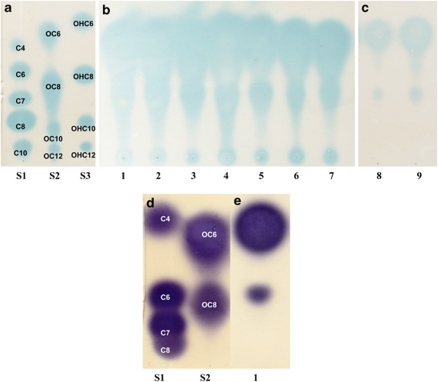
First, it was of interest to establish if the Psv pathogen and olive knot residents produce AHL QS signaling molecules. All strains that we tested (Table 1) produced AHL compounds as detected initially by plate T-streak assays of C. violaceum CV026 and A. tumefaciens NTL4 (pZLR4), and E. coli MT102 (pJBA132), biosensors. By TLC analysis we initially assigned tentatively the type of the AHLs produced by Psv, P. agglomerans and E. toletana (Figure 1 and Table 2). The results showed that all Psv and E. toletana strains produced two AHL molecules tentatively identified as C6-3-oxo-HSL and C8-3oxo-HSL (Figure 1). P. agglomerans on the other hand most probably produced C4-HSL and C6-HSL. In order to unequivocally confirm the AHLs produced by the three stains, we used C18 reverse-phase high-performance liquid chromatography and mass spectrometry, and were able to verify the production of 3-oxo-C6-HSL and 3-oxo-C8-HSL by Psv and E. toletana, and C6-HSL and C4-HSL by P. agglomerans (Supplementary Figure 1).
Figure 1.
AHL production of Psv isolated from olive and oleander knots, E. toletana and P. agglomerans. (a) TLC analysis of the synthetic AHL standards using the A. tumefaciens pNTL4 biosensor as overlay. (b) TLC analysis of the AHLs produced by seven Psv strains using the A. tumefaciens pNTL4 biosensor as overlay. Lanes: (1) Strain LMG 2209T; (2) DAPP-PG 536; (3) DAPP-PG 722; (4) DAPP-PG 723; (5) DAPP-PG 725; (6) DAPP-PG 726; (7) DAPP-PG 728. (c) TLC analysis of the AHLs produced by two E. toletana strains using the A. tumefaciens pNTL4 biosensor as overlay. Lanes: (8) Strain CFBP 6631T; (9) DAPP-PG 735. (d) TLC analysis of the synthetic AHL standards using the C. violaceum CV026 biosensor as overlay. (e) TLC analysis of the AHLs produced by P. agglomerans DAPP-PG 734 (Lane 1) using the C. violaceum CV026 biosensor as overlay. AHL, acyl homoserine lactone; Psv, Pseudomonas pv. savastanoi; TLC, thin-layer chromatography.
Table 2. Phenotypes tested for AHL QS regulation in Psv DAPP-PG 722, P. agglomerans DAPP-PG 734 and E. toletana DAPP-PG 735.
| Psv DAPP-PG 722 |
P. agglomerans DAPP-PG 734 |
E. toletana DAPP-PG 735 |
|||||||
|---|---|---|---|---|---|---|---|---|---|
| WT | pssI− | pssR− | WT | pagI− | pagR− | WT | etoI− | etoR− | |
| Phenotype | |||||||||
| AHL | + | − | + | + | − | + | + | − | + |
| IAA | + | + | + | + | + | + | + | + | + |
| Siderophore | + | + | + | + | + | + | + | + | + |
| Protease | - | − | − | − | − | − | − | − | − |
| Lipase | + | + | + | + | + | + | + | + | + |
| Swarming | + | + | + | - | − | − | − | − | − |
| Swimming | + | + | + | + | + | + | + | + | + |
| EPS | + | − | − | + | − | + | + | − | +++ |
Abbreviations: AHL, acyl homoserine lactone; EPS, exoploysaccahride; IAA, indole-3-acetic acid; Psv, Pseudomonas pv. savastanoi; QS, quorum sensing; WT, wild type.
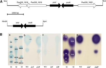
The AHL QS system of Psv DAPP-PG 722 was identified and cloned, as explained under Materials and methods, consisting of a luxI homolog designated pssI and a luxR homolog designated pssR (Figure 2a). The PssI/R system shows a very high degree of homology (over 90%) to the AhlI/R AHL QS system of P. syringae, which responds to C6-3-oxo-HSL and is involved in virulence (Quinones et al., 2005). Similarly, we identified and cloned the AHL QS system of E. toletana DAPP-PG 735 (see section Materials and methods) consisting of a luxI homolog designated etoI and a luxR homolog designated etoR (Figure 2a). The EtoI/R system shows significant homology (approximately 60%) to the ExpI/R system of Erwinia chrysanthemi pv. zeae (=Dickeya zeae), which is responsible for not only producing but also responding to C6-3-oxo-HSL (Nasser et al., 1998; Hussain et al., 2008). It was established by PCR, cloning and sequencing that the P. agglomerans AHL QS system of strain DAPP-PG 734 was orthologous (over 95% identity) to the PagI/R system of P. agglomerans pv. gypsophilae (Chalupowicz et al., 2009).
Figure 2.
Gene maps and AHL production by AHL QS-knockout mutants of the olive knot isolates of Psv, E. toletana and P. agglomerans. (A) Gene maps of the cloned ppsI/R and etoI/R loci of Psv and E. toletana, respectively. Neighboring the ppsI/R system, genes are shown that are orthologs in sequenced Psv NCPPB 3335. In fact the region sequenced here in strain DAPP-PG 722 is orthologous to the one present in strain NCPPB 3335. (B) (a) TLC analysis of the synthetic AHL standards using the A. tumefaciens pNTL4 biosensor as overlay. (b) TLC analysis of the AHLs produced by the parent Psv DAPP-PG 722 strain and mutant derivatives using the A. tumefaciens pNTL4 biosensor as overlay. pssI− is DAPP-PG 722PSSI; pssR− is DAPP-PG 722PSSI. (c) TLC analysis of the AHLs produced by the parent strain E. toletana DAPP-PG 735 and mutant derivatives using the A. tumefaciens pNTL4 biosensor as overlay. tolI- is DAPP-PG 735TOLI; tolR- is DAPP-PG 735TOLR. (d) TLC analysis of the synthetic AHL standards using the C. violaceum CV026 biosensor as overlay. (e) TLC analysis of the AHLs produced by the parent P. agglomerans DAPP-PG 734 strain and mutant derivatives using the C. violaceum CV026 biosensor as overlay. pagI− is DAPP-PG 734PAGI; pagR− is DAPP-PG 734PAGR. AHL, acyl homoserine lactone; Psv, Pseudomonas pv. savastanoi; QS, quorum sensing; savastanoi; TLC, thin-layer chromatography.
Psv, P. agglomerans and E. toletana knockout mutants were generated in both the luxI- and the luxR-family QS genes as described under Materials and methods. None of the luxI-family mutants of the three species (DAPP-PG 722PSSI, DAPP-PG 734PAGI and DAPP-PG 738ETOI; Table 1) produced detectable levels of AHLs when purified from spent supernatants (Figure 2b). This result indicated that all three species most probably possess only one AHL QS system; for Psv, further support for this conclusion is provided by the fact that only one luxI/R family pair is present in its genome, which has been sequenced recently (Rodriguez-Palenzuela et al., 2010). The luxR-family mutants on the other hand (DAPP-PG 722PSSR, DAPP-PG 734PAGR and DAPP-PG 738ETOR; Table 1) produced amounts of AHLs similar to the amounts produced by the wild type (Figure 2b), indicating that for all three systems there was no signal amplification loop through the regulation of the luxI-family gene. These conclusions were based on the TLC analysis of spent supernatant extracts purified from equal amounts of culture at the same growth phase. This is a very good indication, however measurements throughout the growth phase would probably confirm this conclusion further.
QS in Psv regulates virulence in planta
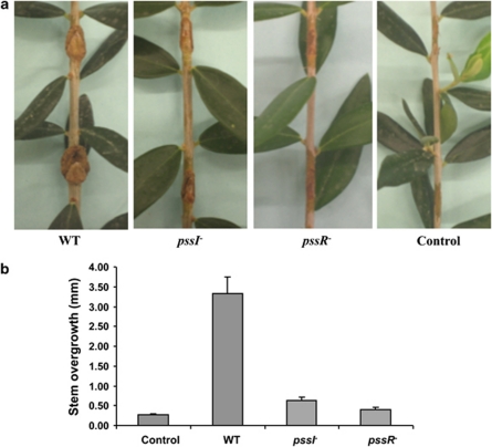
To examine whether the PssI/R AHL QS system of Psv was involved in pathogenicity and virulence, the Psv DAPP-PG 722 parental strain and the ppsI and ppsR derivative knockout mutants were inoculated on 1-year-old olive plants and disease severity was evaluated after 60 days. The results showed clearly that mutations in pssI and pssR drastically reduced knot size and therefore the virulence of the mutants (Figure 3a). Knot depth was reduced significantly (P⩽0.01) in the stems inoculated with the mutants DAPP-PG 722PSSI and DAPP-PG 722PSSR as compared with those inoculated with the wild type, showing reductions of approximately 80% and 88%, respectively (Figure 3b). Importantly, it was therefore concluded, to our knowledge, that that AHL QS, for the first time, had been observed to have a central role in the virulence of Psv.
Figure 3.
Role of AHL QS on olive knot formation induced by Psv. (a) The stems of 1-year-old olive plants (cv. Frantoio) were inoculated with 108 c.f.u. ml−1 Psv DAPP-PG 722 (wild type: WT), pssI− and pssR− derivative mutants, and with control plants. Disease symptoms were observed 60 days after the inoculation. Large raised knots were observed in stems inoculated with DAPP-PG 722 (wild type). The AHL QS deficiency mutants pssI− and pssR− induced only limited cell proliferation, which remained restricted to the wound margins. (b) Knot size was estimated by measuring the depth of the tissue overgrowths in five plants per each treatment, and for three knots per plant. Statistically significant differences in knot size between the wild type and the QS mutants were determined by analysis of variance. Each value is the mean of five replicates±s.d. The means are not significantly different (P=0.01) according to Duncan's multiple-range test. AHL, acyl homoserine lactone; Psv, Pseudomonas pv. savastanoi; QS, quorum sensing; savastanoi.
Psv AHL synthase mutants can be rescued by the presence of E. toletana and in part by P. agglomerans
The presence of E. toletana and P. agglomerans in olive knots could result in the formation of a poly-microbial community with the Psv pathogen. As E. toletana and P. agglomerans both produce AHLs and as Psv AHL-negative mutants have very low virulence (see above), this allows an elegant setup in order to establish if AHL signals are shared and if interspecies communities form in the olive knot.
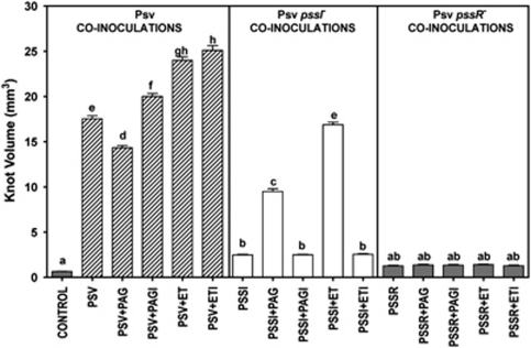
As E. toletana produces structurally the same AHLs as Psv (see above), an in planta co-inoculation was initially set up, where the pssI AHL synthase mutant was used for co-infection of olive plants together with the E. toletana DAPP-PG 735 wild-type strain. This co-inoculation resulted in the Psv ppsI mutant being able to induce knot formation just like the wild-type strain (Figure 4). This can most probably be attributed to the two strains making a consortium, where E. toletana provides the AHLs to DAPP-PG 722PSSI, which is then able to induce AHL QS target gene expression resulting in olive knot formation. Similar pairwise inoculations were also performed with the P. agglomerans DAPP-PG 734 wild-type strain and here only partial restoration of knot formation was observed (Figure 4). This could be because of P. agglomerans producing different AHLs than Psv, which the PssI/R system, most probably responded to with less efficiency. Importantly, pairwise in planta inoculations with either the E. toletana or the P. agglomerans AHL synthase mutant, etoI or pagI, respectively, did not restore olive knot formation. This clearly indicates that interspecies communication through sharing of AHLs was key in the previous co-inoculation experiments where the Psv AHL synthase mutants were co-inoculated independently with the two wild-type strains (Figure 4).
Figure 4.
Effect of co-inoculation of olive plants (cv. Frantoio) with Psv and E. toletana, P. agglomerans and the respective AHL QS mutants. Psv: The columns indicate effect on disease severity, expressed as knot volume (60 days after the inoculation), of inoculation of the WT Psv (hatched columns), of QS-impaired mutants of Psv (white columns represent PSSI mutant and black columns PSSR mutants) alone or in combination with P. agglomerans (Pag) or E. toletana (Et) and their relative QS-impaired mutants, PagI, PagR, EtoI and EtoR. Olive control plants were inoculated with distilled water (gray column). Each value is the mean of five replications±s.e. Mean followed by the same letters are not statistically different at P=0.05 (Duncan's multiple-range test). AHL, acyl homoserine lactone; Psv, Pseudomonas pv. savastanoi; QS, quorum sensing.
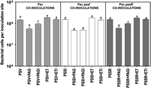
The Psv pssR mutant could not be complemented for olive knot formation by E. toletana and P. agglomerans; the most likely reason being that the the ppsR Psv mutant cannot respond to AHLs. These results also indicate that E. toletana and P. agglomerans wild-type strains cannot cause disease alone in the absence of wild-type Psv. Importantly, in all co-inoculation experiments, comparable amounts of Psv colony-forming units (c.f.u.) were present at the inoculation site (Figure 5).
Figure 5.
Effect on Psv levels of co-inoculation of olive plants (cv. Frantoio) with Psv and E. toletana, P. agglomerans and the respective AHL QS mutants. Psv: The black columns indicate effect on bacterial growth in planta (60 days after the inoculation) of inoculation of the QS-impaired mutants of Psv of the same bacterium PssI (white columns) or PssR (hatched columns) alone or in combination with P. agglomerans (Pag) or E. toletana (Et) and their relative QS-impaired mutants, PagI, PagR, EtI and EtR. Each value is the mean of five replications±s.e. Mean followed by the same letters are not statistically different at P=0.05 (Duncan's multiple-range test). Psv, Pseudomonas pv. savastanoi.
It was concluded that both E. toletana and P. agglomerans can form stable interspecies communities with Psv and that communication through sharing of AHL signals was taking place in planta.
Role of interspecies signaling and pairwise consortia in olive knot disease
As evident from the results described above, Psv forms bacterial communities with E. toletana and P. agglomerans. In order to begin to understand the role and outcome of possible interactions among these three bacterial species, the virulence of Psv in the presence of the other two olive knot-resident bacteria was tested in planta. One-year-old olive plants were inoculated for each bacterial strain with 10 μl of a bacterial suspension having 108 c.f.u. ml−1. As shown in Figure 4, significant enhancement of Psv virulence in the presence of E. toletana was observed as indicated by a considerable increase (almost 30%) of knot volume. This indicated that E. toletana contributes to the pathogenicity of Psv; the increase in virulence was not because of changes in the levels of Psv in the co-inoculations as there was no significant difference in bacterial load (Figure 5). The same contribution to Psv virulence was observed when co-inoculating with the E. toletana AHL synthase mutant; one cannot exclude, however, that Psv AHL signals are perceived by E. toletana. No increase in virulence, on the other hand, was observed when P. agglomerans was co-inoculated with Psv, and in fact a slight decrease of knot volume was observed. When co-inoculating with the P. agglomerans AHL synthase mutant, however, a significant increase in knot volume formation was detected, meaning that QS regulates some factor(s) in P. agglomerans, which limit Psv growth and/or gene expression.
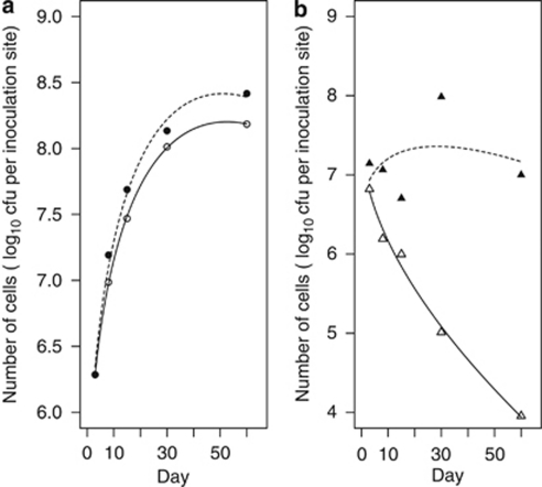
Following the evident results on the poly-bacterial nature of the disease and of the sharing of AHL signals between Psv and E. toletana, it was of interest to gain more insight into this bacterial community. The impact of co-inoculation on the growth dynamics of these bacteria was determined in two separate experiments. As the results of these experiments were similar, only data from one of the experiments were reported. Co-inoculations were therefore performed and bacterial populations were determined until 60 dpi. When Psv DAPP-PG 722 was inoculated alone, a 10-fold increase could be detected in the bacterial population isolated from inoculated tissue 8 dpi (appreciatively 107 c.f.u. per site) (Figure 6a, open circles). The population of Psv progressively increased to reach about 1.5 × 10⁸ c.f.u. per site at 60 dpi. When mixed inoculation with E. toletana DAPP-PG 735 (Figure 6a, filled circle) was performed, Psv growth was significantly increased (likelihood ratio test with P<0.0001), whereas when E. toletana was inoculated alone in olive plants, a decline was observed in its population sizes (Figure 6b, open triangle). On the other hand, the growth of E. toletana could not be described adequately by using a second-order polynomial model, but nonetheless it was stimulated significantly at 30 dpi (the standard error of difference was 0.018) in the presence of Psv (Figure 6b, filled triangle), and after 60 dpi, the population size from the mixed infection was approximately 103-fold when compared with results obtained from the single inoculations. These results are a further indication of the intimate and mutualistic relationship between Psv and E. toletana in the olive knot, leading to enhanced disease symptoms, sharing of AHL QS signals and increasing bacterial growth of both species.
Figure 6.
The population dynamics of Psv DAPP-PG 722 and E. toletana DAPP-PG 735 strains inoculated in olive (cv. Frantoio) stems alone or in combination. (a) Growth of Psv alone (solid line and open circle) or in combination with E. toletana (dotted line and filled circle). (b) Growth of E. toletana alone (solid line and open triangle) or in combination with Psv (dotted line and filled triangle). The symbols indicate observed counts and the lines indicate the linear mixed model fit. The standard error of a mean was 0.013.
Testing the AHL QS-dependent regulation of several phenotypes in P. savastanoi, P. agglomerans and E. toletana
In order to establish the possible role of the AHL QS systems in the olive knot-associated bacteria, several important phenotypes were tested for AHL QS regulation in all three species. A list of all phenotypes tested for AHL QS regulation is summarized in Table 2.
Interestingly, we determined that P. agglomerans DAPP-PG 734 and E. toletana DAPP-PG 735 also produce IAA. We tested wild-types Psv (DAPP-PG 722), P. agglomerans (DAPP-PG 734), E. toletana (DAPP-PG 735) and their derivative mutants (DAPP-PG 722PPSI, DAPP-PG 722PSSR, DAPP-PG 734PAGI, DAPP-PG 734PAGR, DAPP-PG 735ETOI and DAPP-PG 735ETOR) for the production of IAA. Similarly, we tested whether AHL QS was involved in siderophore production, EPS production, secreted proteolytic and lipolytic activities, and motility by swimming and swarming. The results of these investigations are summarized in Table 2. Surprisingly, in most tests no significant differences were observed between the parent strains and their QS mutant derivatives under the conditions tested. EPS production was tested qualitatively in a plate assay in all wild-type and mutant strains. Both AHL QS mutants of Psv strain DAPP-PG 722 showed a clear reduction in EPS production when compared with the wild type (data not shown); EPS production was restored when mutant DAPP-PG 722PSSI was grown in the presence of exogenous C6-3-oxo- or C8-3oxo-HSL (data not shown). Similarly, for P. agglomerans, inactivation of pagI resulted in a reduction of EPS production; however, no reduction was observed in the pagR mutant when compared with the wild type. EPS production was restored when mutant DAPP-PG 734PAGI was grown in the presence of C4-HSL or C6-HSL (data not shown). EPS production was also reduced in the E. toletana DAPP-PG 735ETOI mutant when compared with that in the wild-type, and complementation through exogenous addition C6-3-oxo-HSL or C8-3oxo-HSL restored EPS production. In E. toletana, however, inactivation of the etoR significantly increased EPS production (data not shown).
Discussion
Many studies thus far have shown that plant bacterial diseases are a result of complex interactions between the pathogen and host. The interaction of the pathogen with the resident bacterial flora is however much less studied and understood. Do plant pathogenic bacteria form stable bacterial communities with other resident, non-pathogenic bacteria? Are these association networks resulting in a poly-microbial infection? Does intercellular signaling have a role in the establishment of these bacterial consortia? These are the questions we are beginning to address here using the bacterial community present in the olive knot disease. This niche is created by the Psv pathogen; however, several other resident, non-pathogenic bacterial species have been isolated in this environment, most commonly E. toletana (Rojas et al., 2004) and P. agglomerans (Marchi et al., 2006). The major conclusions of this work are that (i) the three different species isolated from the olive knot produce AHL signals and possess one AHL QS system; notably E. toletana and Psv produce the same AHLs, (ii) AHL QS in Psv is essential for virulence as knockout mutants are basically not virulent, (iii) Psv AHL synthase mutants can be rescued by E. toletana and in part by P. agglomerans, evidencing the sharing of AHL signals and the formation of stable bacterial communities, and (iv) E. toletana forms communities in planta, acting as an associated pathogen synergistically with the principal pathogen Psv as co-inoculations increase the bacterial count of both species and the severity of disease. All the bacterial mutants constructed in this study were produced through homologous recombination events and also involved plasmid insertions. There is, therefore, a possibility that our experiments performed in planta without antibiotic selection could lead to mutants reverting to the wild type. As all the various control experiments in our study did not result in a wild-type behavior, we concluded that no significant reversion took place.
Currently, the only known molecular determinants of knot development by Psv are the type-III secretion system (Sisto et al., 2004) and the phytohormones cytokinins and IAA (Surico et al., 1985; Glass and Kosuge, 1988; Rodriguez-Moreno et al., 2008). To this list, AHL QS can now be added and to our knowledge this is the first regulatory system that has been linked to Psv virulence. AHL QS is likely not affecting initial colonization but rather the gene expression of virulence-associated factors at high population densities. It would now be relevant to determine whether AHL QS is involved in the regulation of the three virulence determinants identified thus far. Most likely that AHL QS is also regulating many other virulence factors. The recent sequencing of the Psv genome highlighted the presence of many loci, which could be involved in virulence, including five different secretion systems, type-III effectors, catabolic systems of phenolic compounds, chemotaxis, motility, adhesion and toxin genes (Rodriguez-Palenzuela et al., 2010). We have reported that EPS production is regulated by the PssI/R system in Psv; however, it is not known yet if EPS is involved in the disease process. EPS, for example, is a virulence factor and is regulated by AHL QS is closely related P. syringae (Quinones et al., 2005).
The life of bacteria inside the knot is unknown at large. A recent study, however, has shown that Psv creates the knot by forming bacterial aggregates, microcolonies and multilayer biofilms (Rodriguez-Moreno et al., 2009; Perez-Martinez et al., 2010). Little is known about the effects of P. agglomerans and E. toletana on knot development and the possible formation and stability of the multispecies bacterial consortia formed in the olive knot. P. agglomerans is widespread in many diverse natural and agricultural habitats; in particular, it is associated with many plants as a common epiphyte and endophyte (Lindow and Brandl, 2003). In addition, P. agglomerans can be transformed from a commensal and epiphytic bacterium associated with many plants to a host-specific tumorigenic pathogen, through acquisition of a plasmid-borne pathogenicity island (Barash and Manulis-Sasson, 2007).
The AHLs produced by E. toletana (C6-3-oxo-HSL and C8-3oxo-HSL) are the same as the ones produced by Psv. This is evidence that there could be interspecies signaling through AHLs taking place between the two species when they occupy the same niche. P. agglomerans on the other hand produces C4-HSL and C6-HSL; it cannot be excluded that PssR and/or EtoR are able to respond to these AHLs. Similarly, it is not known if PagR can also respond to the AHLs produced by E. toletana and Psv. Our studies here involving binary in planta co-inoculations have shown that E. toletana can rescue the Psv pssI AHL synthase mutants for their ability to induce olive knot formation. As the symptoms take at least 25–30 days to develop, this result has the connotation that E. toletana and Psv are able to form a stable consortium, where the two species undergo interspecies signaling and AHL sharing. P. agglomerans can also rescue, in part, the Psv ppsI mutant; the reason for this partial rescue is either that, the AHLs produced by P. agglomerans are not recognized well by PssR, or the consortium is not as stable as it is for E. toletana. It is possible that in this niche, which allows such a consortium to grow, could then encourage the appearance of bacterial cheaters (for example, signal blind cheaters, which do not respond to AHL signals), which do not contribute to the community, but can benefit from the ‘factors' or ‘public goods' produced by the QS cooperators (Diggle et al., 2007; Venturi et al., 2010). Many more bacterial isolates need to be isolated from this niche in order to determine this possibility.
The ability of Psv to induce olive knot formation relies on its ability to regulate gene expression in response to the host environment. A question that we also began to address here is, whether the indigenous avirulent microflora contributes to the disease caused by Psv. The co-inoculation of E. toletana with Psv resulted in a significant enhancement of olive knot volume, indicating that this consortium is probably very stable and Psv benefits from the presence of E. toletana and possibly vice versa. Importantly, in addition, co-inoculation studies determined that growth of Psv in the olive knot was significantly stimulated by E. toletana and vice versa, indicating a close association, possible metabolic, nutrient and signal sharing, between the two bacterial species. This synergistic effect of E. toletana with P. savastanoi clearly indicates a mutual benefit for both species when growing in and sharing the same niche. Understanding the mechanisms of cooperation and communication of these two bacteria will shed light on the process of interspecies communication. E. toletana is not pathogenic when inoculated with the AHL QS Psv pssR mutant, but can act as an accessory pathogen when in consortium with wild-type Psv. It is possible that gene expression of Psv can be influenced by the presence of bacterial residents, or that the microbial consortium stabilizes Psv metabolically. The co-inoculation of P. agglomerans with Psv, on the other hand, resulted in a reduction of knot volume, which increased when the co-inoculant was the AHL synthase mutant pagI. P. agglomerans most likely produces and AHL QS regulates the factor, which affects Psv, and when the production is relieved, P. agglomerans can also act as an accessory pathogen.
There are very few reports on the contribution/role of the host's resident indigenous microbial flora on the action of an incoming pathogen. Interestingly, in 1998 it was reported that in the rhizosphere of wheat, there is an inter-population of bacterial species, which can possibly share AHL molecules (Pierson et al., 1998). Recently, two studies have shown that virulence of P. aeruginosa is influenced by the indigenous microflora, and have also shown that it affects the gene expression patterns of virulence-associated factors (Duan et al., 2003; Sibley et al., 2008). It is currently unknown if there is sharing of signals among these bacteria; it has been observed that the AI (autoinducer)-2 signal produced by both Gram-positive and Gram-negative bacteria could be an interspecies signal in this community (Duan et al., 2003; Sibley et al., 2008). Interestingly however, Dulla and Lindow (2009) have reported very recently that indigenous epiphytic bacteria producing the same AHLs of pathogenic P. syringae pv. syringae can suppress and interfere with the disease-causing process. This work provided evidence for an AHL-mediated cross-talk in the plant phyllosphere, which resulted in changes of behavior (mainly motility, which meant less invasion) and consequently virulence of P. syringae pv. syringae. The occurrence of disease in co-inoculations with AHL-producing epiphytic bacteria was reduced by 50%. This is believed to be because of premature and untimely induction of AHL QS affecting the expression of virulence-associated factors. This example shows that interspecies communication with indigenous bacterial flora can also have negative consequences for plant pathogens.
The data presented here further enhance the apparent complexity of poly-microbial infections; our results indicate a role of AHL sharing and interspecies signaling, which provides an excellent working model to further study bacterial community interactions. This study focuses on AHL-mediated interactions in natural habitats; so far the role of QS in plant-associated bacteria has been studied almost exclusively using single strains despite the possibility of interspecies signaling that can be mediated through sharing of AHL signal molecules in mixed microbial communities. The interactions between bacteria in natural settings are complicated by a number of factors, including host response, environmental parameters and the species composition of the community. It is suggested that future studies will need to develop in vitro systems for a convenient and detailed understanding of interspecies interactions. We are only just beginning to develop tools for studying microbial interactions in the natural environment.
We will focus our future studies on the stability and the role played by each species in this microbial consortium. Psv can be regarded as the niche maker or leader in this consortium, whereas P. agglomerans and E. toletana as niche occupants or residents, which, however, can become auxiliary pathogens in the presence of a niche maker. At present it is unclear whether or not such pathogenic consortia have a common rationale. We speculate that the olive knot consortium evolved to be stable and cooperative because the augmented gene pool and the combined metabolic repertoire increases the survival chances of the participants under a variety of conditions (Venturi et al., 2010). This work suggests that complex interspecies communication processes may be necessary for the formation of such consortia.
Acknowledgments
We thank Iris Bertani and Giuliano Degrassi for interest and useful discussions. TH and ZRSM benefited from an ICGEB fellowship. This work was supported in part by ICGEB core funding. This research was also supported in part by ‘Fondazione Cassa di Risparmio di Perugia' (Italy), project ‘Indagini sui batteri endofiti associati a piante di olivo affette da rogna, volte all'individuazione di nuove strategie di lotta alla malattia,' to RB.
Footnotes
Supplementary Information accompanies the paper on The ISME Journal website (http://www.nature.com/ismej)
Supplementary Material
References
- Alexeyev MF.1999The pKNOCK series of broad-host-range mobilizable suicide vectors for gene knockout and targeted DNA insertion into the chromosome of Gram-negative bacteria Biotechniques 26824–826.828. [DOI] [PubMed] [Google Scholar]
- Andersen JB, Heydorn A, Hentzer M, Eberl L, Geisenberger O, Christensen BB, et al. gfp-based N-acyl homoserine-lactone sensor systems for detection of bacterial communication. Appl Environ Microbiol. 2001;67:575–585. doi: 10.1128/AEM.67.2.575-585.2001. [DOI] [PMC free article] [PubMed] [Google Scholar]
- Barash I, Manulis-Sasson S. Virulence mechanisms and host specificity of gall-forming Pantoea agglomerans. Trends Microbiol. 2007;15:538–545. doi: 10.1016/j.tim.2007.10.009. [DOI] [PubMed] [Google Scholar]
- Bassler BL. Small talk. Cell-to-cell communication in bacteria. Cell. 2002;109:421–424. doi: 10.1016/s0092-8674(02)00749-3. [DOI] [PubMed] [Google Scholar]
- Better M, Lewis B, Corbin D, Ditta G, Helinski DR. Structural relationships among Rhizobium meliloti symbiotic promoters. Cell. 1983;35:479–485. doi: 10.1016/0092-8674(83)90181-2. [DOI] [PubMed] [Google Scholar]
- Caballero-Mellado J, Onofre-Lemus J, Estrada-de Los Santos P, Martinez-Aguilar L. The tomato rhizosphere, an environment rich in nitrogen-fixing Burkholderia species with capabilities of interest for agriculture and bioremediation. Appl Environ Microbiol. 2007;73:5308–5319. doi: 10.1128/AEM.00324-07. [DOI] [PMC free article] [PubMed] [Google Scholar]
- Chalupowicz L, Barash I, Panijel M, Sessa G, Manulis-Sasson S. Regulatory interactions between quorum-sensing, auxin, cytokinin, and the Hrp regulon in relation to gall formation and epiphytic fitness of Pantoea agglomerans pv. gypsophilae. Mol Plant Microbe Interact. 2009;22:849–856. doi: 10.1094/MPMI-22-7-0849. [DOI] [PubMed] [Google Scholar]
- Diggle SP, Griffin AS, Campbell GS, West SA. Cooperation and conflict in quorum-sensing bacterial populations. Nature. 2007;450:411–414. doi: 10.1038/nature06279. [DOI] [PubMed] [Google Scholar]
- Duan K, Dammel C, Stein J, Rabin H, Surette MG. Modulation of Pseudomonas aeruginosa gene expression by host microflora through interspecies communication. Mol Microbiol. 2003;50:1477–1491. doi: 10.1046/j.1365-2958.2003.03803.x. [DOI] [PubMed] [Google Scholar]
- Duan K, Sibley CD, Davidson CJ, Surette MG. Chemical interactions between organisms in microbial communities. Contrib Microbiol. 2009;16:1–17. doi: 10.1159/000219369. [DOI] [PubMed] [Google Scholar]
- Dulla GF, Lindow SE. Acyl-homoserine lactone-mediated cross talk among epiphytic bacteria modulates behavior of Pseudomonas syringae on leaves. ISME J. 2009;3:825–834. doi: 10.1038/ismej.2009.30. [DOI] [PubMed] [Google Scholar]
- Feil H, Feil WS, Chain P, Larimer F, DiBartolo G, Copeland A, et al. Comparison of the complete genome sequences of Pseudomonas syringae pv. syringae B728a and pv. tomato DC3000. Proc Natl Acad Sci USA. 2005;102:11064–11069. doi: 10.1073/pnas.0504930102. [DOI] [PMC free article] [PubMed] [Google Scholar]
- Fernandes A, Marcelo M. A possible synergistic effect of Erwinia sp. on the development of olive knot symptoms caused by Pseudomonas syringae pv. savastanoi in Olea europaea. Acta Horticulturae. 2002;586:729–731. [Google Scholar]
- Fuqua C, Greenberg EP. Listening in on bacteria: acyl-homoserine lactone signalling. Nat Rev Mol Cell Biol. 2002;3:685–695. doi: 10.1038/nrm907. [DOI] [PubMed] [Google Scholar]
- Fuqua C, Parsek MR, Greenberg EP. Regulation of gene expression by cell-to-cell communication: acyl-homoserine lactone quorum sensing. Annu Rev Genet. 2001;35:439–468. doi: 10.1146/annurev.genet.35.102401.090913. [DOI] [PubMed] [Google Scholar]
- Gardan L, Bollet M, Abughorrah F, Grimont F, Grimont PAD. DNA relatedness among the pathovar strains of Pseudomonas syringae subsp. savastanoi Janse (1982) and proposal of Pseudomonas savastanoi sp. nov. Int J Syst Bacteriol. 1992;42:606–612. [Google Scholar]
- Garrett KA, Madden LV, Hughes G, Pfender WF. New applications of statistical tools in plant pathology. Phytopathology. 2004;94:999–1003. doi: 10.1094/PHYTO.2004.94.9.999. [DOI] [PubMed] [Google Scholar]
- Gavini F, Mergaert J, Beji A, Mielcarek C, Izard D, Kersters K, et al. Transfer of Enterobacter agglomerans (Beijerinck 1888) Ewing and Fife 1972 to Pantoea gen. nov. as Pantoea agglomerans comb. nov. and description of Pantoea dispersa sp. nov. Int J Syst Bacteriol. 1989;39:337–345. [Google Scholar]
- Glass NL, Kosuge T. Role of indoleacetic acid–lysine synthetase in regulation of indoleacetic acid pool size and virulence of Pseudomonas syringae subsp. savastanoi. J Bacteriol. 1988;170:2367–2373. doi: 10.1128/jb.170.5.2367-2373.1988. [DOI] [PMC free article] [PubMed] [Google Scholar]
- Gordon SA, Weber RP. Colorimetric estimation of indoleacetic acid. Plant Physiol. 1951;26:192–195. doi: 10.1104/pp.26.1.192. [DOI] [PMC free article] [PubMed] [Google Scholar]
- Gould TA, Herman J, Krank J, Murphy RC, Churchill ME. Specificity of acyl-homoserine lactone synthases examined by mass spectrometry. J Bacteriol. 2006;188:773–783. doi: 10.1128/JB.188.2.773-783.2006. [DOI] [PMC free article] [PubMed] [Google Scholar]
- Hirano SS, Upper CD. Bacteria in the leaf ecosystem with emphasis on Pseudomonas syringae—a pathogen, ice nucleus, and epiphyte. Microbiol Mol Biol Rev. 2000;64:624–653. doi: 10.1128/mmbr.64.3.624-653.2000. [DOI] [PMC free article] [PubMed] [Google Scholar]
- Hofte M, De Vos P.2006Plant pathogenic Pseudomonas speciesIn: Gnanamanickam SS (ed.).Plant-Associated Bacteria Springer: The Netherlands; 507–533. [Google Scholar]
- Huber B, Riedel K, Hentzer M, Heydorn A, Gotschlich A, Givskov M, et al. The cep quorum-sensing system of Burkholderia cepacia H111 controls biofilm formation and swarming motility. Microbiology. 2001;147:2517–2528. doi: 10.1099/00221287-147-9-2517. [DOI] [PubMed] [Google Scholar]
- Hussain MB, Zhang HB, Xu JL, Liu Q, Jiang Z, Zhang LH. The acyl-homoserine lactone-type quorum-sensing system modulates cell motility and virulence of Erwinia chrysanthemi pv. zeae. J Bacteriol. 2008;190:1045–1053. doi: 10.1128/JB.01472-07. [DOI] [PMC free article] [PubMed] [Google Scholar]
- Joardar V, Lindeberg M, Jackson RW, Selengut J, Dodson R, Brinkac LM, et al. Whole-genome sequence analysis of Pseudomonas syringae pv. phaseolicola 1448A reveals divergence among pathovars in genes involved in virulence and transposition. J Bacteriol. 2005;187:6488–6498. doi: 10.1128/JB.187.18.6488-6498.2005. [DOI] [PMC free article] [PubMed] [Google Scholar]
- Kennelly MN, Cazorla FM, de Vicente A, Ramos C, Sundin GW. Pseudomonas syringae diseases of fruit trees: progress toward understanding and control. Plant Dis. 2007;91:4–17. doi: 10.1094/PD-91-0004. [DOI] [PubMed] [Google Scholar]
- King EO, Ward MK, Raney DE. Two simple media for the demonstration of pyocyanin and fluorescin. J Lab Clin Med. 1954;44:301–307. [PubMed] [Google Scholar]
- Kovach ME, Elzer PH, Hill DS, Robertson GT, Farris MA, Roop RM, II, et al. Four new derivatives of the broad-host-range cloning vector pBBR1MCS, carrying different antibiotic-resistance cassettes. Gene. 1995;166:175–176. doi: 10.1016/0378-1119(95)00584-1. [DOI] [PubMed] [Google Scholar]
- Lindow SE, Brandl MT. Microbiology of the phyllosphere. Appl Environ Microbiol. 2003;69:1875–1883. doi: 10.1128/AEM.69.4.1875-1883.2003. [DOI] [PMC free article] [PubMed] [Google Scholar]
- Marchi GA, Sisto A, Cimmino A, Andolfi A, Cipriani MG, Evidente A, et al. Interaction between Pseudomonas savastanoi pv. savastanoi and Pantoea agglomerans in olive knots. Plant Pathol. 2006;55:614–624. [Google Scholar]
- McClean KH, Winson MK, Fish L, Taylor A, Chhabra SR, Camara M, et al. Quorum sensing and Chromobacterium violaceum: exploitation of violacein production and inhibition for the detection of N-acylhomoserine lactones. Microbiology. 1997;143 (Part 12:3703–3711. doi: 10.1099/00221287-143-12-3703. [DOI] [PubMed] [Google Scholar]
- Moretti C, Ferrante P, Hosni T, Valentini F, D'Onghia A, Fatmi M, et al. 2008Characterization of Pseudomonas savastanoi pv. savastanoi strains collected from olive trees from different countriesIn: M'Barek Fatmi AC, Sante Iacobellis N, Mansfield JW, Murillo J, Schaad NW, Ullrich M (eds).Pseudomonas Syringae Pathovars and Related Pathogens—Identification, Epidemiology and Genomics Springer: The Netherlands [Google Scholar]
- Nasser W, Bouillant ML, Salmond G, Reverchon S. Characterization of the Erwinia chrysanthemiexpI–expR locus directing the synthesis of two N-acyl-homoserine lactone signal molecules. Mol Microbiol. 1998;29:1391–1405. doi: 10.1046/j.1365-2958.1998.01022.x. [DOI] [PubMed] [Google Scholar]
- Onofri A.2007Routine statistical analysis of field experiments by using an Excel extension 6th National Conference of the Italian Biometric Society 20–22.June; Pisa, Italy.
- Onofri A. Current statistical issues in weed research. Weed Res. 2010;50:5–24. [Google Scholar]
- Perez-Martinez I, Rodriguez-Moreno L, Lambertsen L, Matas IM, Murillo J, Tegli S, et al. Fate of a Pseudomonas savastanoi pv. savastanoi type III secretion system mutant in olive plants (Olea europaea L.) Appl Environ Microbiol. 2010;76:3611–3619. doi: 10.1128/AEM.00133-10. [DOI] [PMC free article] [PubMed] [Google Scholar]
- Pierson EA, Wood DW, Cannon JA, Blachere FM, Pierson LS. Interpopulation signaling via N-acyl-homoserine lactones among bacteria in the wheat rhizosphere. Mol Plant Microbe Interact. 1998;11:1078–1084. [Google Scholar]
- Piper KR, Von Bodman SB, Farrand SK. Conjugation factor of Agrobacterium tumefaciens regulates Ti plasmid transfer by autoinduction. Nature. 1993;362:448–450. doi: 10.1038/362448a0. [DOI] [PubMed] [Google Scholar]
- Quinones B, Dulla G, Lindow SE. Quorum sensing regulates exopolysaccharide production, motility, and virulence in Pseudomonas syringae. Mol Plant Microbe Interact. 2005;18:682–693. doi: 10.1094/MPMI-18-0682. [DOI] [PubMed] [Google Scholar]
- Riedel K, Hentzer M, Geisenberger O, Huber B, Steidle A, Wu H, et al. N-acylhomoserine-lactone-mediated communication between Pseudomonas aeruginosa and Burkholderia cepacia in mixed biofilms. Microbiology. 2001;147:3249–3262. doi: 10.1099/00221287-147-12-3249. [DOI] [PubMed] [Google Scholar]
- Rodriguez-Moreno L, Barcelo-Munoz A, Ramos C. In vitro analysis of the interaction of Pseudomonas savastanoi pvs. savastanoi and nerii with micropropagated olive plants. Phytopathology. 2008;98:815–822. doi: 10.1094/PHYTO-98-7-0815. [DOI] [PubMed] [Google Scholar]
- Rodriguez-Moreno L, Jimenez AJ, Ramos C. Endopathogenic lifestyle of Pseudomonas savastanoi pv. savastanoi in olive knots. Microb Biotechnol. 2009;2:476–488. doi: 10.1111/j.1751-7915.2009.00101.x. [DOI] [PMC free article] [PubMed] [Google Scholar]
- Rodriguez-Palenzuela P, Matas IM, Murillo J, Lopez-Solanilla E, Bardaji L, Perez-Martinez I, et al. Annotation and overview of the Pseudomonas savastanoi pv. savastanoi NCPPB 3335 draft genome reveals the virulence gene complement of a tumour-inducing pathogen of woody hosts. Environ Microbiol. 2010;12:1604–1620. doi: 10.1111/j.1462-2920.2010.02207.x. [DOI] [PubMed] [Google Scholar]
- Rojas AM, de Los Rios JE, Fischer-Le Saux M, Jimenez P, Reche P, Bonneau S, et al. Erwinia toletana sp. nov., associated with Pseudomonas savastanoi-induced tree knots. Int J Syst Evol Microbiol. 2004;54:2217–2222. doi: 10.1099/ijs.0.02924-0. [DOI] [PubMed] [Google Scholar]
- Ryan RP, Dow JM. Diffusible signals and interspecies communication in bacteria. Microbiology. 2008;154:1845–1858. doi: 10.1099/mic.0.2008/017871-0. [DOI] [PubMed] [Google Scholar]
- Sambrook J, Fritsch EF, Maniatis T.1989Molecular Cloning: a Laboratory Manual2nd edn.Cold Spring Harbor: NY, USA [Google Scholar]
- Schwyn B, Neilands JB. Universal chemical assay for the detection and determination of siderophores. Anal Biochem. 1987;160:47–56. doi: 10.1016/0003-2697(87)90612-9. [DOI] [PubMed] [Google Scholar]
- Shaw PD, Ping G, Daly SL, Cha C, Cronan JE, Jr, Rinehart KL, et al. Detecting and characterizing N-acyl-homoserine lactone signal molecules by thin-layer chromatography. Proc Natl Acad Sci USA. 1997;94:6036–6041. doi: 10.1073/pnas.94.12.6036. [DOI] [PMC free article] [PubMed] [Google Scholar]
- Sibley CD, Duan K, Fischer C, Parkins MD, Storey DG, Rabin HR, et al. Discerning the complexity of community interactions using a Drosophila model of polymicrobial infections. PLoS Pathog. 2008;4:e1000184. doi: 10.1371/journal.ppat.1000184. [DOI] [PMC free article] [PubMed] [Google Scholar]
- Sisto A, Cipriani MG, Morea M. Knot formation caused by Pseudomonas syringae subsp. savastanoi on olive plants is hrp-dependent. Phytopathology. 2004;94:484–489. doi: 10.1094/PHYTO.2004.94.5.484. [DOI] [PubMed] [Google Scholar]
- Staskawicz B, Dahlbeck D, Keen N, Napoli C. Molecular characterization of cloned avirulence genes from race 0 and race 1 of Pseudomonas syringae pv. glycinea. J Bacteriol. 1987;169:5789–5794. doi: 10.1128/jb.169.12.5789-5794.1987. [DOI] [PMC free article] [PubMed] [Google Scholar]
- Steindler L, Venturi V. Detection of quorum-sensing N-acyl homoserine lactone signal molecules by bacterial biosensors. FEMS Microbiol Lett. 2007;266:1–9. doi: 10.1111/j.1574-6968.2006.00501.x. [DOI] [PubMed] [Google Scholar]
- Surico G, Iacobellis NS, Sisto A. Studies on the role of indole-3-acetic acid and cytokinins in the formation of knots on olive and oleander plants by Pseudomonas syringae pv. savastanoi. Physiol Plant Pathol. 1985;26:309–320. [Google Scholar]
- Vasanthakumar A, McManus PS. Indole-3-acetic acid-producing bacteria are associated with cranberry stem gall. Phytopathology. 2004;94:1164–1171. doi: 10.1094/PHYTO.2004.94.11.1164. [DOI] [PubMed] [Google Scholar]
- Venables WN, Ripley BD.2002Modern Applied Statistics with S4th edn.Springer-Verlag: NY, USA [Google Scholar]
- Venturi V, Bertani I, Kerenyi A, Netotea S, Pongor S. Co-swarming and local collapse: quorum sensing conveys resilience to bacterial communities by localizing cheater mutants in Pseudomonas aeruginosa. PLoS One. 2010;5:e9998. doi: 10.1371/journal.pone.0009998. [DOI] [PMC free article] [PubMed] [Google Scholar]
- Vivian A, Mansfield J. A proposal for a uniform genetic nomenclature for avirulence genes in phytopathogenic pseudomonads. Mol Plant Microbe Interact. 1993;6:9–10. [Google Scholar]
- Von Bodman SB, Bauer WD, Coplin DL. Quorum sensing in plant-pathogenic bacteria. Annu Rev Phytopathol. 2003;41:455–482. doi: 10.1146/annurev.phyto.41.052002.095652. [DOI] [PubMed] [Google Scholar]
Associated Data
This section collects any data citations, data availability statements, or supplementary materials included in this article.


