Abstract
An international group of clinicians working in the field of Dysmorphology has initiated the (re-) definition of all terms used to describe the external human phenotype. The goal is that through standardization of all terms and consensus regarding their definitions the reliability of description of features in humans will increase, comparisons of findings between patients will become more reliable, and discussions with other workers in the field such as developmental biologists and molecular geneticists will become more accurate. Here we report on the (re-) definition of terms needed to describe the major characteristics of the hands and feet. We provide a limited description of the anatomy of this region, limited background anthropometry, and an illustrated list of definitions.
Keywords: nomenclature, definitions, feet, hands, limbs, malformations, anomalies, multiple anomalies, anatomy
INTRODUCTION
This paper is part of a series of six papers defining the morphology of regions of the human body [Allanson et al, 2009a;Carey et al., 2009;Hall et al., 2009;Hennekam et al., 2009; Hunter et al., 2009]. The series is accompanied by an introductory article describing general aspects of this study [Allanson et al., 2009b]. The reader is encouraged to consult the introduction when using the definitions.
An early classification of limb anomalies is that of Isidore Geoffrey Saint-Hilaire (secondary citation, not reviewed). This classification divided anomalies into three categories (phocomèle, hemimèle, and ectromèle). A more recent classification is that by Frantz and O’Rahilly [Frantz and O’Rahilly, 1961]. Modifications of this scheme were implemented by others [Swanson et al., 1968; Swanson, 1976]. These schemes are hierarchical classifications and most do not include definitions of the terms.
The similar, but distinct, system of Temtamy and McKusick was developed [Temtamy and McKusick, 1969]. Most of the seven categories that they defined were subdivided into syndromic and non-syndromic forms. While this approach is clearly useful when the goal is to define distinct diagnostic entities, it is not appropriate for a purely descriptive endeavor. The Temtamy and McKusick system focuses on terms for commonly observed patterns of anomalies, not individual structures. Findings that do not conform to commonly recognized patterns cannot be described by their system.
Poznanski lists a glossary of terms used to describe abnormalities and variants of the hands (his text does not address the foot) [Poznanski, 1984]. These definitions are insufficient for a terminological effort such as is undertaken here. However, some are a starting point and others are listed below as being replaced by a more precise term. Finally, the textbook by Dr. Jon Aase includes a number of definitions that were used as starting points [Aase, 1990].
General Descriptors and Background for the Terms
The use of the word “digit” versus “finger” and “toe” is problematic. “Finger” is usually specific to digits 2–5 of the hand and “toe” to digits 2–5 of the foot whereas “digit” is a more general term that can be used to describe any finger or toe. We generally use the word “finger” to be distinct from “thumb” (pollex, although this latter term is used rarely in English) and “toe” is distinct from “hallux” (which, oddly enough, is more commonly used in English than is great toe). It is important for the user to read the definition to understand the context in which the term “finger” or “toe” is used. We recognize that some languages do not use a term equivalent to “finger” and “toe” and instead use a modifier of digit (e.g., dita della mano). The term “ray” is a yet more general term that can refer to a finger, toe, thumb, or hallux, but it also includes metacarpals/tarsals, whereas “digit”, “thumb”, “hallux”, “finger”, and “toe” only refers to the phalangeal segments of the ray. The term “ray” is little used in this terminology.
The proximo-distal axis of the digits is specified according to the name of the underlying bones e.g., the portion of the finger overlying the distal phalangeal bone is the distal phalanx of the finger.
The digits are numbered 1 to 5 (or 6 or more in patients with polydactyly) from the radial/tibial side of the limb, starting with thumb/pollex (great toe/hallux) to little finger/toe; “F” denotes that they are fingers, “T” denotes toes. It is acknowledged that identifying the thumb as “F1” contradicts the general notion that this digit is designated as “thumb” instead of “first finger”, but for simplicity this has been used. In the case of polydactyly, if one can determine the origin of the duplicated digit (e.g., for a partially duplicated thumb where a relatively small digit emanates from the distal first metacarpal, and it is clearly a partially duplicated thumb and not an index finger, the duplicated digit is sub-labeled A and B, with A used for the more anterior (radial/tibial) sub-digit. In this case, the six digits are numbered F1A, F1B, F2, F3, F4, and F5. If the origin of the supernumerary hand digit cannot be clearly ascertained, the digits are labeled F1, F2… recognize that the latter scheme leads to issues comparing pre- and postoperative digit identifiers.
Joint specification: interphalangeal joints = IPJ (which is a general term for fingers and toes but is a specific term for the normal thumb and hallux, as they have only a single IPJ) with use of the more specific terms distal IPJ (DIPJ) for the joint between the middle and distal phalanges and proximal IPJ (PIPJ) for the joint between the middle and proximal phalanges of any triphalangeal digit. The metacarpal-phalangeal joint is abbreviated as the MCPJ and MTPJ is used for the metatarsal-phalangeal joint.
Fingertip. The distal segment of the digit overlying the distal phalanx of the finger and including the nail, and dorsal and ventral surfaces.
Descriptions should specify bilateral vs. right or left for laterally paired structures. We do not specify terms for findings of asymmetry. Instead, we endorse the approach of defining a body part as small or large, which is less ambiguous and reduces the number of terms that must be defined.
The axes of the limb are specified as per embryological terminology. Anterior-Posterior (A/P) (from the thumb/great toe to the little finger/small toe), Dorsal-Ventral (D/V) (from the back of the hand/top of the foot to the palm/sole), and Proximo-Distal (P/D) (from the shoulder/hip to the fingers/toes). Note that common medical usage often substitutes the terms “preaxial” for anterior and “postaxial” for posterior.
There are some definitions where one term applies only to the feet and a similar second term applies to the hands. For others, a single term for all limbs may be used with a modifier for “feet” or “hands”, as appropriate. The terms were set out this way primarily for convenience, as some of the terms, definitions, and comments are cumbersome or awkward if one tries to define them in terms of both hand and foot features. In some cases, the definitions were substantially different owing to distinct anatomic features of the hand or foot.
For a few of the features, the position of the patient is important (e.g., pes planus). In those cases, the positioning of the patient is discussed in the comments for the term.
Note that radiographs are not used in this terminology. See Allanson et al [2009a] for a discussion of this issue.
The terms are organized alphabetically in three sections (“Hands and Feet”, “Creases”, and “Nails”). We use the singular form as the default for all terms. For some terms, the singular form cannot exist (e.g., Cutaneous syndactyly of the toes). In such cases, the singular and plural forms of a word are mixed (e.g., “Toe” and “Toes”), alphabetized by the next word of the term. No attempt was made to hierarchically organize the terms. The primary or preferred form of each term appears in the paper in bold and italic font, except within the comments section for that term.
DEFINITIONS
Acheiria: See Hand, absent
Acromicria: See Hand, small
Adactyly
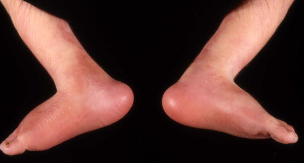
Definition: The absence of all phalanges of all digits of a limb and the associated soft tissues (Fig. 1). objective
Fig. 1.
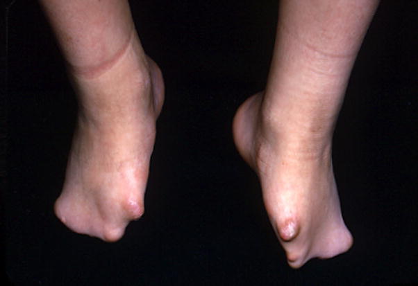
Adactyly of the feet, bilateral.
Comment: This descriptor does not require absence of the metacarpal or metatarsal bones. A qualifying phrase is added to specify which limb has the attribute of adactyly.
Replaces: Aphalangy
Amniotic band: See Digital constriction ring
Aphalangy: See Adactyly
Apodia: See Foot, absent
Arachnodactyly: See Finger, long; Toe, long; Finger, slender; Toe, slender
Arch, dropped or fallen: See Pes planus
Arch, high: See Pes cavus
Brachytelephalangy: See Finger, short distal phalanx of; Toe, short distal phalanx of
Camptodactyly
Definition: The DIP and/or PIP joints of the fingers cannot be extended to 180 degrees by either active or passive extension (Fig. 2). subjective
Fig. 2.
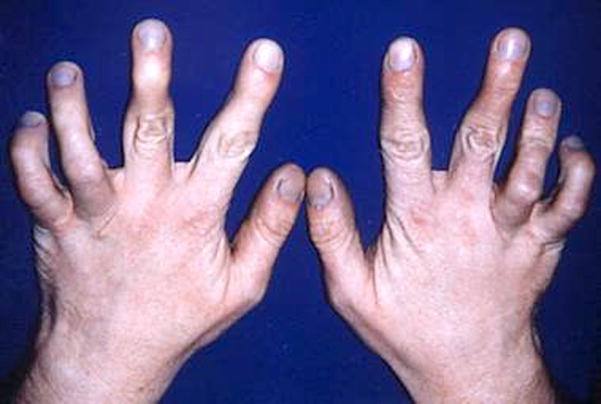
Camptodactyly of F45, bilateral.
Comment: Note that some restrict the use of the term to inability to extend the PIP joint of the fifth finger. We do not restrict the term in this way. This term should not be used if the patient has Clenched hand. A similar effect can be created by radial angulation within the distal phalanx with thickening of the epiphysis, which is called Kirner deformity or dystelephalangy [Ozonoff, 1992]. The affected digits should be specified.
Replaces: Streblodactyly
Clinodactyly
Definition: A digit that is laterally curved in the plane of the palm (Fig. 3). subjective
Fig. 3. Clinodactyly, radial, F5, bilateral.
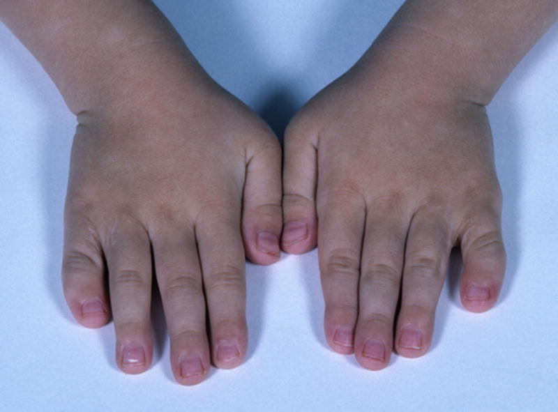
This person also has Short finger, F5, bilateral, subjective. See also Figs. 13, 20.
Comment: The curvature in this term is restricted to the phalanges and does not refer to deviation at the MCPJ/MTPJ (see Radial deviation of the fingers). Typically involves an abnormally shaped middle phalanx, but this is not obligate. The identity of the finger/toe should be specified and the directionality of the deviation. This finding is most common in F5, and is almost always radial, but can be seen in any digit and can be in either direction. A similar effect can be created by radial angulation within the distal phalanx with epiphyseal thickening, which is called Kirner deformity or dystelephalangy [Ozonoff, 1992].
Replaces: Incurved finger
Clubbing
Definition: Broadening of the soft tissues (non-edematous swelling of soft tissues) of the digital tips in all dimensions associated with an increased longitudinal and lateral curvature of the nails (Fig. 4). subjective
Fig. 4. Clubbing.
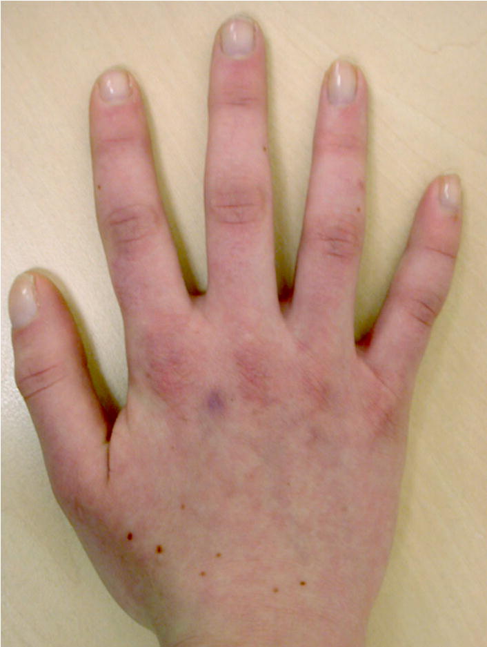
Note that the clubbing is best viewed laterally (e.g., the thumb) whereas it is difficult to appreciate in the other digits viewed dorsally. See also Figs. 5 and 63.
Comment: This definition does not use specification of nail plate angles for several reasons. First, there is no agreement on which angle should be measured. Second, there are no known data to establish the range of normality. Third, clinicians rarely perform such measurements. Clubbed fingers are often described as resembling the end of drumsticks. The affected digits should be specified as described in the introductory comments. If no subtype identifier is specified, it is assumed that all digits are involved. Although increased soft tissue is the sole or major component of clubbing, some cases may be associated with bone spurs or modest overgrowth of the distal phalanx. As this is not relevant to the clinical (non-radiographic) assessment of clubbing, it is not specified in the definition of this term.
Clubhand: See Hand, radial deviation of; Hand, ulnar deviation of
Digit: See various terms under “finger” or “toe” instead of digit
Digit pad, prominent
Definition: A soft tissue prominence of the ventral aspects of the fingertips or toe tips (Fig. 5). subjective
Fig. 5. Prominent digit pads, right hand, F3,4.
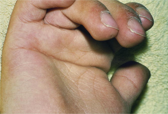
Note that this patient also has Clubbing and a Single transverse palmar crease.
Comment: The prominence of the finger or toe pads varies throughout life, being greater in neonates and dependent on the state of hydration. This term should not be used if the digit has clubbing. Note that the synonym “fetal fingertip pads” should not be interpreted to mean that the prominent pad necessarily was present in this individual since fetal life. We do not endorse the use of the term “Persistent fetal pads” as this term implies knowledge of the natural history of this finding, which may or may not be known.
Synonym: Fetal finger/toe tip pads; Fetal finger/toe tip pads
Replaces: Persistent fetal pads
Digital constriction ring
Definition: A narrow segment of significantly reduced circumference of a digit (Fig. 6). subjective
Fig. 6.
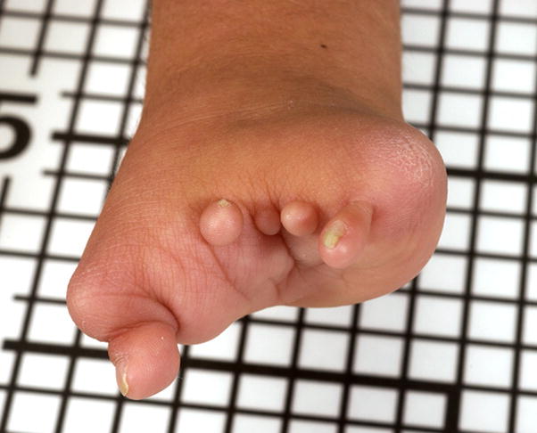
Digital constriction rings, left hand, F1-5, near MCPJ.
Comment: The description should specify the hand and digit that is affected and the approximate location of the band relative to the phalanges. It may be described as partial, if it does not involve the entire circumference of the digit.
Replaces: Amniotic bands
Ectrodactyly: See Foot, split; Hand, split
Fetal fingertip pads: See Digit pad, prominent
Finger, absent
Definition: The absence of all phalanges of a digit of the hand and the associated soft tissues (Fig. 7). objective
Fig. 7. Finger, absent, right hand.
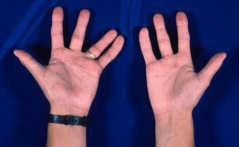
Note here that the identity of the missing digit is not specified, as there are no clinical data to allow this to be determined. See also Fig. 68.
Comment: This term does not require absence of the metacarpal. The affected digits should be specified. This definition excludes Partial absence of the finger. Note that it can be challenging to number the missing digit if it is a central digit that is missing. In these cases, it is appropriate to modify the term by adding the phrase “non-thumb”. In contrast, it will be obvious that the thumb is present or absent. It may also be difficult to distinguish an absent finger from two digits with an extreme degree of osseous and cutaneous syndactyly. If a patient is missing all digits of a limb, the term Adactyly should be used. The term Oligodactyly may be used as a synonym, but only in situations where the missing digit is not specified. For example, to state that a patient has Oligodactyly F5 is awkward because the oligodactyly refers to the digits that are present and the F5 refers to the digit that is absent, which is awkward and could become confusing with higher order deficiencies. If a digit that cannot be identified is missing, the term can be modified with a cardinal number, e.g., “Absent fingers, two”.
Replaces: Hypodactyly
Synonym: Oligodactyly
Finger, broad
Definition: Increased width of a non-thumb digit of the hand (Fig. 8). subjective
Fig. 8. Broad fingers, right hand.
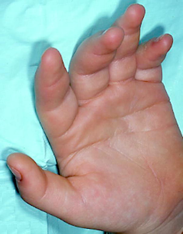
Appreciate that the dorsal-ventral dimension of these digits is not increased, whereas the lateral (proximo-distal) dimension is increased. See also Fig. 99.
Comment: Note that the girth may be increased in a broad finger, but this must be distinguished from Macrodactyly, because there the length is also increased. This distinction can be subtle. This term should not be used when the increased width is limited to the distal phalanges, instead use Broad fingertips. The affected digit should be specified by the numbering scheme in the introduction. This term is not used for the first digit, see Broad thumbs. When a thumb and one or more fingers are affected, it may be more economical to specify “Broad fingers, F1-5” instead of separately specifying “Broad thumb” and “Broad fingers F2-5”..
Synonym: Wide fingers
Replaces: Pachydactyly; Thick fingers
Fingers, cutaneous syndactyly of
Definition: A soft tissue continuity in the A/P axis between two fingers that extends distally to at least the level of the PIPJ (Fig. 9). objective OR
Fig. 9.
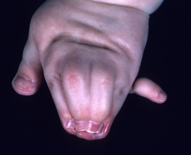
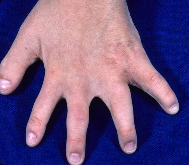
A. Cutaneous syndactyly of F2-5, left hand, complete, objective. Note that this patient also has a Broad thumb, and Postaxial polydactyly of the hand, type B and Fused nails F2- 5. B. This patient has Cutaneous syndactyly of F2-4, partial, subjective. See also Fig. 14.
A soft tissue continuity in the A/P axis between two fingers that lies significantly distal to the flexion crease that overlies the metacarpophalangeal joint of the adjacent fingers. subjective
Comment: We have set an arguably arbitrary threshold to distinguish the object form the subjective finding. While severe degrees of cutaneous syndactyly are clearly objective, more subtle degrees are subjective. We set this threshold to distinguish these two situations. The digits (or parts of) are joined together by tissue that is not normally present between the digits at that point in the P/D axis. A modifier of “complete” may be used if the cutaneous syndactyly extends to the distal end of the nail bed of the digits. The affected digits should be specified. Note that the unqualified term “syndactyly” is no longer allowed as it is unclear whether this refers to bony or cutaneous syndactyly.
Replaces: Syndactyly (without adjective); Zygodactyly
Finger, hypoplastic: See Finger, small
Finger, incurved: See Clinodactyly
Finger, long
Definition: The middle finger is more than 2 SD above the mean for newborns 27 to 41 weeks EGA or above the 97th centile for children from birth to 16 years of age AND the five digits retain their normal length proportions relative to each other (i.e., it is not the case that the middle finger is the only lengthened digit) (Fig. 10). subjective OR
Fig. 10.
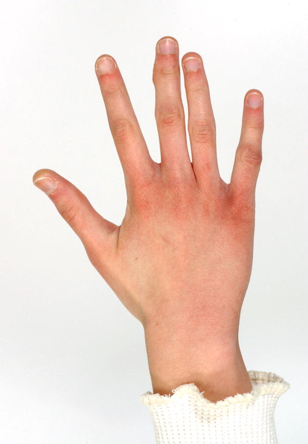
Long fingers, right hand, subjective.
Fingers that appear disproportionately long compared to the palm of the hand. subjective
Comment: The first definition is recognized to be problematic, because it implies that the other fingers are all as relatively long as is the middle finger. As the determination of the proportionality of the other four digits is clearly subjective, the term must be regarded as subjective. The term arachnodactyly has been abandoned as it is bundled (narrow and long digits) and is sometimes used for only narrow or only long digits. The term “long hand” should not be used, as it is a bundled definition of two readily separable terms, Long fingers, and Long palm. If only a subset of the digits of a limb is lengthened, the affected digits should be specified.
Replaces: Arachnodactyly
Finger, narrow: See Finger, slender
Fingers, overlapping
Definition: A finger resting on the dorsal surface of an adjacent digit when the hand is at rest (Fig. 11). objective
Fig. 11.
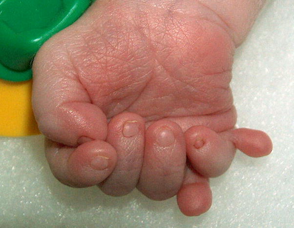
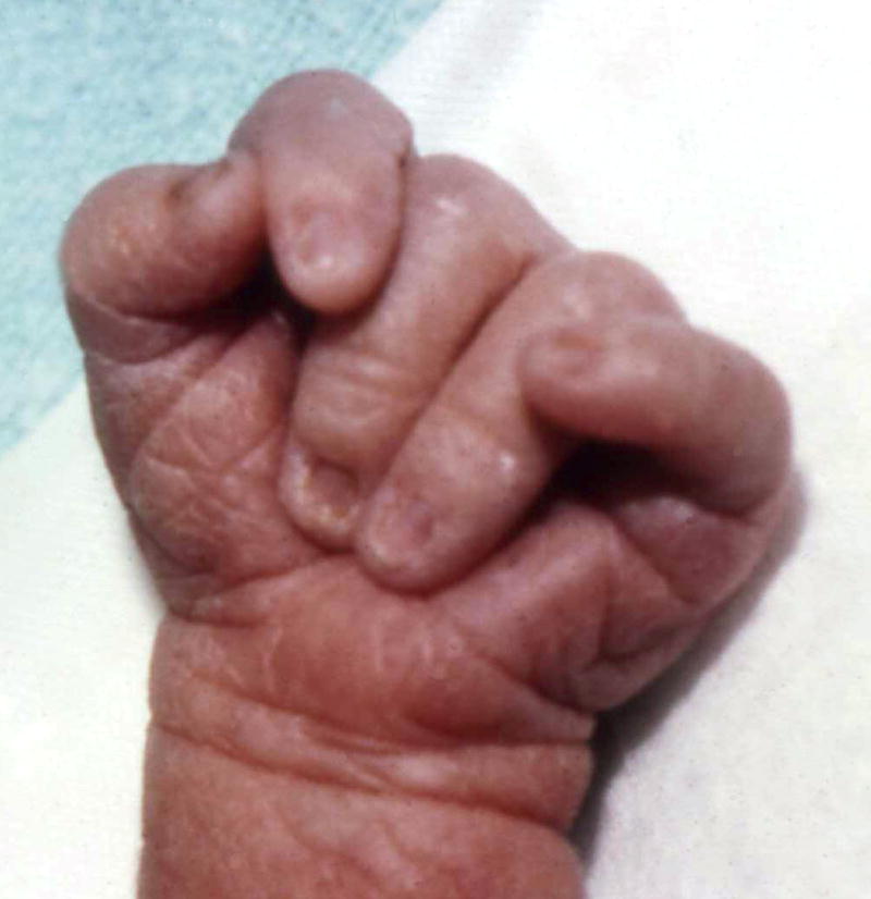
A. Overlapping fingers, right hand, F54, F56. Note that this patient also has Mesoaxial polydactyly and Postaxial polydactyly, type B. B. Overlapping fingers, right hand, F23, F54. See also Figs. 37, 45, and 58A.
Comment: This descriptor is ordered depending on which digits are involved (see figure legend for examples). The affected digits should be specified. The ordering of the numbers specifies which digit is dorsal, i.e., with dorsum of the hand facing upward the digit on top is/are recorded first separated by a comma from the digit that is/are overlapped. Fingers that are laterally deviated, but do not rest on top of adjacent fingers should be coded as Clinodactyly.
Finger, partial absence of
Definition: The absence of a phalangeal segment of a finger (Fig. 12). objective
Fig. 12. Partial absence of fingers, left hand, F2-5, proximal.
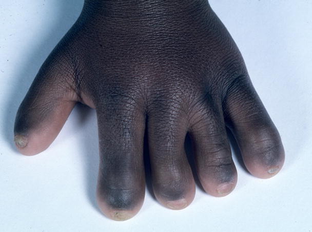
Note that this patient also has Small nails, F1-5.
Comment: The part that is absent may be specified. The “distal” modifier specifies the loss of the distal phalanx; clinically this is defined by the absence of the nail. The “proximal” modifier specifies the loss of the proximal or middle phalanx with the nail still present and/or the radiographic finding of a remaining phalanx that is similar to a distal phalanx. It may be difficult to determine which phalanx is absent without x-rays and even then, there are circumstances where the missing bone may not be exactly identified (note that no attempt is made to distinguish missing middle from proximal phalanges). In this situation the location adjective will have to be removed. This finding is distinct from Short fingers.
Replaces: Hypophalangy
Finger, radial deviation of
Definition: Angulation of a digit toward the anterior axis (radial side) of the limb (Fig. 13). subjective
Fig. 13. Radial deviation of the finger, left hand, F2.
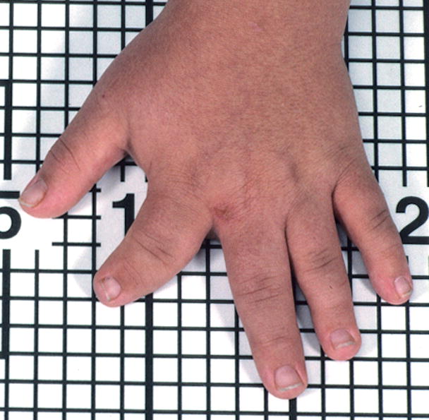
Note that this patient also has Clinodactyly, F2, radial and that these two findings are distinct. See also Fig. 47.
Comment: The deviation is at the MCPJ. The affected digits should be specified. This finding is distinct from Clinodactyly.
Finger, short
Definition: The middle finger is more than 2 SD below the mean for newborns 27 to 41 weeks EGA or below the 3rd centile for children from birth to 16 years of age AND the five digits retain their normal length proportions relative to each (i.e., it is not the case that the middle finger is the only shortened digit) (Fig. 14). subjective OR
Fig. 14. Short fingers, subjective.
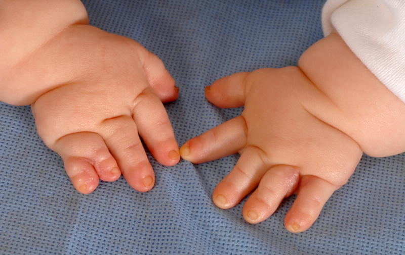
See also Figs. 3, 47, 69, and 99. Note that this patient also has Short palms, subjective and cutaneous syndactyly of F4,5, right hand, objective.
Fingers that appear disproportionately short compared to the hand. Subjective
Comment: This is an acknowledged bundled term as the definition in most anthropometric sources assumes that the other fingers are all as relatively short as is the middle finger. As the determination of the proportionality of the other four digits is clearly subjective, the term must be regarded as subjective. The Bell classification of brachydactyly (summarized in [Temtamy and McKusick, 1969]) is a complex assessment of hand patterning – that usage of the word “brachydactyly” is independent of the use here. When “Brachydactyly type x” is used, this refers to the Bell classification patterns. When Brachydactyly of the hand is used, it solely refers to reduced length of the specified digits. “Short hand” should not be used as it is a bundled definition of Short fingers and Short palm. If the distal phalanges are judged to be proportionately shortened, the additional term of Short distal phalanges of the fingers should not be used. If the digit is short overall and the distal phalanx is disproportionately shorter than are the finger overall, then both terms should be used.
Synonym: Brachydactyly of the hand
Finger, short distal phalanx of
Definition: Short distance from the end of the finger to the most distal interphalangeal crease or DIPJ flexion point (Fig. 15). subjective
Fig. 15. Short distal phalanx of the finger, left hand, F1.
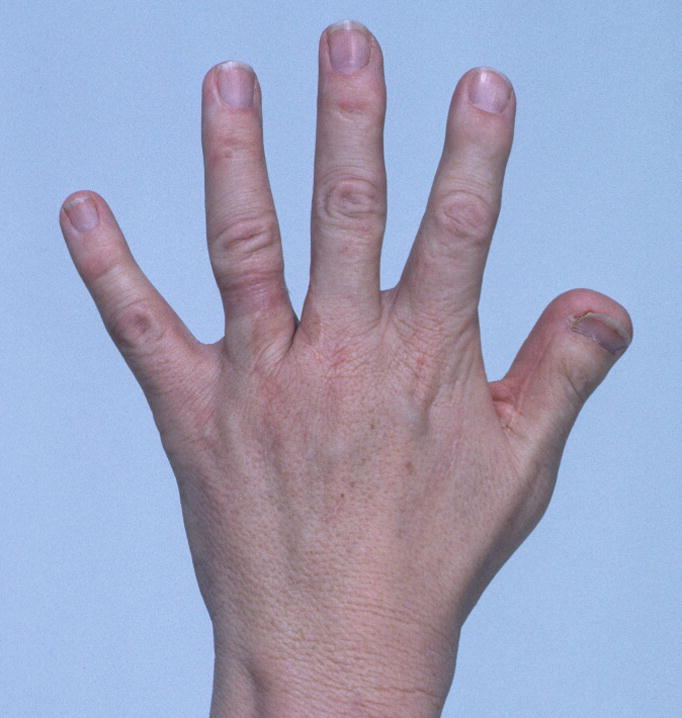
Note that this patient also has a Short nail, F1. See also Fig. 60A.
Comment: This term differs from Partial absence of the finger because in that term, the phalanx must be missing, whereas in this term it may be small, but present. Distal phalangeal lengths can be assessed subjectively by comparing that digit segment to the rest of the digit, to other normal digits in that patient, or to typical patients of that age or build. Regarding the subjective definition, for individuals who do not have flexion creases, one may determine this by flexing the DIP joint and estimating the length of the terminal segment of the digit. Alternatively, one may be able to palpate the joint.
Synonym: Brachytelephalangy
Replaces: Short terminal phalanges; Short terminal finger
Finger, slender
Definition: Digits are disproportionately narrow (reduced girth) for the hand/foot size or build of the individual (Fig. 16). subjective
Fig. 16. Slender fingers, right hand, F2-5.
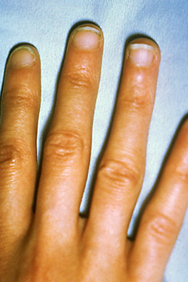
Note that only some of the digits are clearly shown in this figure.
Comment: The affected digits should be specified as described. The assessment of this finding is difficult when the digits are long. The bundled and pejorative term “arachnodactyly” is replaced by the separate descriptors Long finger and Slender finger.
Synonym: Narrow digits; Narrow fingers
Replaces: Thin digits; Arachnodactyly
Finger, small
Definition: Significant reduction in both length and girth of the finger compared to the contralateral finger, or alternatively, compared to a typical finger size for an age-matched individual (Fig. 17). subjective
Fig. 17. Fingers, small, left hand, F3,4.
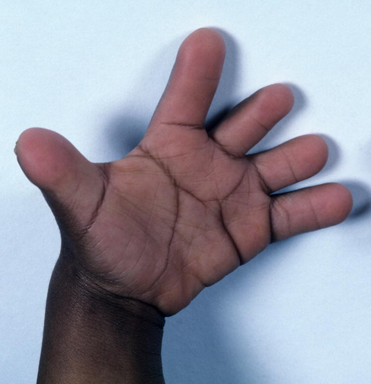
This patient also has Deep palmar creases.
Comment: This is a bundled term, comprising Short finger and Slender digit, but it is so widely used that it is included. This term is only used if the finger has the normal number of phalangeal segments. An appropriate alternative term for the first digit is Small thumb, when it is the only digit affected. When a thumb and one or more fingers are affected, it may be more economical to specify “Small fingers, F1-5” instead of separately specifying “Small thumb” and “Small fingers F2-5”.
Replaces: Underdeveloped finger; Hypoplastic finger
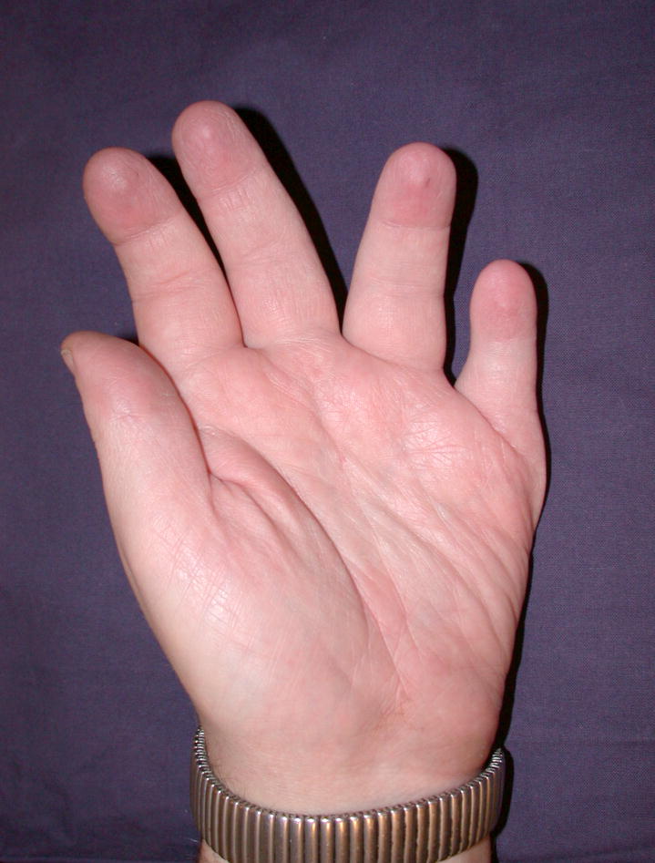
Fingers, splayed
Definition: Divergence of digits along the A/P axis (in the plane of the palm) (Fig. 18). subjective
Fig. 18. Splayed fingers, left hand, F23, F34.
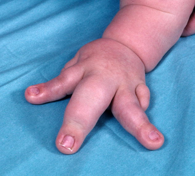
Note that this patient also has Macrodactyly F2-4
Comment: This may be associated with Macrodactyly, but this should be assessed and coded separately. The affected digits should be specified.
Synonym: Spreading of the fingers
Fingers, spreading of: See Fingers, splayed
Finger, tapered
Definition: The gradual reduction in girth of the digit from proximal to distal (Fig. 19). subjective
Fig. 19.
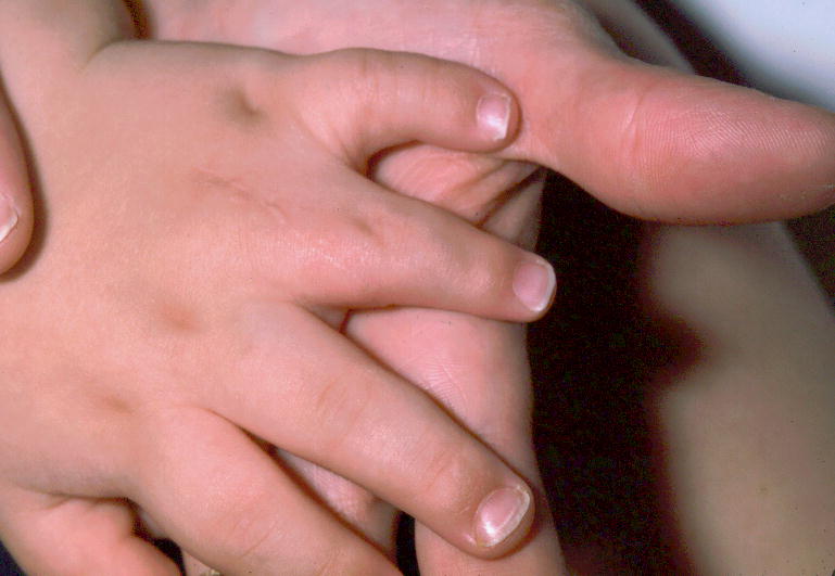
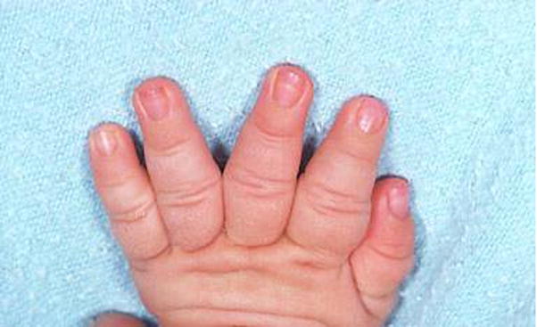
A. Tapered finger, left hand, F4. B. Tapered fingers, right hand, F2-5. See also Figs. 42, 44, and 93.
Comment: If the digits are not uniformly affected, the affected fingers should be specified. If not specified, it refers to all the digits of the hand.
Finger, thick: See Finger, broad
Finger, thin: See Finger, slender
Finger, ulnar deviation of
Definition: Angulation of the digit towards the posterior or postaxial axis (ulnar) of the limb (Fig. 20). subjective
Fig. 20. Ulnar deviation of finger, right hand, F4.
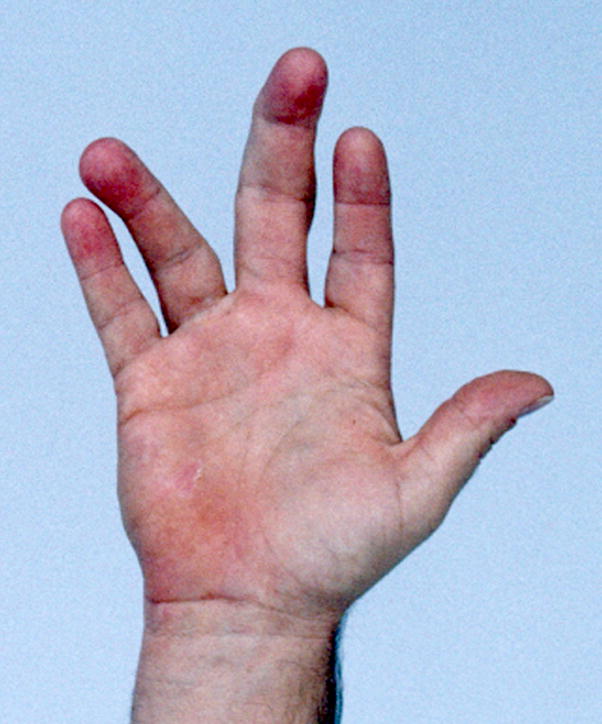
Note that this patient also has Clinodactyly of F4, ulnar. Note that his middle finger manifests Clinodactyly radial, F3, but that this finger is not deviated, demonstrating the distinction of these two features. See also Fig. 45.
Comment: The deviation is at the MCPJ. The affected digits should be specified. This finding is independent of Clinodactyly.
Finger, underdeveloped: See Finger, small
Finger, wide: See Finger, broad
Finger-like thumb: See Thumb, triphalangeal
Fingertip, broad
Definition: Increased width of the distal segment of a finger (Fig. 21). subjective
Fig. 21.
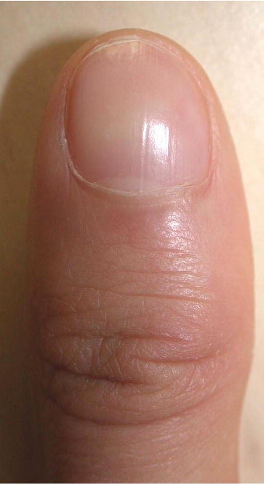
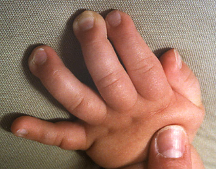
A. Broad fingertip, right hand, F1. Note how the digit widens at the IPJ. B. Broad fingertips, left hand, F3,4. See also Fig. 93.
Comment: This term should be reserved for use when the distal digit is significantly broader than the middle part. It should not be used if the digit has Clubbing or Macrodactyly or if the entire finger is broad; instead use Finger, broad.
Synonym: Wide fingertips
Replaces: Spatulate fingertips
Fingertip, spatulate: See Fingertip, broad
Fingertip, wide: See Fingertip, broad
Fingertip pads, fetal: See Digit, prominent pad
Foot, absent
Definition: The total absence of the foot, with no bony elements distal to the tibia or fibula (Fig. 22). objective
Fig. 22. Absent foot, right.
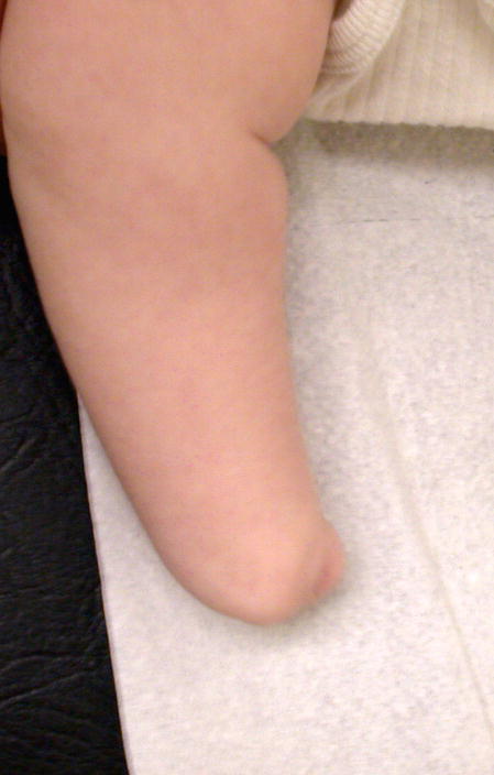
Note that this is the same patient as is shown in Fig. 27. One limb has partial absence of the foot and the other complete.
Comment: See Foot, partial absence of, for a related definition.
Synonym: Apodia
Foot, brachydactyly of: See Toe, short
Foot, broad
Definition: A foot for which the measured width is above the 95th centile for age (Fig. 23). objective
Fig. 23. Broad foot, left.
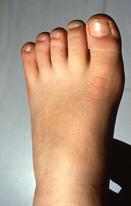
In this example, the forefoot appears more broad than does the midfoot, but this distinction is not noted in the nomenclature.
OR A foot that appears disproportionately wide for its length. subjective
Comment: Note that the normative data [Malina et al., 1973; Hall et al., 2007] only include 4 – 16 year olds. Since foot length is still increasing at the 16 year old age point on the graphs, extrapolation is not recommended. Therefore, persons more than 16 years of age or less than 4 years of age can only be assessed subjectively (until appropriate norms are identified or generated). The Hall et al [2007] handbook (pp. 238-9) states that foot width should be measured from the medial aspect of the first MTPJ to the lateral aspect of the fifth MTPJ. The subjective definition should be used with caution in patients with Short feet. In persons with polydactyly that includes a supernumerary metatarsal, that should be separately coded and the measurement definition from Hall et al, [2007] would need to be modified to account for the supernumerary digit (i.e., if the patient had postaxial polydactyly, measure from the sixth MTPJ). It may be argued that this is double counting polydactyly as a wide foot, as the norms are derived from those with pentadactylous limbs.
Synonym: Wide foot
Foot, cleft: See Foot, split
Foot, long
Definition: Foot length more than 2 SD above the mean for a newborn of 27 – 41 weeks gestation (Fig. 24). objective
Fig. 24. Long feet, subjective.
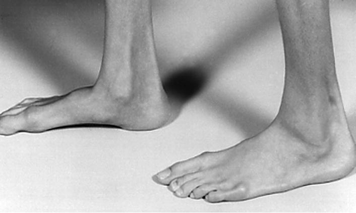
Note that this patient also has Long toes.
Foot length above the 97th centile for individuals from birth to 16 years of age objective OR A foot that appears longer than expected for age. subjective
Comment: Note that for females over the age of 16 years, the curves are essentially flat by 15 years of age, so the 16-year-old norms and distribution can be used for older individuals [Anderson et al., 1956; Hall et al., 2007]. However, the male norms still have significant growth at 16 years, where the graph ends. Therefore, only subjective assessments can be made for males over the age of 16 years. For infants, the norms are specified in SD, per the primary source [Merlob et al., 1984]. Because of the wide range of normal for foot measurements, it may be hard to discriminate a long foot of normal width from that of a narrow foot of normal length, thus the subjective definition is substantially inferior. If the toes are also long, that should be coded separately.
Foot, narrow
Definition: A foot for which the measured width is below the 5th centile for age (Fig. 25). objective OR
Fig. 25. Narrow foot, left, subjective.
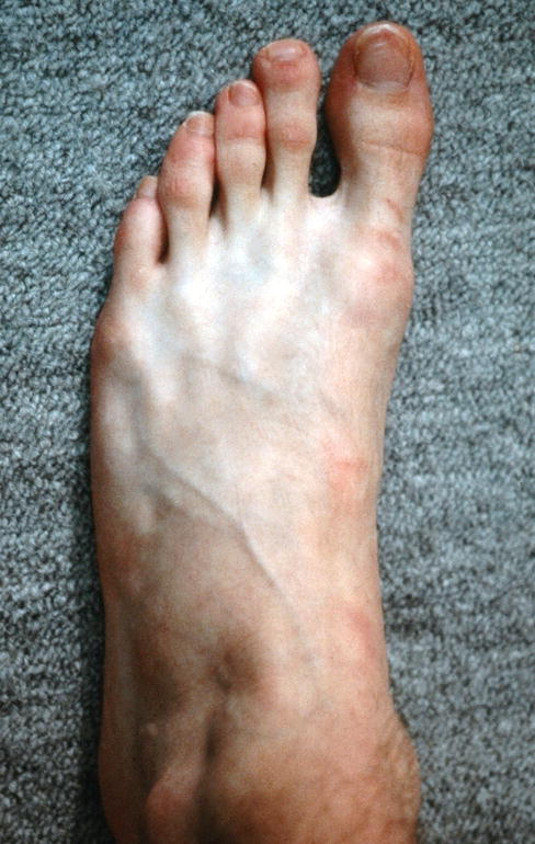
A foot that appears disproportionately narrow for its length. subjective
Comment: The normative data [Hall et al., 2007] only include 4 – 16 year olds. Since the foot width is still increasing at the 16 year old age point on the graphs, extrapolation is not recommended. Therefore, persons more than 16 years or less than 4 years of age can only be assessed subjectively (until appropriate norms are identified or generated). Foot width should be measured from the medial aspect of the first MTP joint to the lateral aspect of the fifth MTP joint [Hall et al., 2007]. It does not seem sensible to apply this term to individuals with polydactyly that includes a duplicated metatarsal.
Foot, osseous syndactyly of
Definition: Lateral (A/P) fusion of the digits (phalanges and/or metatarsals) by hard tissue (cartilage and/or bone) (Fig. 26). objective
Fig. 26. Foot, osseous syndactyly of.
Comment: Metatarsal syndactyly can be demonstrated by the inability to independently manipulate the two metatarsals (see Polydactyly, mesoaxial for a description of this maneuver) or the continuity of two metatarsals is demonstrated [Biesecker, 2007]. The description should include the bones that are fused. Osseous syndactyly is distinct from Symphalangism, where the phalanges are fused at the joint longitudinally (P/D axis). The exact nature of the bony anomaly may not be definable without x-ray evidence. Note that the unqualified term “syndactyly” is no longer allowed as it is unclear whether this refers to bony or cutaneous syndactyly.
Replaces: Syndactyly (without adjective)
Foot, partial absence of
Definition: An incomplete absence of the foot, with no bony elements distal to the tarsals, but with preservation of some or all of the tarsals (Fig. 27). objective
Fig. 27. Foot, partial absence of, left.
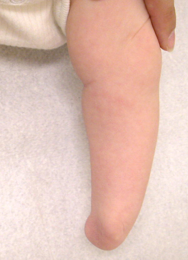
Note that this is the same patient as is shown in Fig. 22. One limb has complete absence of the foot and the other partial.
Comment: See Foot, absent for a related term.
Foot, postaxial polydactyly of
Definition: Presence of a supernumerary digit that is not a hallux (Fig. 28). objective
Fig. 28. Postaxial polydactyly of the left foot.
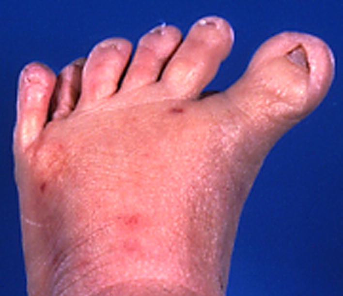
Note that this patient also has Small nails.
Comment: Although it is appealing to believe in many cases that the supernumerary (non-hallux) digit is the most fibular, there may be no evidence for this. When the digit is de minimus, this seems reasonable by parsimony. When it is fully formed, associated with a supernumerary metatarsal, and fully functional, it may be impossible to determine which toe is supernumerary. Nevertheless, the designation as postaxial is reasonable given the tradition of this designation. Postaxial polydactyly has been divided into two types: A (a fully formed digit) and B (digitus minimus, or a pedunculated, non-articulating, non-functional appendage). We recognize these subtypes but note that post-axial polydactyly actually represents a spectrum from type A to type B. When the type is indeterminate, no subtype is specified. The term uses the word “postaxial” instead of the embryologic “posterior” because the former is established in clinical medicine.
Synonym: Posterior polydactyly
Replaces: Posterior duplication of the limb/foot; fibular polydactyly
Foot, preaxial polydactyly of
Definition: Duplication of all or part of the first ray (Fig. 29). objective
Fig. 29.
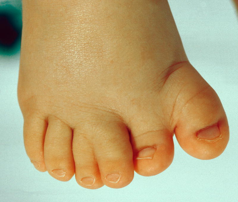
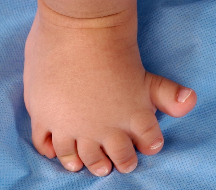
A. Preaxial polydactyly of the right foot. Note that this duplication is nearly complete. B. Preaxial polydactyly of the right foot. This patient has the same finding, but the duplicated digits are more separated than in Fig. 29A.
Comment: There is a wide spectrum of this malformation. The mild end of the spectrum is a bifid (not cleft) nail or a distal phalanx of the hallux with a central lacuna or bifid tip. Broadened halluces without a recognizable anterior/posterior cleft should be coded as Broad hallux. Note that toe number may be affected by the finding of preaxial polydactyly. See the introductory comments for guidance on digit numbering. It is optional to modify the term by adding “partial” to a duplicated hallux that involves less than the entire ray (metatarsal and both phalanges) and “complete” for a digit that has complete duplication of all three bones.
Synonym: Anterior polydactyly
Replaces: Anterior duplication of the limb; Tibial polydactyly
Foot, rocker bottom
Definition: The presence of both a ‘prominent heel’ and a ‘convex contour of the sole’ (Fig. 30). subjective
Fig. 30.
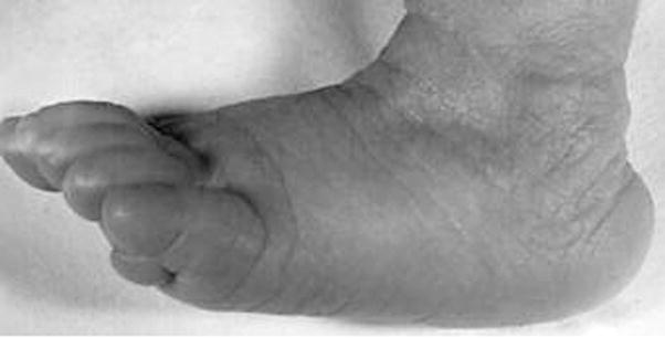
Rocker bottom foot, left.
Comment: The term is a bundled definition but is in common usage. For that reason, it was included by consensus of the group.
Foot, short
Definition: A measured foot length that is more than 2 SD below the mean for a newborn of 27 – 41 weeks gestation (Fig. 31). objective OR
Fig. 31.
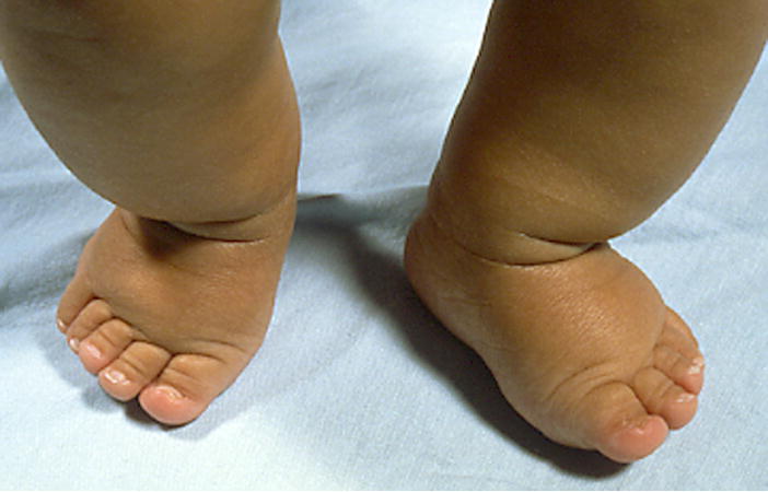
Short feet, subjective.
A foot that is less than the 3rd centile for individuals from birth to 16 years of age. objective OR A foot that appears disproportionately short. subjective
Comment: Note that for females over the age of 16 years, the curves are essentially flat by 15 years of age, so the 16-year-old norms and distribution can be used for older individuals [Anderson et al., 1956; Hall et al., 2007]. However, the male norms still show significant growth at 16 years, where the graph ends. Therefore, only subjective assessments can be made for older males. For infants, the norms are specified in SD, per the primary source [Merlob et al., 1984]. Foot length includes the relatively long calcaneus and tarsal bones as well as the metatarsals and first toe, and so the hand and foot measurements may reflect different growth abnormalities. Foot length should be interpreted with caution if the individual has either Pes cavus or Pes planus.
Foot, split
Definition: Longitudinal deficiency of a digital ray of the foot except rays 1 or 5 (Fig. 32). subjective
Fig. 32.
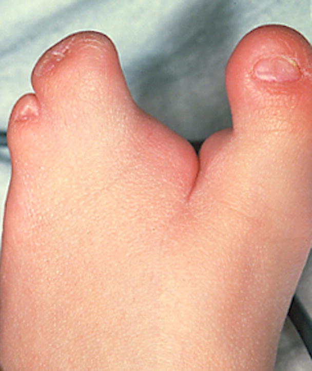
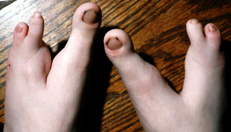
A. Split foot, left. B. Split feet. Note that this patient has a more severe, deeper notch in the feet than does the patient in A. The shape and numbers of other affected digits is highly variable.
Comment: The threshold for this deficiency should be that all of the phalanges in the ray are absent and at least part of the metatarsal. If just the phalanges that are missing, this should be coded as Absent toe. The absence of a central ray or rays frequently yields a cleft appearance of the foot. This term is preferred to “ectrodactyly of the foot” as this has been used (more appropriately) for various forms of phalangeal hypoplasia or amputation. “Lobster claw deformity” is neither descriptive nor acceptable. There is wide variation in the affected feet, but the common factor is the absent central ray, justifying Split foot as a clinical designation.
Synonym: Cleft foot
Replaces: Ectrodactyly; Lobster claw deformity
Foot, wide: See Foot, broad
Hallux, absent
Definition: The absence of both phalanges of a hallux and the associated soft tissues (Fig. 33). objective
Fig. 33.
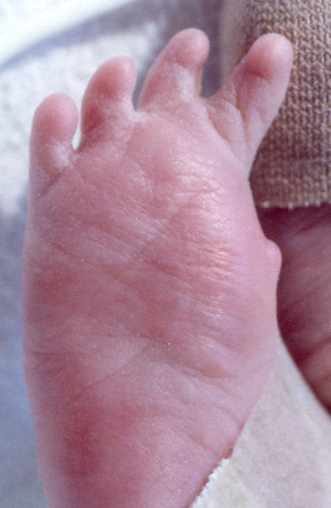
Absent hallux, right.
Comment: This descriptor does NOT require absence of the metatarsal. The definition excludes partial absent hallux. Oligodactyly and hypodactyly are replaced with more specific terms to allow the distinction of loss of a preaxial digit (thumb or great toe) from loss of digits 2–5. When a great toe and one or more other toes are absent, it may be more economical to specify “Absent toes, T1-3” instead of separately specifying “Absent hallux” and “Absent toes T2-3”.
Synonym: Absent great toe
Replaces: Hypodactyly; Oligodactyly
Hallux, broad
Definition: Visible increase in width of the hallux without an increase in the dorso-ventral dimension (Fig. 34). subjective
Fig. 34. Broad hallux, right.
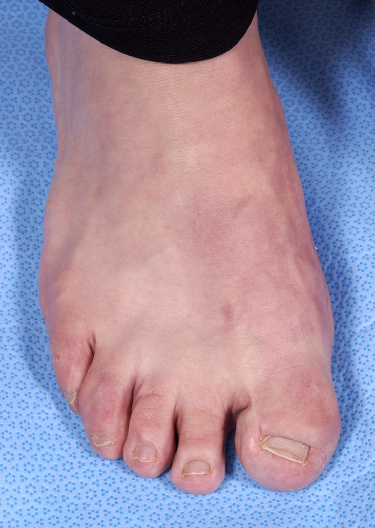
See also Fig. 60B.
Comment: Note that girth may be increased in a broad hallux, but this must be distinguished from Macrodactyly because there the length is also increased. This assessment may be difficult when the hallux is short. Note the separate definition for Toes, broad. When a great toes and one or more other toes are affected, it may be more economical to specify “Broad toes, T1-5” instead of separately specifying “Broad hallux” and “Broad toes T2-5”.
Synonym: Broad great toe
Hammertoe
Definition: Hyperextension of the MTP joint with hyperflexion of the proximal interphalangeal (PIP) joint (Fig. 35). subjective
Fig. 35. Hammertoe, T3, left.
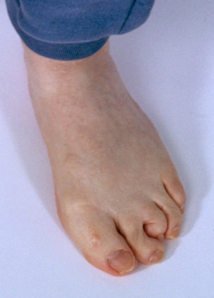
See also Fig. 57.
Comment: This term replaces the phrase “dorsiflexed toe” as that term is an incomplete description of the abnormality [Young et al., 2005]. Note that this is not a synonym of mallet toe.
Replaces: Dorsiflexed toe
Hand, absent
Definition: The total absence of the hand, with no bony elements distal to the radius or ulna (Fig. 36). objective
Fig. 36. Absent hand, left.
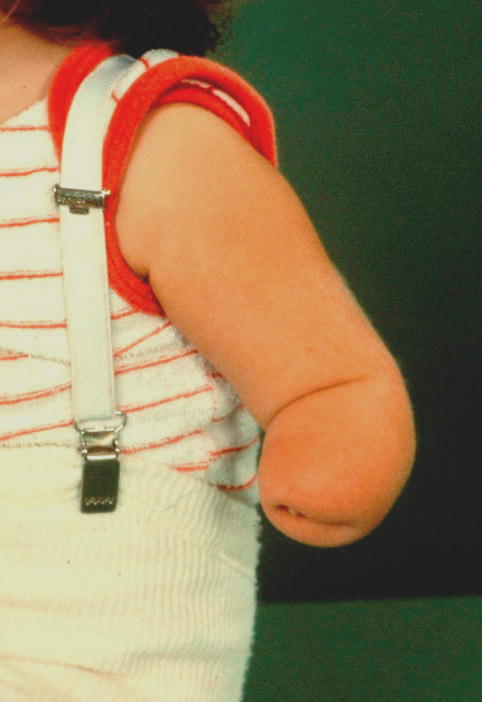
Note that this patient also has a shortened radius and ulna, although that finding is not required for this finding.
Comment: See Hand, partial absence for a related term.
Synonym: Acheiria
Hand, brachydactyly of: See Finger, short
Hand, cleft: See Hand, split
Hand, clenched
Definition: All digits held completely flexed at the metacarpophalangeal and interphalangeal joints (Fig. 37). subjective
Fig. 37. Clenched hand, right.
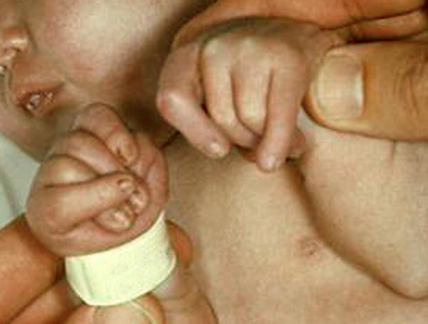
Note that the left hand does not warrant the term because not all of the digits are completely flexed at the MCPJ and IPJ. The left hand would warrant the finding of Overlapping fingers F23, F54, left.
Comment: Is distinguished from Camptodactyly, as that term may describe fewer than five digits of a eudactylous hand and does not involve the MCPJ. The digits may overlap when they lie flexed in the palm. It is not necessary to specify the overlapping fingers finding separately.
Hand, long: See Palm, long
Hand, narrow: See Palm, narrow
Hand, osseous syndactyly of
Definition: Lateral (A/P) fusion of the digits (phalanges and/or metacarpals) by hard tissue (cartilage and/or bone) (Fig. 38). objective
Fig. 38. Osseous syndactyly of the hand.
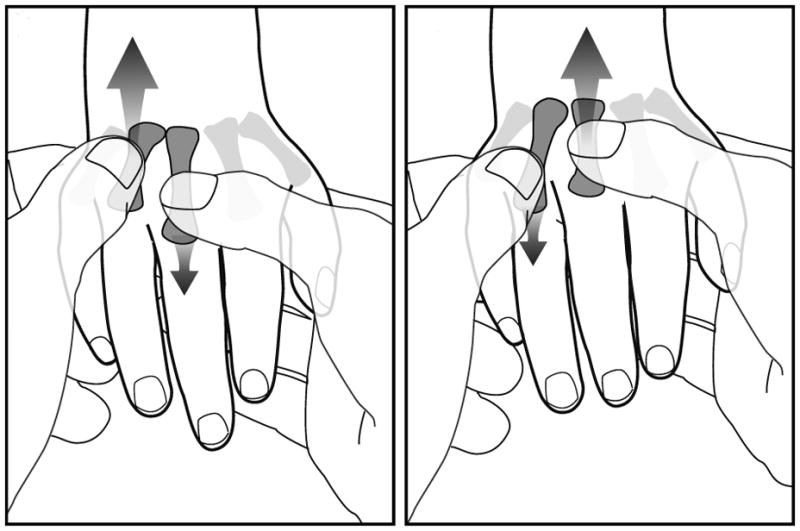
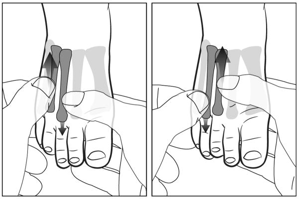
This figure shows the maneuver used to detect this finding. The abnormal finding is not shown. The examiner grasps two adjacent metacarpals and alternately moves them to determine if they are fused or independent.
Comment: Metacarpal syndactyly can be demonstrated by either a maneuver whereby the two metacarpals cannot be independently manipulated by the examiner (see Polydactyly, mesoaxial for a description of this maneuver). Phalangeal syndactyly (typically seen in what used to be called a “mitten hand”) may be difficult to demonstrate without X-rays. The description may include the bones that are fused. Osseous syndactyly is distinct from Symphalangy where the phalanges are fused at the joint longitudinally (P/D axis). See Polydactyly, mesoaxial for discussion of the use of this term with polydactyly.
Replaces: Syndactyly (without adjective)
Hand, postaxial polydactyly of
Definition: Presence of a supernumerary digit that is not a thumb (Fig. 39). objective
Fig. 39.
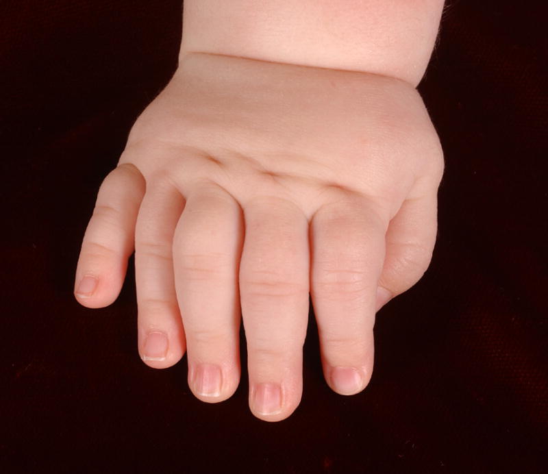
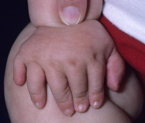
A. Postaxial polydactyly of the right hand, type A. B. Postaxial polydactyly of the right hand. Note that this patient has a digit that is intermediate between type A and type B, so that is not specified. See also Figs. 49A, 58A, and 73A. See Figs. 9A and 11A for examples of Postaxial polydactyly, type B.
Comment: Although it is appealing to believe in many cases that the supernumerary (non-thumb) digit is the most ulnar, there may be no evidence for this. When the digit is de minimus, this seems reasonable by parsimony. When it is fully formed with a supernumerary metacarpal and functional, it may be impossible to determine which of the fingers is supernumerary. Nevertheless, the designation as postaxial is reasonable given the tradition of this designation. Postaxial polydactyly has been divided into two types: A (a fully formed digit) and B (digitus minimus, or a pedunculated, non-articulating, non-functional appendage). We recognize these subtypes but note that post-axial polydactyly actually represents a spectrum from type A to type B. When the type is indeterminate, no subtype is specified.
Synonym: Posterior polydactyly
Replaces: Ulnar polydactyly; Posterior duplication of the limb/hand
Hand, preaxial polydactyly of
Definition: Duplication of all or part of the first ray (Fig. 40). objective
Fig. 40.
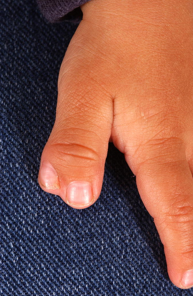
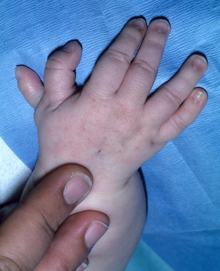
A Preaxial polydactyly of the left hand, partial. B. Preaxial polydactyly of the right hand
Comment: There is a wide spectrum of this malformation. The mild end of the spectrum is a bifid (not cleft) nail or a distal phalanx of the thumb with a central lacuna or bifid tip. Broadened thumbs without a recognizable line of anterior/posterior clefting should be coded as Thumb, broad. Note that finger numbering may be affected by the finding of preaxial polydactyly. See the introductory comments for guidance on digit numbering. It is optional to modify the term by adding “partial” to a duplicated thumb that involves less than the entire ray (metacarpal and both phalanges) and “complete” for a digit that has complete duplication of all three bones.
Synonym: Anterior polydactyly
Replaces: Anterior duplication of the limb/hand; Radial polydactyly
Hand, radial deviation of
Definition: Divergence of the longitudinal axis of the hand at the wrist in a anterior (radial) direction (Fig. 41). subjective
Fig. 41.
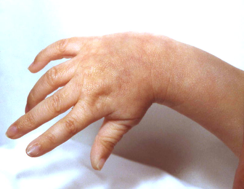
Radial deviation of the hand, right.
Comment: This finding may be associated with a short or absent radius, but that is not required and should be coded separately. The term “clubhand” is replaced because it was felt to be pejorative.
Replaces: Radial clubhand; Clubhand
Hand, short: See Palm, short; Finger, short
Hand, small
Definition: A normally proportioned hand (i.e., the various elements of the hand are in proportion to each other) that is overall small for age or overall body size (Fig. 42). subjective
Fig. 42. Small hands.
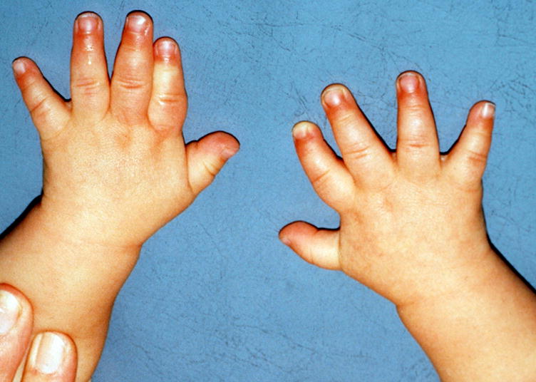
Note that this patient also has Tapered fingers.
Comment: This is acknowledged to be a bundled term, but it is sufficiently common that it was felt to be too cumbersome to describe the component findings. It is arguably superior to code a patient as having objectively determined Short fingers, Short palms, and Narrow palms instead of this bundled term. Note that there are no objective standards that can be used for this assessment, only for middle finger and palm length and palm width. The term acromicria is discouraged because in some definitions, this term also includes small head or facial features; in others it only includes the fingers and toes.
Replaces: Acromicria
Hand, split
Definition: Longitudinal deficiency of a digital ray of the hand except rays 1 or 5 (Fig. 43). subjective
Fig. 43. Split hand, right.
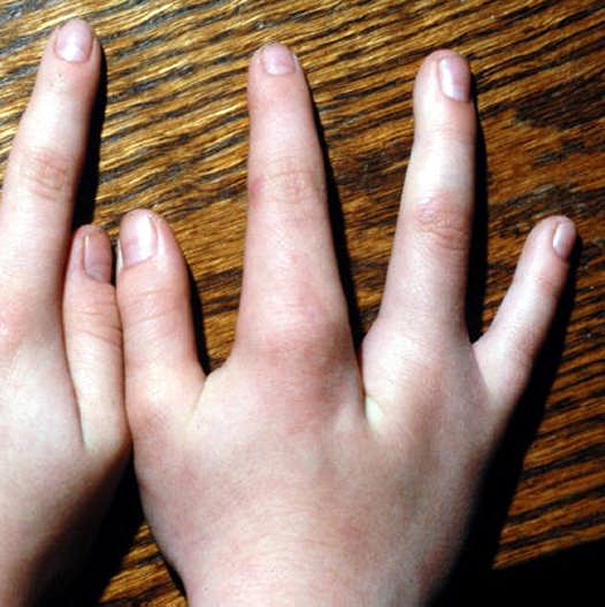
Comment: The threshold for this deficiency should be that all of the phalanges of a central ray (not F1 or F5) are absent and at least part of the associated metacarpal (if it is just the phalanges that are missing, this should be coded as Absent finger). The absence of a central ray or rays frequently yields a cleft appearance of the hand. This term is preferred to “ectrodactyly of the hand” as this has been used (more appropriately) for forms of phalangeal hypoplasia or amputation but it is acknowledged that this is a commonly used term. “Lobster claw deformity” is neither descriptive nor acceptable due to its pejorative nature. There is wide variation in the hands of affected individuals, but the common factor seems to be the absence of the central ray, justifying the use of Split hand as a clinical designation.
Synonym: Cleft hand
Replaces: Ectrodactyly; Lobster claw deformity
Hand, trident
Definition: Splaying of F2-4 along the A/P axis (in the plane of the palm) with relatively normal positioning of F1 and F5 (Fig. 44). subjective
Fig. 44. Trident hand, left.
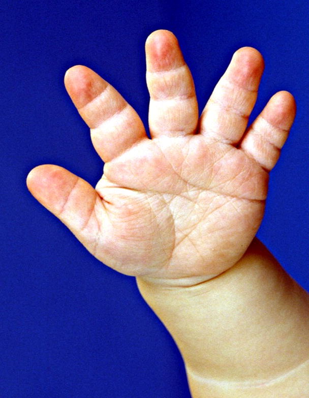
Note that this patient also has Tapered fingers, but that is not required for the finding.
Comment: This term is a subtype of splaying of the digits, but its diagnostic utility warrants a separate definition. Note is made that there were a number of different definitions of this term among the terminology group members. However, this definition was the consensus.
Hand, ulnar deviation of
Definition: Divergence of the longitudinal axis of the hand at the wrist in a posterior (ulnar) direction (Fig. 45). subjective
Fig. 45. Ulnar deviation of the right hand.
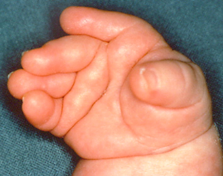
Note that this patient also has Ulnar deviation of the fingers, F2-3 and Overlapping fingers F45.
Comment: May be associated with a short or absent ulna, but that is not required.
Replaces: Ulnar clubhand; Clubhand
Hand, wide: See Hand, broad
Heel, prominent
Definition: Exaggerated or marked projection of the posterior pole of the heel (Fig. 46). subjective
Fig. 46.
Prominent heels.
Comment: No sensible thresholds or landmarks could be identified for this finding so the examiners must rely on their experience.
Hypodactyly: See Finger, absent; Hallux, absent; Thumb, absent; Toe, absent
Hypophalangy: See Finger, partial absence of; Toe, partial absence of
Hypothenar eminence, small
Definition: Reduced muscle mass on the ulnar side of the palm (Fig. 47). subjective
Fig. 47. Hypothenar hypoplasia, left hand.
Note that this patient also has Short fingers F2-5 and Radial deviation of fingers F2-3.
Comment: The reduced soft tissue is typically abductor digiti minimi and flexor digiti minimi brevis. This may be even more difficult to discern than is Small thenar eminence, but the same considerations outlined in that definition apply.
Limb, anterior duplication of: See Foot, preaxial polydactyly of; Hand, preaxial polydactyly of
Lobster claw deformity: See Foot, split; Hand, split
Macrodactyly
Definition: Significant increase in the length and girth of most or all of a digit compared to its contralateral digit (if unaffected) or compared to what would be expected for age/body build. The increased girth is accompanied by an increase in the dorso-ventral dimension AND the lateral dimension of the digit (Fig. 48). subjective
Fig. 48.
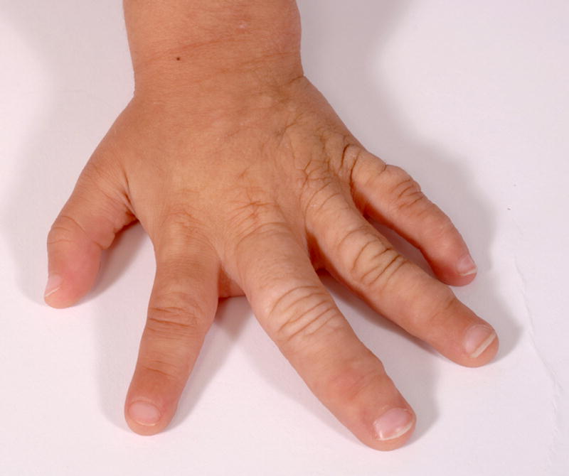
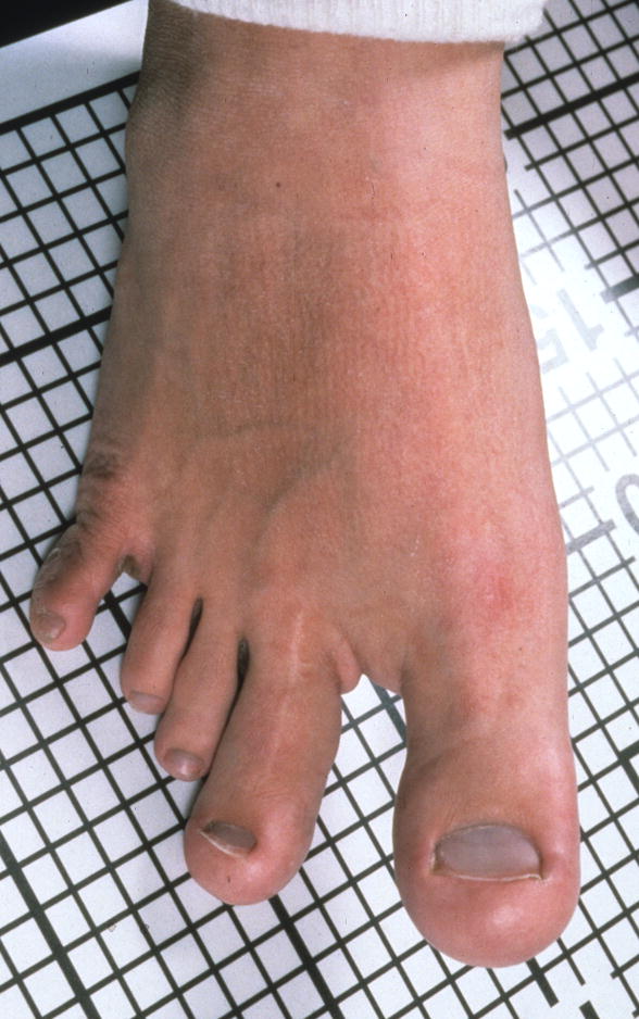
A. Macrodactyly of F2-3, left hand. Note that this person also has Clinodactyly, but that finding should be coded separately. B. Macrodactyly of T1-2, right foot. This patient also has a Sandal gap. See also Figs. 18, 81, and 83.
Comment: There are no recognized standards for the girth of digits and this finding can vary substantially in the population. This assessment relies on the judgment of the examiner to recognize when the difference between the expected and the observed is significant. The affected digits should be specified. The definition does not mandate the component of the increased size (bone, connective tissue, etc.), but should exclude edema. The requirement that the girth is most, or all, of the digit is intended to distinguish this from broad fingertips. This should be distinguished from Broad fingers or Broad fingertips as the girth is circumferential in macrodactyly, which is not the case for broad fingers or broad fingertips, which are increased only (or predominantly) in their lateral (A/P) width.
Replaces: Megalodactyly; Pachydactyly
Megalodactyly: See Macrodactyly
Metacarpal, short
Definition: Diminished length of one or more metacarpal bones in relation to the others of the same hand or to the contralateral metacarpal (Fig. 49). subjective
Fig. 49.
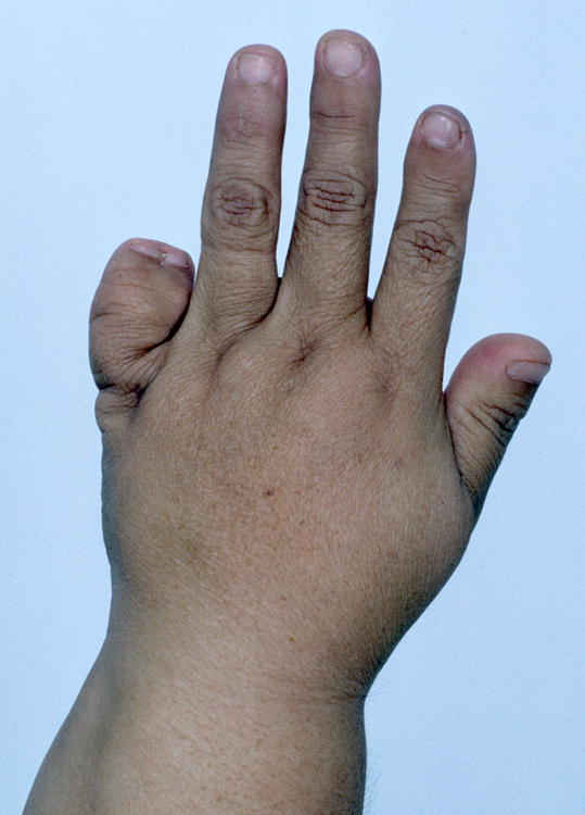
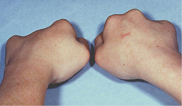
A. Short metacarpal, F5, left hand. Note that this patient also has a Postaxial polydactyly, partial. B. Short metacarpals, F34, left hand F4, right hand. Note that this patient’s hands are shown in dorsal view, with the fingers flexed, which can facilitate the recognition of this finding.
Comment: Short metacarpals can involve any of the metacarpal bones, and the affected ray should be specified. The assessment of isolated short metacarpal can be made by viewing the dorsum of the hand when clenched. Note that if metacarpals F2-5 are affected, the correct term is Short palm. The affected digits should be specified.
Metatarsal, short
Definition: Diminished length of a metatarsal, with resultant proximal displacement of the associated toe (Fig. 50). subjective
Fig. 50.
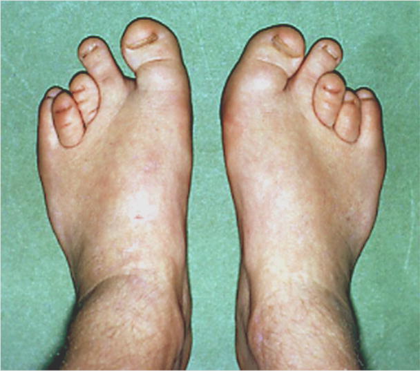
Short metatarsals, T3,4, bilateral.
Comment: This is a subjective assessment and one generally compares the position of the MTP joint to that of the contralateral digit or the putatively shortened ray in proportion to the other rays. The affected digits should be specified.
Metatarsus adductus
Definition: The metatarsals are deviated medially (tibially) (Fig. 51). subjective
Fig. 51. Metatarsus adductus, left.
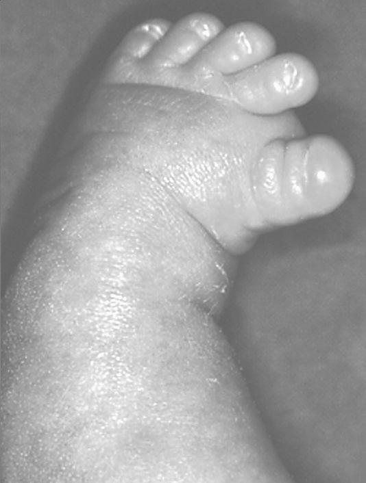
Note that this patient also has a Sandal gap.
Comment: This is considered subjective, as there is no objective threshold for the finding.
Oligodactyly: See Finger, absent; Toe, absent
Pachydactyly: See Finger, broad; Macrodactyly
Palm, broad
Definition: For children from birth to 4 years of age the palm width is more than 2 SD above the mean; for children from 4 to 16 years of age the palm width is above the 95th centile (Fig. 52). objective OR
Fig. 52. Broad palm, left, subjective.
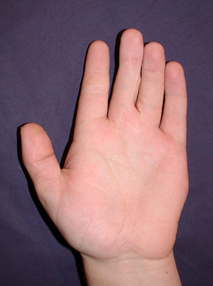
Note that this patient had normal palm length, so that this is not an example of a short palm with only an apparently broad palm.
The width of the palm appears disproportionately wide for the length. subjective
Comment: Hand width is measured across the palm at the level of the MCPJ (radial aspect of the second MCPJ to the ulnar aspect of the fifth MCPJ) [Hall et al., 2007]. Caution is advised with the subjective assessment as short metacarpals can mimic a broad palm. In persons with polydactyly that includes a supernumerary metacarpal, that should be separately coded and the measurement technique from Hall et al, [2007] would need to be modified to account for the supernumerary digit (i.e., with postaxial polydactyly, measure to the sixth MCPJ).
Synonym: Wide palm
Palm, long
Definition: For children from birth to 16 years of age the length of the palm is more than the 97th centile (Fig. 53). objective OR
Fig. 53. Long palms.
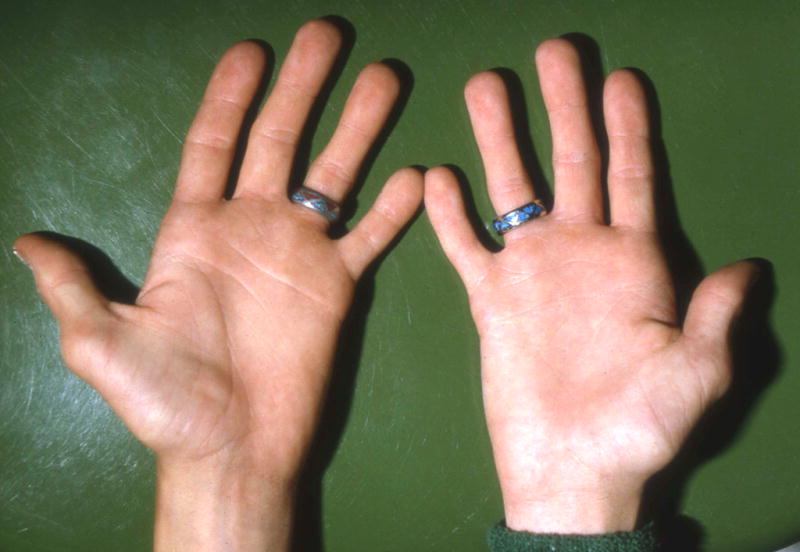
Note that this patient had normal palm width, so that this is not an example of a narrow palm with only an apparently broad palm. Note that this image illustrates a number of other features, which are not delineated. See also Figs. 63 and 85.
The length of the palm appears relatively long compared to the finger length or the limb length. subjective
Comment: Palm length standards are found in [Feingold and Bossert, 1974; Hall et al., 2007]. The palm is measured from the distal wrist flexion crease to the flexion crease of the F3 MCPJ. The term “long hand” should not be used as it is a bundled definition of two readily separable terms, Fingers, long (which, as noted above, can itself be a bundled term) and Palm, long.
Replaces: Long hand
Palm, narrow
Definition: For children from birth to 4 years of age, the palm width is more than 2 SD below the mean; for children from 4 to 16 years of age the palm width is below the 5th centile (Fig. 54). objective OR
Fig. 54. Narrow palms, subjective.
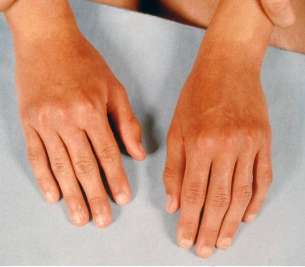
Note that this patient had normal palm width, so that this is not an example of a long palms with only apparently narrow palms.
The width of the palm appears disproportionately narrow for its length. subjective
Comment: Palm width is measured across the palm at the level of the MCPJ (radial aspect of the F2 MCPJ to the ulnar aspect of the F5 MCPJ). Norms are specified in [Hall et al., 2007]. Caution is advised for the subjective assessment as the breadth may be in the normal range with disproportionately increased length, which appears narrow. This finding may be associated with elongated/slender limbs in general, but that finding does not bear on the coding of this feature. Proximal narrowing may indicate small thenar or hypothenar eminences. This term replaces “narrow hands” as that term may leave the impression that it includes the thumb, which it does not.
Replaces: Narrow hand
Palm, short
Definition: For children from birth to 16 years of age the length of the palm is less than the 3rd centile (Fig. 55). objective OR
Fig. 55. Short palms, subjective.
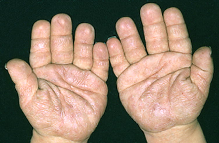
Note that this patient also has Short fingers. This patient does not warrant a finding of Small hands, because the fingers are of normal caliber. See also Fig. 14.
The length of the palm appears relatively small compared to the finger length or the limb length. subjective
Comment: Palm length is measured as described in [Feingold and Bossert, 1974; Hall et al., 2007]. This term is reserved for individuals with shortening of all four metacarpals 2–5. Individuals with fewer than four shortened metacarpals (in a eudactylous hand, the metacarpals of F2-5) should be coded as Metacarpal, short. See the entry for Hand, small for a discussion of this finding. “Short hand” should not be used as it is a bundle of two readily separable terms, Fingers, short (which, as noted above, can itself be a bundled term) and Palm, short.
Replaces: Short hand
Palm, wide: See Palm, broad
Pes cavus
Definition: The presence of an unusually high plantar arch (Fig. 56). subjective
Fig. 56. Pes cavus.
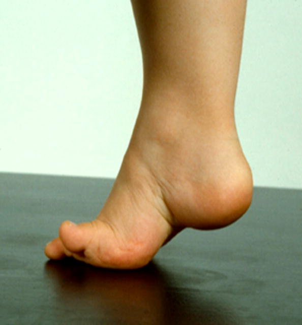
Note that this patient could also be said to have pes equinus, but that term is not included in the current terminology set.
Comment: This is a subjective definition for which we could identify no thresholds or standards. Examiners must rely on their experience to make this assessment.
Replaces: High arch
Pes planus
Definition: A foot where the arch is in contact with the ground or floor when the individual is standing (Fig. 57). objective OR
Fig. 57. Pes planus, left foot, objective.
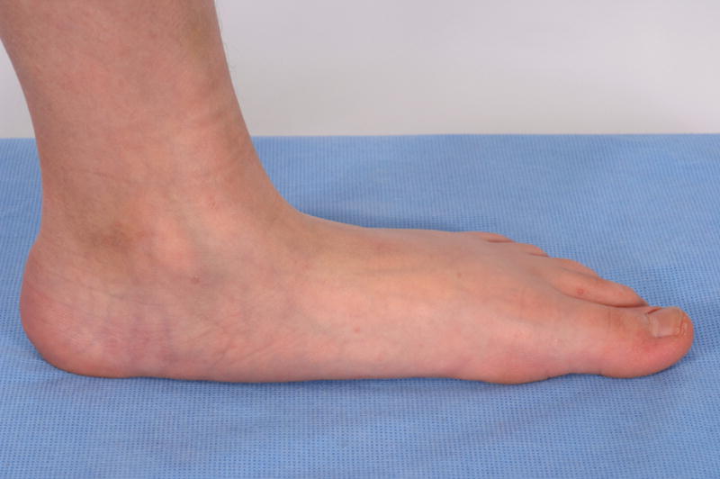
Note that this patient also has Hammertoes bilateral T2.
In a patient lying supine, a foot where the arch is in contact with the surface of a flat board pressed against the sole of the foot by the examiner with a pressure similar to that expected from weight bearing. objective OR
The height of the arch is reduced. subjective
Comment: The second definition was conceived by this working group based on a commonsense analysis of how one could generalize the first definition to a non-ambulatory individual. There are no data on this assessment and we invite feedback on its utility.
Replaces: Dropped arches; Fallen arches
Pollex: See various terms under the heading of “Thumb”
Polydactyly, anterior: See Foot, preaxial polydactyly of; Hand, preaxial polydactyly of
Polydactyly, central: See Polydactyly, mesoaxial
Polydactyly, insertional: See Polydactyly, mesoaxial
Polydactyly, intercalary: See Polydactyly, mesoaxial
Polydactyly, mesoaxial
Definition: The presence of a supernumerary finger or toe (not a thumb or hallux) involving the third or fourth metacarpal/tarsal with associated osseous syndactyly (Fig. 58). objective
Fig. 58.
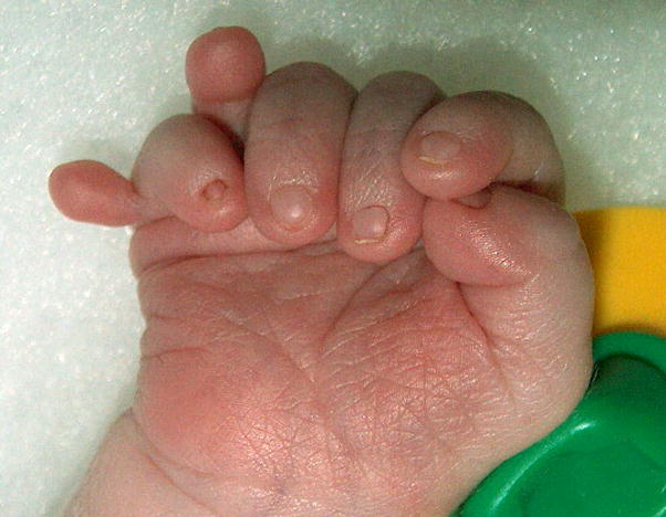
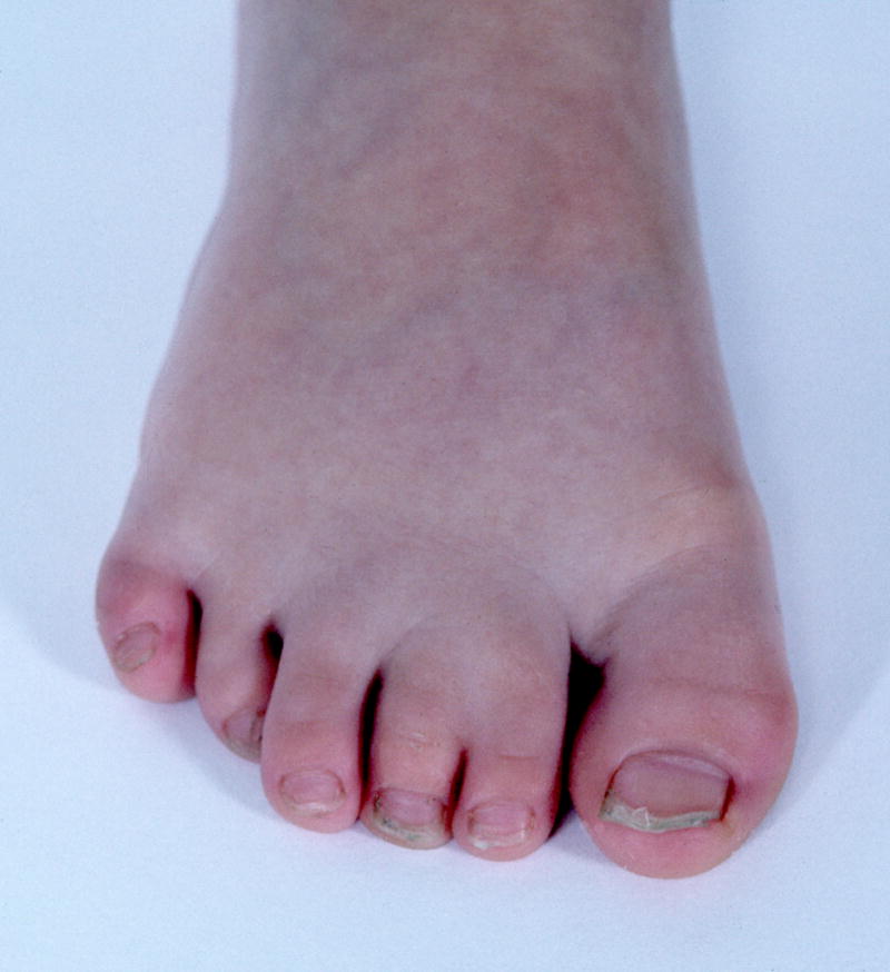
A. Mesoaxial polydactyly of the hand, right. Note that this patient has heptadactyly, manifesting as well Postaxial polydactyly, type B. The figure also shows Small nail, F6 and Overlapping fingers F56. See also Fig. 11A. B. Mesoaxial polydactyly of the right foot. This patient also has Cutaneous syndactyly of the toes, partial, T23.
Comment: To determine this finding in the hand without using a radiograph (see [Allanson et al, 2008b] this issue, for a discussion of the use of radiographs), the examiner grasps two adjacent metacarpals, each with a thumb and index finger on the dorsum and palmar aspect of the hand and attempts to independently maneuver the metacarpals. If they cannot be independently manipulated, osseous syndactyly is present [Biesecker, 2007]. Consensus could not be reached on the utility of the bundled term of Mesoaxial polydactyly, so it is specified in the terminology for the time being. It is arguably superior to describe the two separate terms (e.g., Osseous syndactyly of F3,4 (metacarpal) and Postaxial polydactyly of the hand, type A). The term “insertional polydactyly” may be less desirable because the word “insertion” implies mechanism, which is unlikely to be an insertion. The utility of this term should be reviewed over time.
Synonym: Central polydactyly; Intercalary polydactyly
Replaces: Insertional polydactyly
Polydactyly, mirror image
Definition: A hand or foot with more than five digits that has a recognizable A/P axis of symmetry. The axis can lie within a normally formed or partially duplicated digit resembling a middle finger, index finger, thumb, toe, or hallux. Alternatively, the axis can be in an interdigital space with a flanking pair of digits that resemble a middle finger, index finger, thumb, toe or hallux. The most lateral digits on each side of the hand typically resemble fifth fingers/toes (Fig. 59). subjective
Fig. 59. Mirror image polydactyly, right foot.
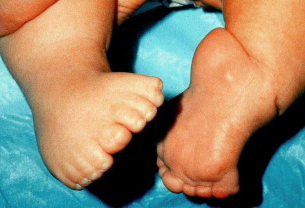
Note that this patient also has Cutaneous syndactyly of numerous digits, notable on the right foot.
Comment: This is considered to be a subjective assessment. Although polydactyly is obviously objective, the assessment of the A/P symmetry component is arguably subjective. It may be associated with a forearm that has two ulnae, but this is not required for the finding to be made.
Polydactyly, fibular: See Foot, postaxial polydactyly of
Polydactyly, posterior: See Foot, postaxial polydactyly of; Hand, postaxial polydactyly of
Polydactyly, radial: See Hand, preaxial polydactyly of
Posterior duplication: See Foot, postaxial polydactyly of; Hand, postaxial polydactyly of
Radial clubhand: See Hand, radial deviation of
Ray, absent
Definition: The absence of all phalanges of a digit and the associated metacarpal/metatarsal (Fig. 60). objective
Fig. 60.
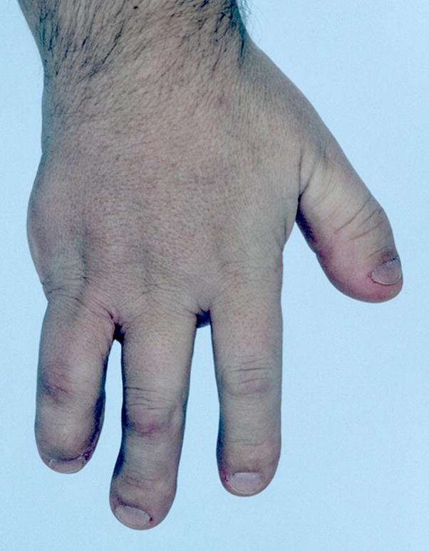
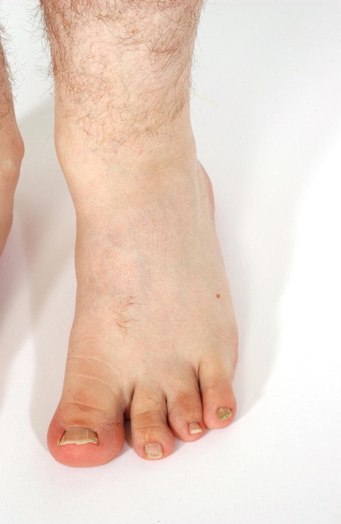
A. Absent ray, right hand. Note that this patient also has Short distal phalanges of the fingers and Short nails. B. Absent ray, left foot. Note that this patient also manifests a Broad hallux, which should be coded separately.
Comment: This descriptor requires, in addition to the absence of the phalanges, absence of the metacarpal or metatarsal. Compare this to Thumb, absent, Hallux, absent, Toes, absent, and Fingers, absent. In most cases, the absent metacarpal or metatarsal can be assessed by palpation, but in some cases radiographs may be useful. This definition excludes Hand, split and Foot, split. If a patient meets the definition of either of those terms, they should be used and Ray, absent should not.
Sandal gap
Definition: A widely spaced gap between the first toe (the great toe) and the second toe (Fig. 61). subjective
Fig. 61.
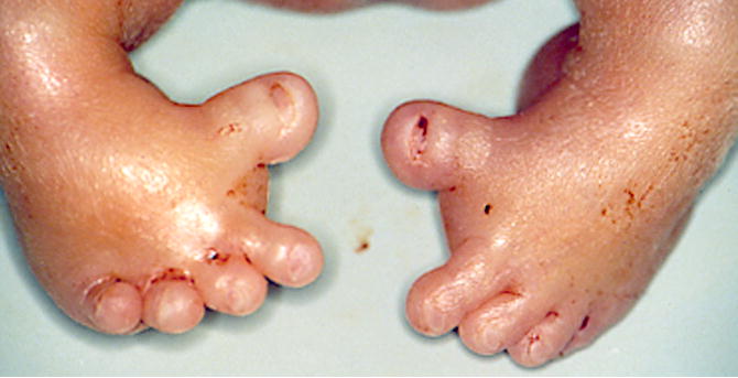
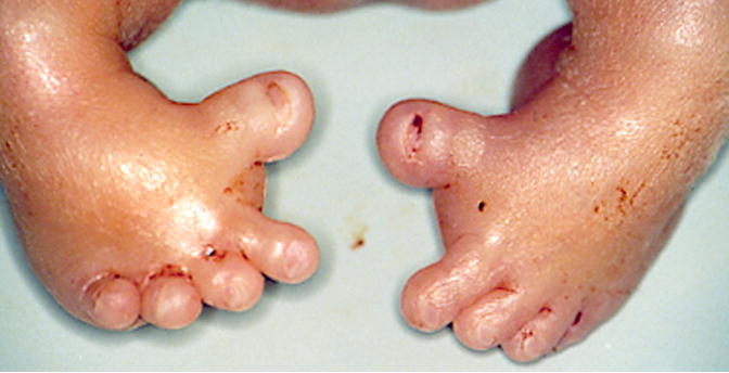
A. Sandal gap, right foot, subjective. B. Sandal gaps, objective. See also Figs. 48 and 51.
Comment: The term is a subjective one but should be used when the gap between the toes is as wide as the second toe is broad.
Sole, convex contour of
Definition: The contour of the foot in lateral profile has a convex shape (Fig. 62). subjective
Fig. 62. Convex contour of the sole right foot.
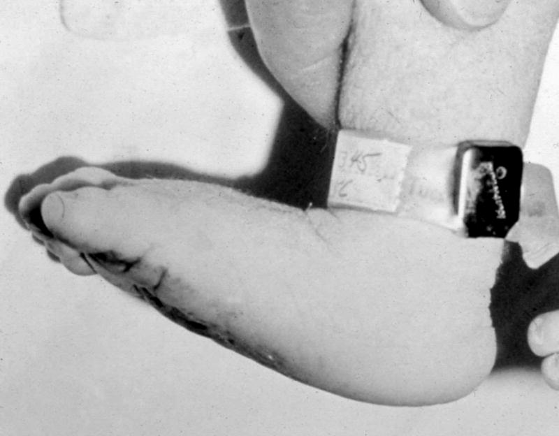
Note the heel in this patient is not sufficiently prominent to warrant the descriptor of Rocker bottom foot.
Comment: This term was established as the convex contour may occur without the prominent heel, which together comprise the bundled term Foot, rocker bottom.
Streblodactyly: See Camptodactyly
Syndactyly; See Fingers, cutaneous syndactyly of; Toes, cutaneous syndactyly of; Foot, osseous syndactyly of; Hand, osseous syndactyly of;
Thenar eminence, small
Definition: Reduced palmar soft tissue mass surrounding the base of the thumb (Fig. 63). subjective
Fig. 63. Thenar hypoplasia right hand.
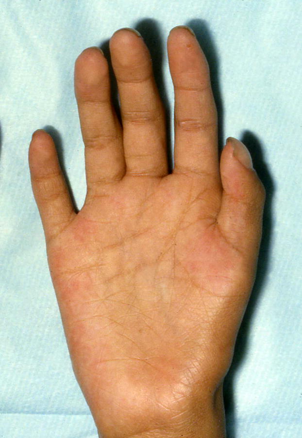
Note that this person also has Clubbing (visible in the thumb only). This patient also has Long palm, right hand, subjective.
Comment: The reduced soft tissue is typically abductor pollicis brevis and flexor pollicis brevis muscle bulk. Detection of this abnormality entails clinical judgment, especially in mild cases. The bulk of the muscle mass around the base of the thumb is diminished, and there may be a mild concavity over the volar aspect of the first metacarpal. When the deficiency is unilateral, comparison between the two hands will point up the often-subtle change in contour of the thenar muscles. If the degree of involvement is severe, the palm may taper in width proximally.
Replaces: Thenar hypoplasia
Terminal phalanx, short: See Finger, short distal phalanx of; Hallux, short distal phalanx of; Thumb, short distal phalanx of; Toe, short distal phalanx of
Thenar hypoplasia: See Thenar eminence, small
Thumb abducted: See Thumb, hitchhiker
Thumb, absent
Definition: The absence of both phalanges of a thumb and the associated soft tissues (Fig. 64). objective
Fig. 64.
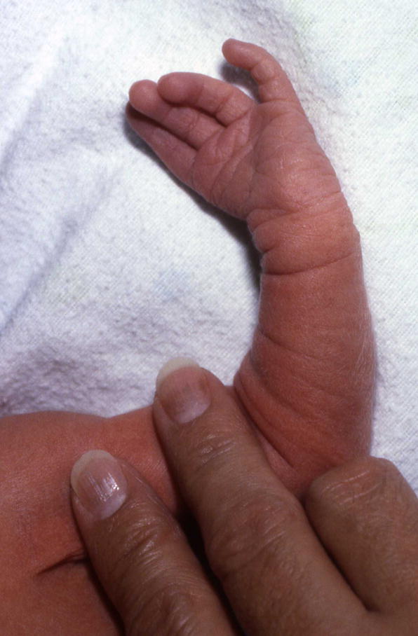
Absent thumb, left.
Comment: This descriptor does not require absence of the metacarpal. This definition excludes Partial absence of the thumb. Oligodactyly and hypodactyly have been replaced with more specific terms to allow the distinction of loss of a thumb from loss of F2-5. The term “oligodactyly” is replaced because oligodactyly refers to the digits that remain, and so “oligodactyly of the thumb” or “oligodactyly F1” is nonsensical.
Replaces: Hypodactyly; Oligodactyly
Thumb, adducted
Definition: In the resting position, the tip of the thumb is on, or near, the palm, close to the base of the fourth or fifth finger (Fig. 65). subjective
Fig. 65.
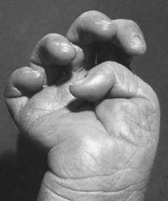
Thumb, adducted, right.
Comment: The thumb is both flexed and adducted. Lesser degrees of adduction than that specified here may warrant the use of this term, for example, when the tip of the thumb lies near the base of F2 or F3.
Thumb, broad
Definition: Increased thumb width without increased dorso-ventral dimension (Fig. 66). subjective
Fig. 66. Broad thumbs.
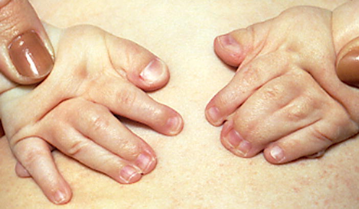
See also Fig. 9A.
Comment: There is substantial variability thumb width and it may be difficult to determine the threshold for this finding. As it is commonly bilateral, comparing to the contralateral digit will not be helpful. Some have suggested using a definition of thumb width measured at the IP joint of >1.5X the width of the index finger measured at the DIP joint, but no data to support this are available. Note that this term should not be used for thumbs that meet the definition for Macrodactyly. There is a standard for thumb width at birth of 9.8 +/− 0.5 mm (1 SD) (see [Saul et al., 1998] p 45), but this is not widely used and does not take into account gestational age, or babies who are small for gestational age. The assessment is difficult when the thumb is short.
Synonym: Broad pollex; Wide thumb
Thumb, digitalized: See Thumb, triphalangeal
Thumb, hitchhiker
Definition: With the hand relaxed and the thumb in the plane of the palm, the axis of the thumb forms an angle ≥ 90 degrees with the long axis of the hand (Fig. 67). objective
Fig. 67.
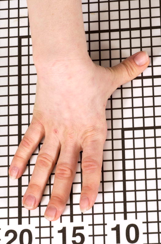
Hitchhiker thumb, right.
Replaces: Abducted thumb
Thumb, partial absence of
Definition: The absence of a phalangeal segment of a thumb (Fig. 68). objective
Fig. 68. Thumb, partial absence of, right.
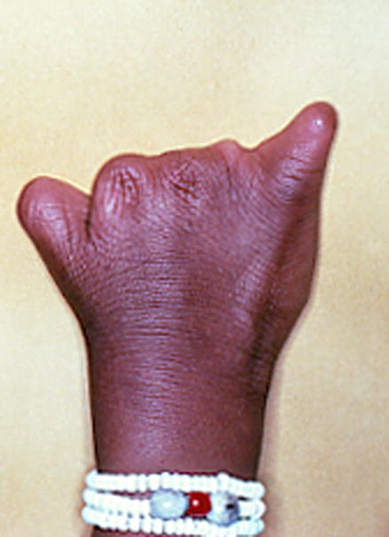
Note that this patient also has Absent fingers, F2-4, and Absent finger F5, partial.
Comment: The part that is absent may be specified. The “distal” modifier specifies the loss of the distal phalanx; clinically this is defined by the absence of the nail. The “proximal” modifier specifies the loss of the proximal phalanx. This finding is distinct from Short thumb.
Replaces: Hypophalangy
Thumb, proximal placement of
Definition: Thumb placement index >0.55 (Fig. 69). Objective OR
Fig. 69. Thumb, proximal placement of, right hand, subjective.
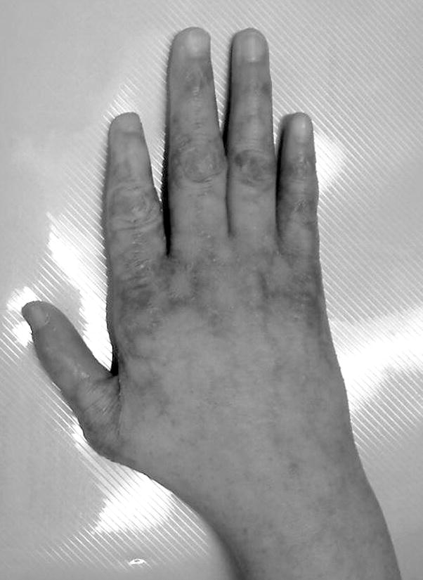
Note that this patient also has Short finger, F2.
The base of the thumb appears closer to the wrist than is typical. Subjective.
Comment: The technique for the thumb placement index is described in detail [Malina et al., 1973; Hall et al., 2007]. Briefly, the thumb placement index is the distance from the proximal crease of the index finger to the angle of the first interdigital space divided by the distance from the proximal crease of the index finger to the wrist flexion crease at the base of the thumb. This term should not be used with Preaxial polydactyly.
Thumb, triphalangeal
Definition: A thumb with three phalanges in a single, proximo-distal axis (Fig. 70). objective
Fig. 70.
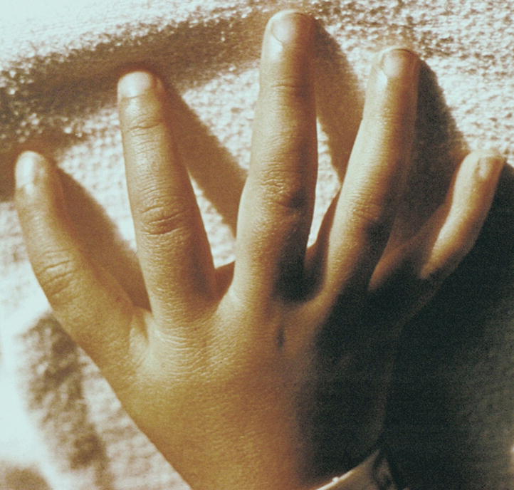
Triphalangeal thumb, right.
Comment: The requirement for a single PD axis relates to the issue that partial forms of Preaxial polydactyly may comprise a partially duplicated thumb with two distal phalanges and a single proximal phalanx. That finding is instead coded as a mild form of thumb polydactyly. Note that this finding can be readily assessed by examination and/or physical manipulation of the thumb.
Replaces: Digitalized thumb; Finger-like thumb
Tibial polydactyly: See Foot, preaxial polydactyly of
Toe, absent
Definition: The absence of all phalanges of a non-hallux digit of the foot and the associated soft tissues (Fig. 71). objective
Fig. 71.
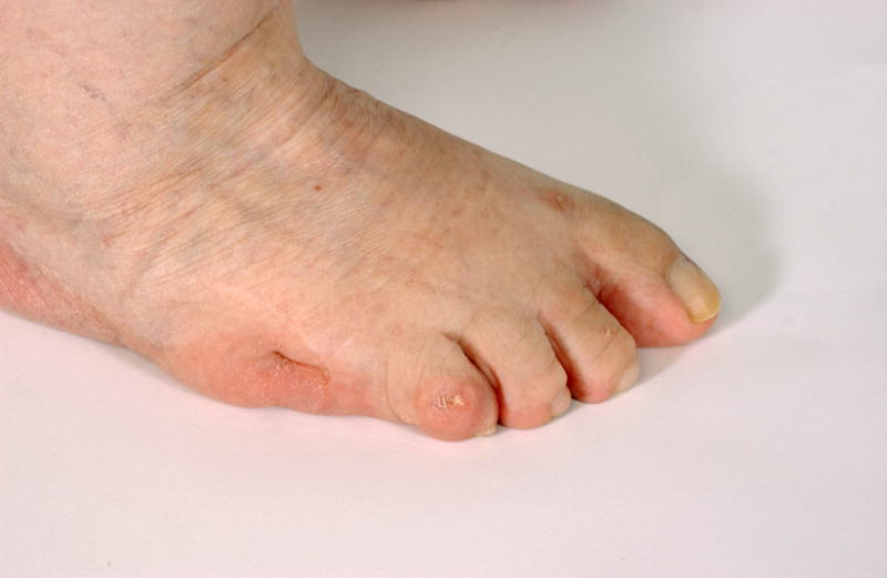
Absent toe, right.
Comment: Note that this descriptor does NOT require absence of the metatarsal. The affected digits should be specified, although this may be difficult. In the latter case, the specified term should be “Absent toe, right foot” (with the identity of the missing toe not specified). This definition excludes Partial absence of the toe. This absent toe definition excludes Absent hallux because it is usually obvious if the missing digit is a hallux. It may be difficult to distinguish an absent toe from two digits with an extreme degree of osseous and cutaneous syndactyly. If all digits are missing, the term Adactyly should be used. The term Oligodactyly may be used as a synonym, but only in situations where the missing digit is not specified. For example, to state that a patient has Oligodactyly F5 is awkward because the oligodactyly refers to the digits that are present and the F5 refers to the digit that is absent, which is awkward and could become confusing with higher order deficiencies.
Replaces: Hypodactyly
Synonym: Oligodactyly
Toe, broad
Definition: Visible increase in width of the non-hallux digit without an increase in the dorso-ventral dimension (Fig. 72). subjective
Fig. 72. Toe, broad, left, T1.
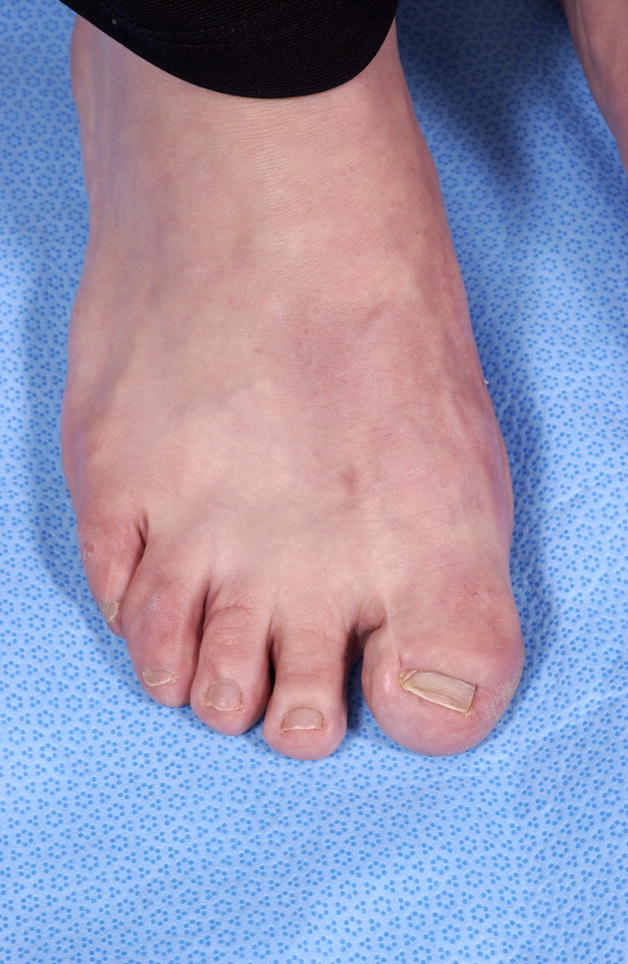
Comment: Note that the girth may be increased in a broad toe, but this must be distinguished from Macrodactyly because in Macrodactyly the length is increased as well. The affected digit should be specified. Note that this assessment may be difficult when the toes are short. This term is not used for the first digit, see Broad hallux. If all five digits are broad, both terms should be used for that patient.
Toes, cutaneous syndactyly of
Definition: A soft tissue continuity in the A/P axis between adjacent foot digits that involves at least half of the P/D length of one of the two involved digits (Fig. 73). objective OR
Fig. 73.
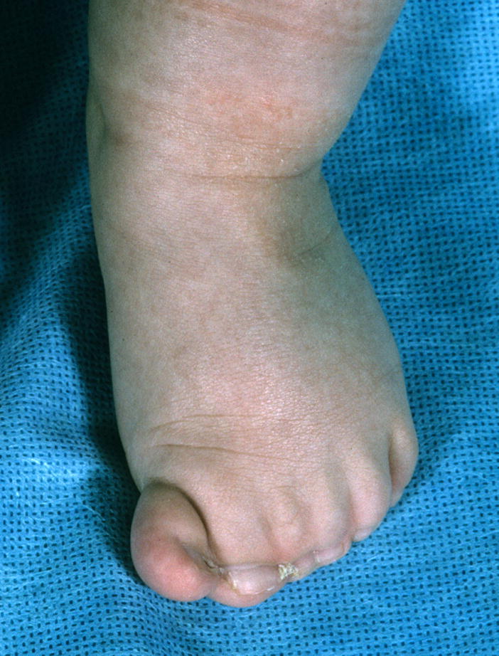
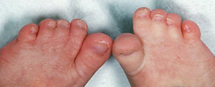
A. Cutaneous syndactyly of the toes, complete, TT1-6, left, objective. See also Figs. 58B, 59, 78, 92, 94, and 101. Note that his patient also has Preaxial polydactyly of the foot, partial, left. B. Cutaneous syndactyly of the toes, partial, T2-4, bilateral, objective. As specified in the definitions, this finding is objective if the syndactyly extends more than half the proximo-distal length of the digits.
A soft tissue continuity in the A/P axis between two digits of the foot that does not meet the prior objective criteria. subjective
Comment: The digits (or part of) are joined together by soft tissue that is not normally present between the digits at that point in the P/D axis. The definition of toe syndactyly is subtly different from that of the hand. The definition used for the hands was thought to be difficult with toes, as the phalangeal lengths are small and an assessment of the degree of syndactyly is impractical. A modifier of “complete” may be used if the syndactyly extends to the distal end of the nail bed. The affected digits should be specified.
Replaces: Syndactyly (without specification); Zygodactyly
Toe, great; all terms referring to the first toe are listed under Hallux or Toe
Toe, hypoplastic: See Toe, small
Toe, long
Definition: Digits that appear disproportionately long compared to the foot (Fig. 74). subjective
Fig. 74. Long toes, left foot.
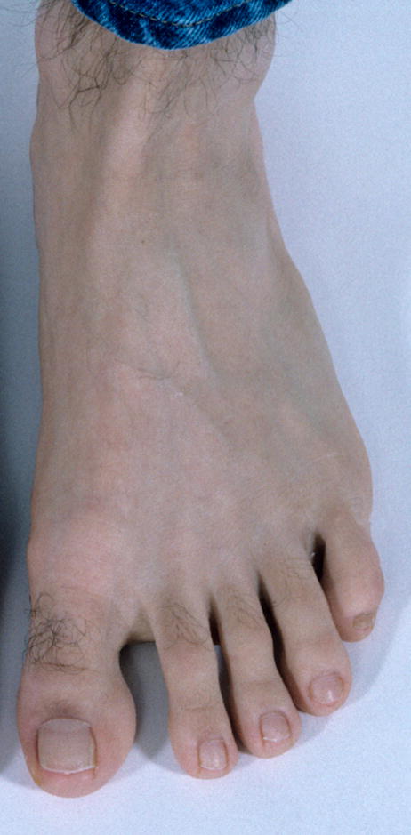
Comment: This finding must be distinguished from digits that are thin but of normal length and that of a short mid and hind foot with normal digit lengths. The affected digits should be specified. The term arachnodactyly should be discontinued as it may be considered disparaging to liken a patient’s digits to that of a spider. If only a subset of the digits of a limb is lengthened, the affected digits should be specified.
Replaces: Arachnodactyly
Toe, narrow: See Toe, slender
Toes, overlapping
Definition: Describes a foot digit resting on the dorsal surface of an adjacent digit when the foot is at rest (Fig. 75). objective
Fig. 75.
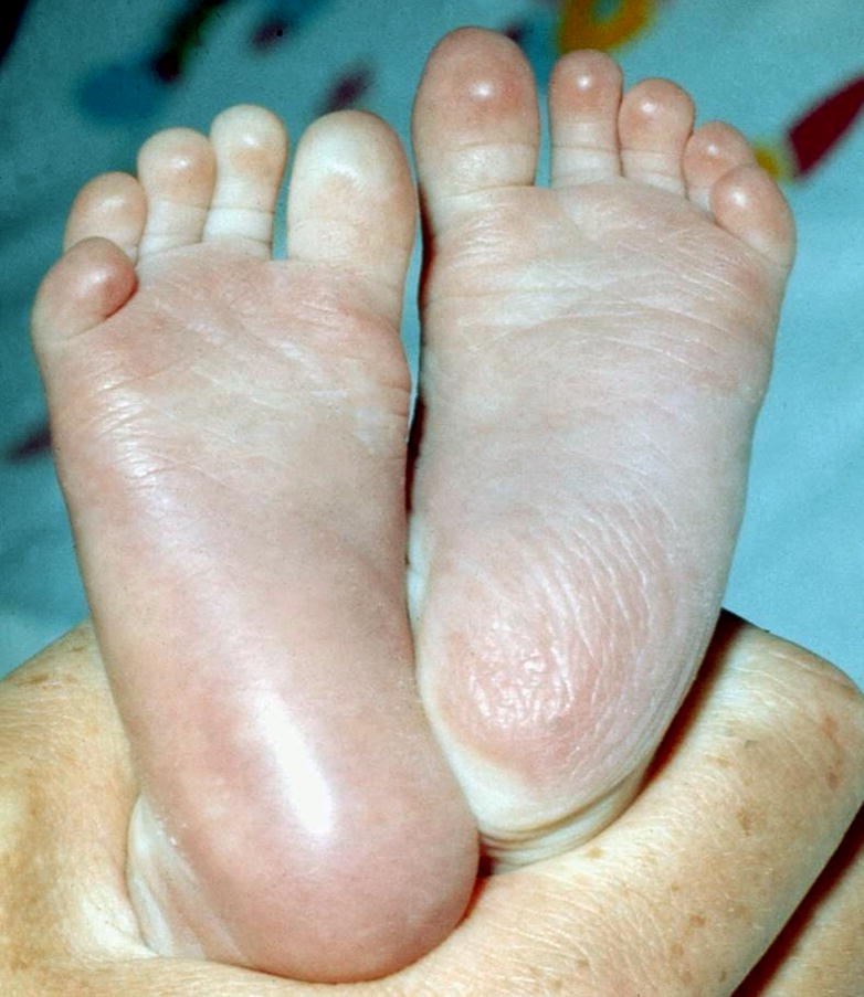
Overlapping toes T45, bilateral.
Comment: This descriptor is ordered depending on which toes are involved. The overriding toe is labeled, as specified in the introduction (item 3): e.g., T3,4. The ordering of the numbers specifies which toe is dorsal, i.e., with dorsum of the foot facing upward the toe on top is/are recorded first separated by a comma from the digit that is/are overlapped. Toes that are laterally deviated, but do not rest on top of adjacent toes should be coded as Clinodactyly.
Toe, partial absence of
Definition: The absence of a phalangeal segment of a toe or hallux (Fig. 76). objective
Fig. 76. Toes, partial absence of, right foot, T2,4.
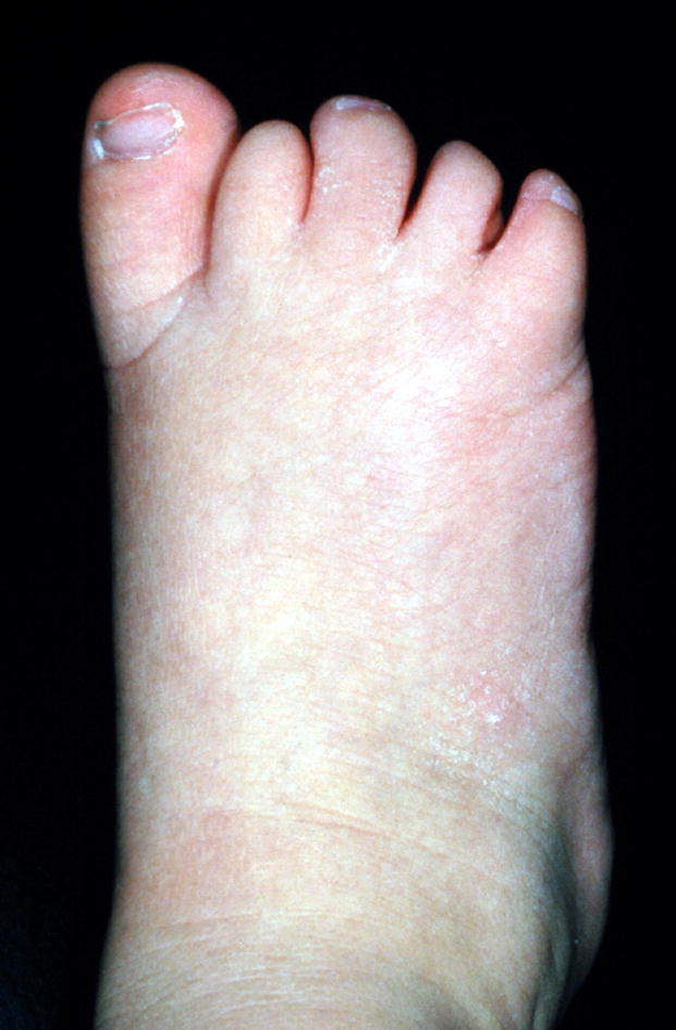
Note that this is the same image as in Fig. 82, Toe, tapered.
Comment: The part that is absent may be specified. The “distal” modifier specifies the loss of the distal phalanx; clinically this is defined by the absence of the nail. The “proximal” modifier specifies the loss of the proximal or middle phalanx with the nail still present. It may be difficult to know which phalanx is absent without X-rays and even then, the missing bone may not be identified (note no attempt is made to distinguish missing middle from proximal phalanges). In this situation the location adjective should be removed. Note that this finding is distinct from Short toes, (which are reduced in length but have the normal number of phalangeal segments). Partial absence of the hallux is an alternative term that may be used for the first toe.
Replaces: Hypophalangy
Toe, short
Definition: Digits that appear disproportionately short compared to the foot (Fig. 77). subjective
Fig. 77. Short toes, T1-5, right foot.
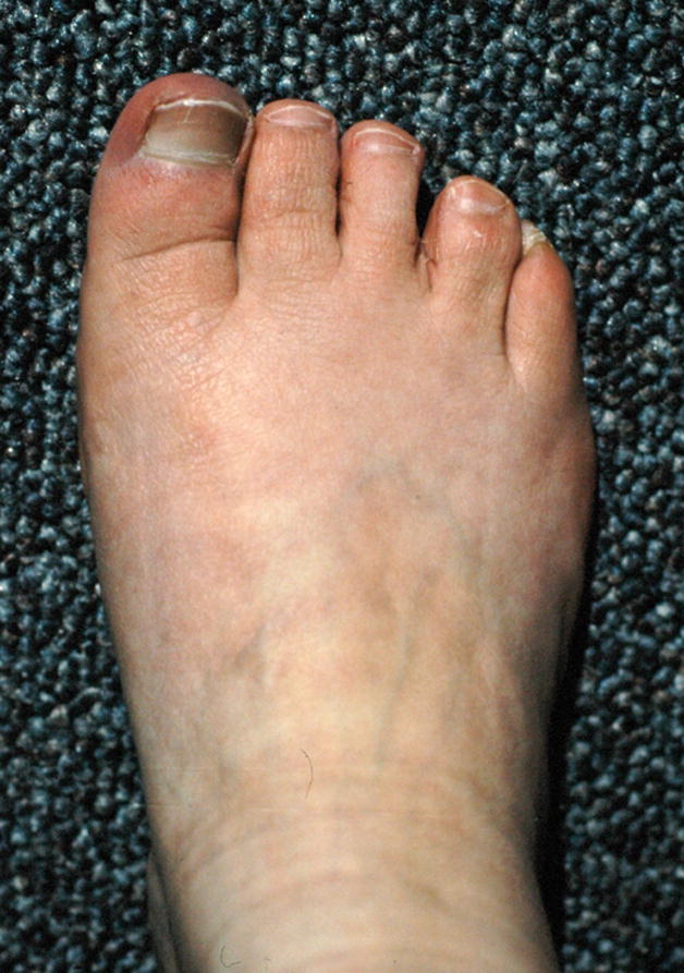
Note that this patient also has Short distal phalanges of toes T2,3.
Comment: This finding must be distinguished from digits that are of increased girth but of normal length and that of a long mid- and hind foot with normal digit lengths. The affected digits should be specified as described in the introductory comments. Note that we designate brachydactyly as a synonym, but this use of the term is distinct from the use of the same word in Bell’s classification (see Short fingers).
Synonym: Brachydactyly of the foot
Toe, short distal phalanx of
Definition: Short distance from the end of the toe to the most distal interphalangeal crease or DIPJ flexion point (Fig. 78). subjective
Fig. 78. Short distal phalanges of toes T2-4, right foot.
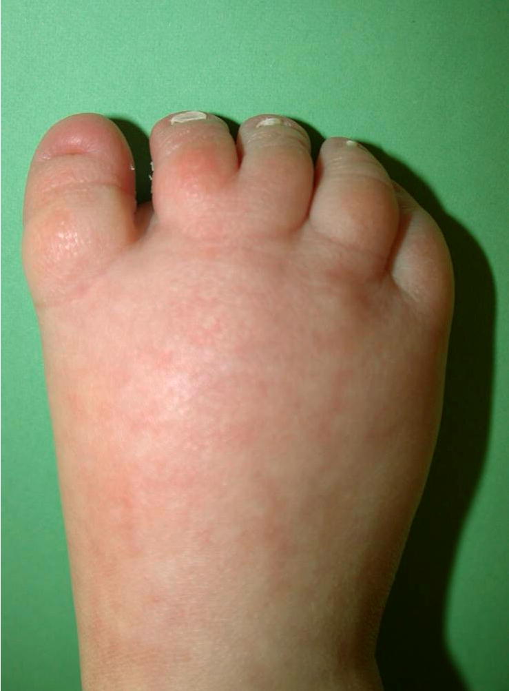
Note that this patient also has Cutaneous syndactyly of toes T23, subjective. See also Fig. 77.
Comment: This term differs from Partial absence of the toe because in that term the phalanx must be missing, whereas here it may be small, but present. Relative shortening of the distal phalanges of the toes can be harder to assess than in the fingers, as they are normally quite short. Distal phalangeal lengths can be assessed subjectively by comparing that digit segment to the rest of the digit, to other normal digits in that patient, or to typical patients of that age or build. Although flexion creases can be useful in the hand (see Short distal phalanx of the finger,), this may not be practical in the foot. Alternatively, one may be able to palpate the joint.
Synonym: Brachytelephalangy
Replaces: Short terminal phalanx of the hallux
Toe, slender
Definition: Digits are disproportionately narrow (reduced girth) for the hand/foot size or build of the individual (Fig. 79). subjective
Fig. 79. Slender toes, T2-5, right foot.
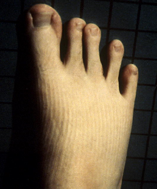
Note that this patient also has Long toes T1-5.
Comment: The affected digits should be specified. The assessment of this finding is difficult when the digits are long. The bundled and pejorative term “arachnodactyly” is replaced by the separate descriptors Long toe and Slender toe.
Synonym: Narrow digit; Narrow toe
Replaces: Thin digits; Arachnodactyly
Toe, small
Definition: Significant reduction in both length and girth of the toe compared to the contralateral toe, or alternatively, compared to a typical toe size for an age-matched individual (Fig. 80). subjective
Fig. 80. Small toe, T1, left foot.
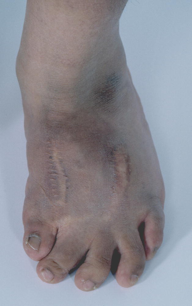
See also Fig. 81.
Comment: This is an acknowledged bundled term. There are no standards for this finding, clinical judgment must be used. The affected toes should be numbered.
Toes, splayed
Definition: Divergence of digits along the A/P axis (in the plane of the sole) (Fig. 81). subjective
Fig. 81. Splayed toes, T23 right foot, T12, left foot.
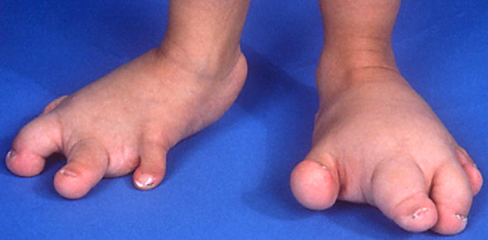
Note that this finding differs from Toes, widely spaced, because in splayed toes the toes have a divergent axis of orientation. This patient also has Small toe, T1, right and Macrodactyly of T2,3, right, and T1,2, left.
Comment: This may be associated with Macrodactyly but this should be recorded separately. The affected digits should be specified.
Synonym: Spreading of the digits
Toe, tapered
Definition: The gradual reduction in girth of the digit from proximal to distal (Fig. 82). subjective
Fig. 82. Toe, tapered, T5.
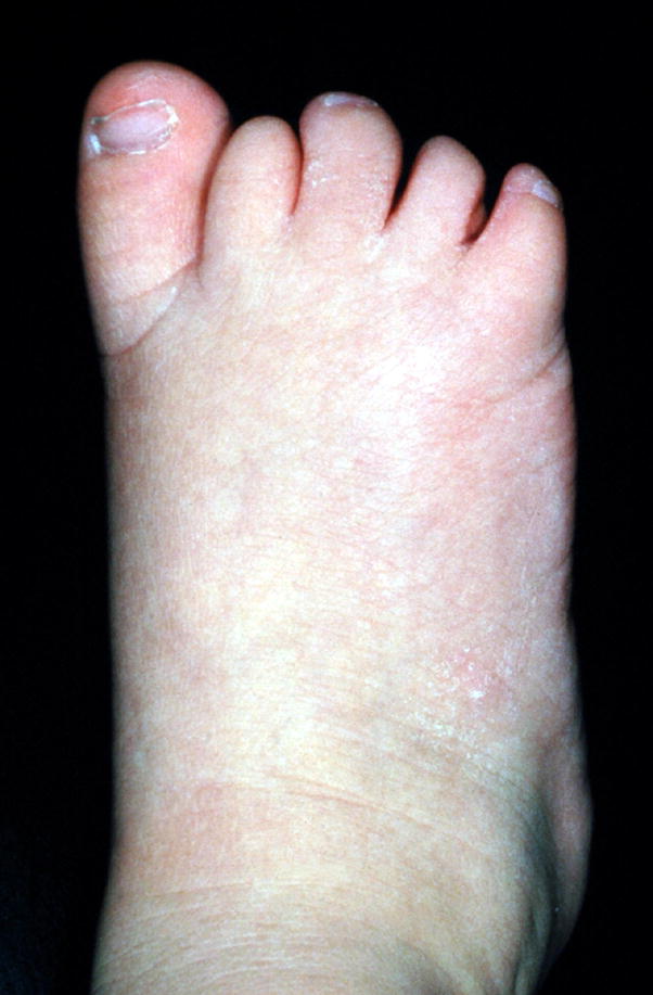
Note that this is the same image as in Fig. 76, Toe, partial absence.
Comment: If the digits are not uniformly affected, they should be specified. If not specified, it refers to all the digits of the foot.
Toe, thick: See Toe, broad
Toe, thin: See Toe, slender
Toe, wide: See Toe, broad
Toes, widely spaced
Definition: An overall widening of the spaces between the digits (Fig. 83). subjective
Fig. 83. Toes, widely spaced, T1,2, T4,5.
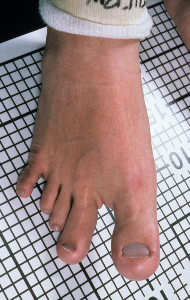
Note that this patient also has Macrodactyly of T1,2.
Comment: This description is based on the width of the gap between the toes. It is usually used when the width of the toes remains normal rather than to describe a situation where the toes are thin or narrow. This term should not be used for the situation where the finding is limited to a gap between T1,2 (see Sandal gap).
Ulnar clubhand: See Hand, ulnar deviation of
Ulnar polydactyly: See Hand, postaxial polydactyly of
Zygodactyly: See Fingers, cutaneous syndactyly of; Toes, cutaneous syndactyly of
CREASES
See Fig. 84 for a diagram of the three major palmar creases. One end of the distal transverse crease is on the radial side of the hand proximal to the base of the index finger or the second interdigital space and extends toward the ulnar side of the palm. One end of the proximal transverse crease begins on the radial (anterior) side of the palm in the first interdigital space and extends across the palm towards, but does not typically reach, the ulnar side of the palm. One end of the thenar crease is typically coincident with the radial part of the proximal transverse crease and extends proximally toward the wrist.
Fig. 84.
Normal palmar creases. See text for explanatory material.
Crease, simian: See Palmar crease, single transverse
Palmar crease, absent
Definition: The absence of a major crease of the palm (distal transverse crease, proximal transverse crease, or thenar crease) (Fig. 85). subjective
Fig. 85. Palmar creases, absent, left hand.
Note that this patient also has Long palm, subjective, left and Single transverse crease, right hand.
Comment: This term is not used to describe the “missing” crease in the case of a patient with a single transverse palmar crease.
Palmar crease, bridged
Definition: A crease that connects the proximal and distal transverse palmar creases (Fig. 86). objective
Fig. 86.
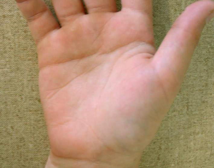
Bridged palmar crease, right hand.
Comment: The crease that connects the two transverse creases should itself be more in the transverse (antero-posterior) than longitudinal (proximo-distal) orientation.
Synonym: Transitional palmar crease
Palmar creases, decreased
Definition: Poorly defined or shallow palmar creases (Fig. 87). subjective
Fig. 87.
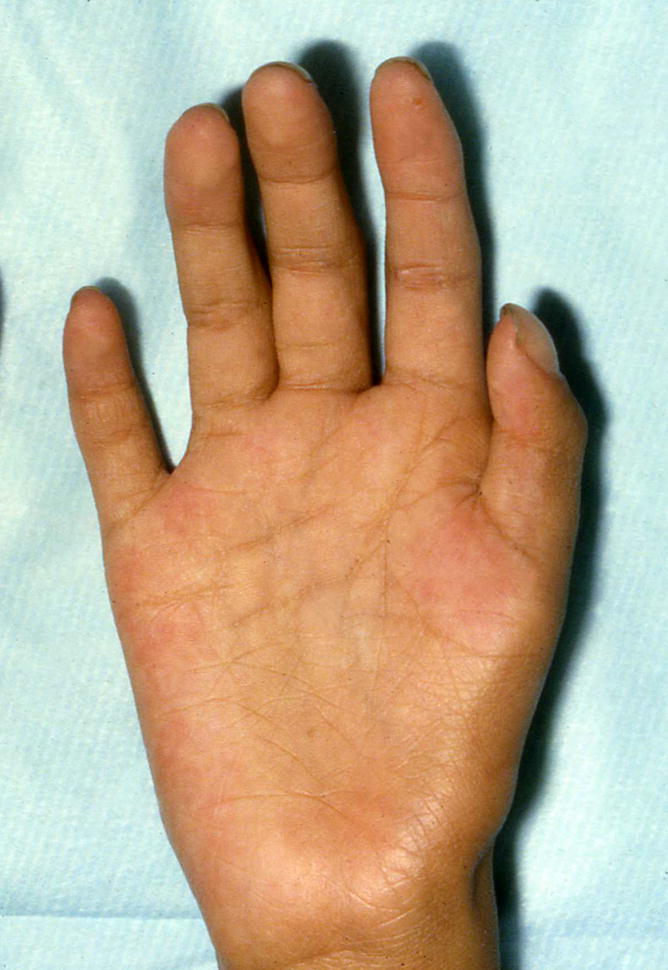
Palmar creases, decreased, right hand.
Comment: This is a completely subjective term, requiring an experienced observer to distinguish this finding from common variation in creases. It refers to an overall diminution of the creases, not to diminution of a subset of the creases.
Replaces: Smooth palms
Palmar crease, deep
Definition: Excessively deep creases of the palm (Fig. 88). subjective
Fig. 88. Palmar creases, deep.
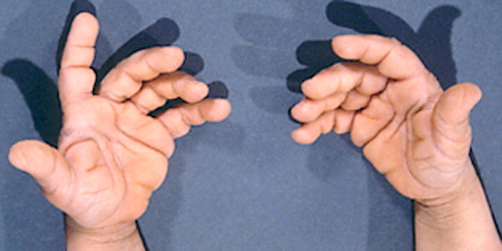
See also Fig. 17.
Comment: Like the term Decreased palmar creases, this assessment requires an experienced observer to distinguish this from common crease variation. One view is that deep creases are those in which lint could still get stuck, even if the palm is fully opened.
Palmar crease, single transverse
Definition: The distal and proximal transverse palmar creases are merged into a single transverse palmar crease (Fig. 89). objective
Fig. 89. Single transverse palmar crease, right hand.
Comment: The term “five finger crease” for the proximal transverse crease is sourced from [Hall et al., 2007]. The subtypes of single transverse palmar crease are not recognized here [Hook et al., 1974]. Replaces the term “Simian crease” as that term is disparaging and less descriptive.
Replaces: Simian crease
Palmar crease, transitional: See Palmar crease, bridged
Palms, smooth: See Palmar creases, decreased
Plantar crease, deep longitudinal
Definition: Narrow, paramedian longitudinal depressions in the plantar skin of the forefoot (Fig. 90). subjective
Fig. 90.
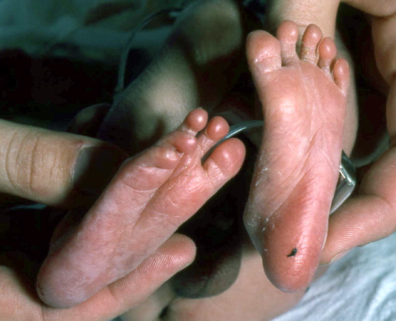
Deep longitudinal plantar crease, right foot.
Comment: As described for a number of other terms, there are no thresholds or guides for the clinician to assess this finding. Instead, the clinician must rely on experience.
Sydney crease
Definition: Extension of the proximal transverse crease (five finger crease) to the ulnar edge of the palm (Fig. 91). objective
Fig. 91.
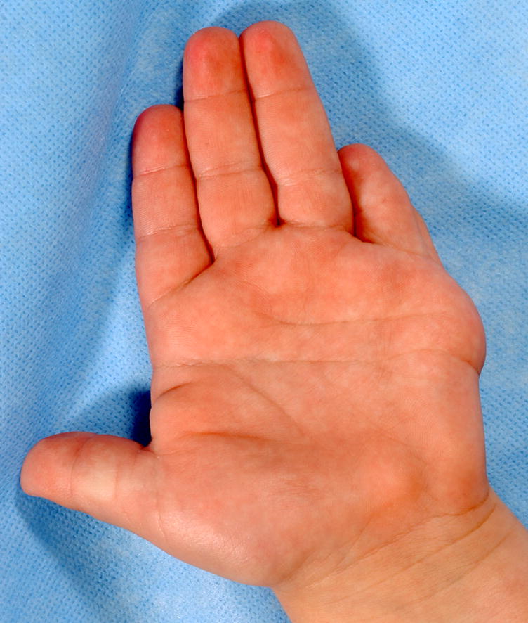
Sydney crease, left hand.
Comment: The proximal transverse (five finger) crease starts on the radial side of the hand near the base of the index finger and extends toward the ulnar side of the palm, but does not reach the ulnar side. In this finding, the crease extends completely to the ulnar margin of the palm.
NAILS
Brachyonychia: See Short nail
Koilonychia: See Nail, concave
Micronychia: See Nail, small
Nail, bifid
Definition: A digit with two nails, with at least some soft tissue between them (Fig. 92). subjective
Fig. 92. Nail, bifid, T1, left foot.
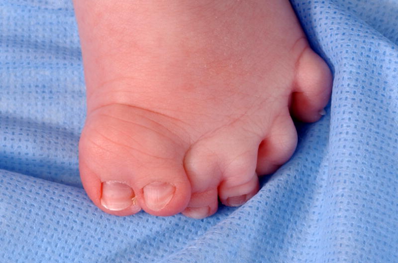
See also Fig. 99. Note that this patient also has a Broad toe, T1 and Cutaneous syndactyly, partial, T23.
Comment: See the Preaxial polydactyly definition for a related point on bifid nails. The affected digits should be specified. If the patient has a partially duplicated digit with two completely separate nails, this term should not be used.
Nail, concave
Definition: The natural longitudinal (P/D) convex arch is not present or is inverted (Fig. 93). subjective
Fig. 93. Nail, concave, left hand, F4.
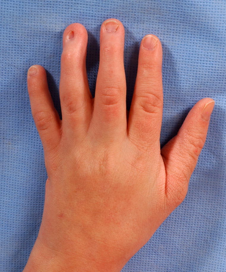
See also Fig. 103. Note that this image also shows Tapered fingers, left, F23, and Broad fingertip, left, F4.
Comment: This often results in a saucer- or spoon-shaped nail and the free edge of the nail is typically everted. The affected digits should be specified. Note that the bundled term koilonychia is an abnormal shape of the fingernail where the nail has raised ridges and is thin and concave.
Replaces: Koilonychia; Spoon shaped nails
Nails, dystrophic: The term “dystrophic” is no longer accepted as a descriptor for nails with an unusual morphology. The specific characteristic should be described.
Nails, fused
Definition: A nail plate that has a longitudinal separation with partially separated nails, each with a separate lateral radius of curvature (Fig. 94). subjective
Fig. 94. Nail, fused, T1, right foot.
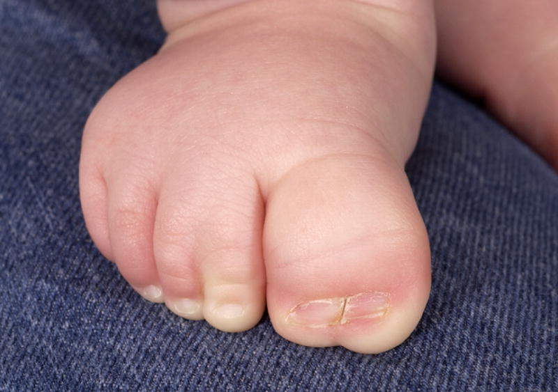
See also Fig. 9A. This patient also has a Broad toe, left, T1 and Cutaneous syndactyly, partial, left, T23.
Comment: The use of the word “fused” is not meant to imply that pathogenetically these nails were separate and merged. This is distinct from a split or cleaved nail, where the two parts of the nails share the same radius of curvature. The involved digits should be specified. It may be associated with underlying syndactylous digits, but these are coded separately.
Nail, hypoplastic: See Nail, small
Nail, hyperconvex
Definition: When viewed on end (with the digit tip pointing toward the examiner’s eye) the curve of the nail forms a tighter curve of convexity (Fig. 95). subjective
Fig. 95. Nails, Hyperconvex, left, F3,4.
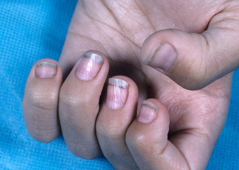
See also Fig. 102.
Comment: No objective standards were identified for this finding. Another way to describe this finding is to say that the observed curve has a smaller radius than does the typical nail. The affected digits should be specified.
Nail narrow
Definition: Decreased width of nail (Fig. 96). subjective
Fig. 96. Nail, narrow, T2, left foot.
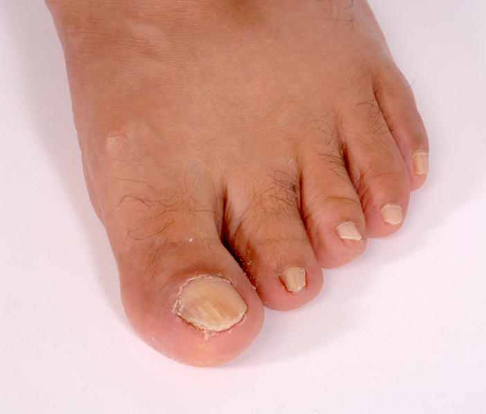
Note that the length of this nail is normal, so it is coded as narrow, not small.
Comment: Standards for newborns were identified [Seaborg and Bodurtha, 1989] but none were identified for older persons. Therefore, for newborns, this could be objective, but this is rarely measured. The affected digits should be specified.
Nail pits
Definition: Small (typically about 1 mm or less in size) depressions on the dorsal nail surface (Fig. 97). subjective
Fig. 97.
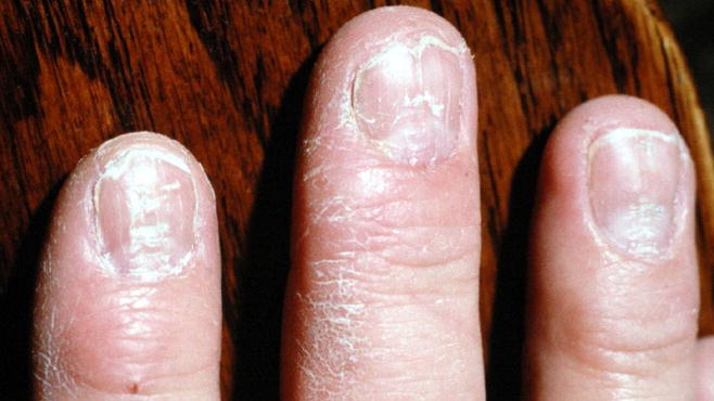
Nails, pitted, F2-4, right hand.
Comment: The affected digits should be specified.
Nail, ridged
Definition: Longitudinal, linear prominences in the nail plate (Fig. 98). subjective
Fig. 98.
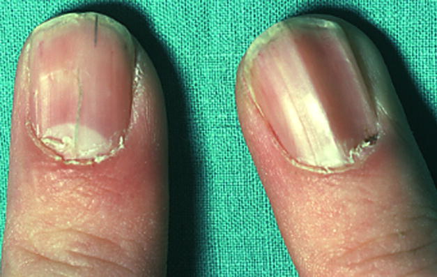
Nails, ridged, F1, bilateral.
Comment: There may be only one, or several ridges. The affected digits should be specified.
Nail, short
Definition: Decreased length of nail (Fig. 99). subjective
Fig. 99. Nails, short, F1,5, left hand.
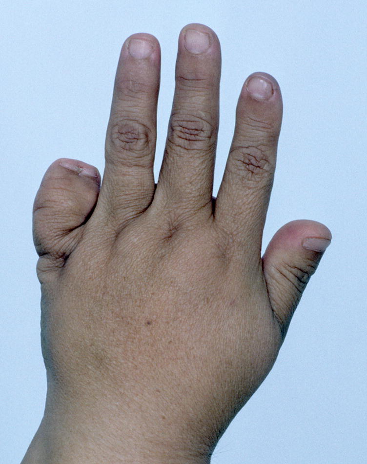
See also Fig. 15 and 60A. Note that this patient also has Bifid nail, F5 left hand; Broad finger, F5, left hand; and Short finger, left hand, F1.
Comment: Use this designation when the length is reduced but the width is normal. Standards for newborns were identified [Seaborg and Bodurtha, 1989] but none were identified for older persons. Therefore, for newborns, this could be objective, but this is rarely measured. The affected digits should be specified.
Synonym: Brachyonychia
Nail, small
Definition: A nail that is diminished in length and width (Fig. 100). subjective
Fig. 100.
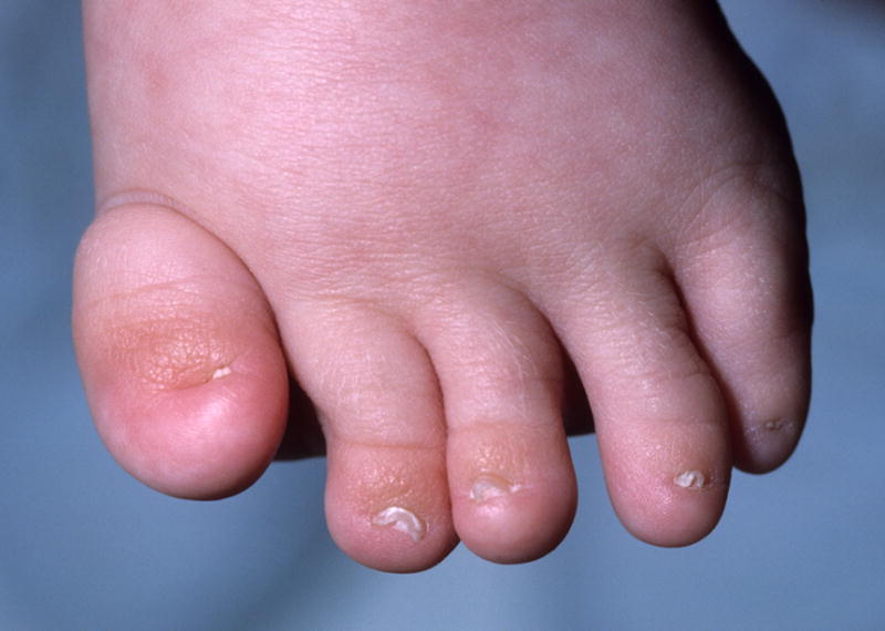
Comment: This is a bundled term but is retained because of its common usage. This term may be used when the width and length are reduced, although it may be preferable to code the patient as having both Short nail and Narrow nail. Standards for newborns were identified [Seaborg and Bodurtha, 1989] but none were identified for older persons. Therefore, for newborns, this could be objective, but this is rarely measured. The affected digits should be specified.
Replaces: Hypoplastic nails
Synonym: Micronychia
Nail, split
Definition: A nail plate that has a longitudinal separation and the two sections of the nail share the same lateral radius of curvature (Fig. 101). subjective
Fig. 101.
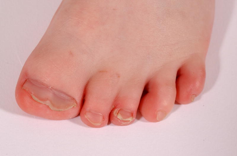
Nail, split, T1, left foot. Note that this image also shows a Broad toe, T1, left foot and Cutaneous syndactyly, partial, T23, left foot.
Comment: This is distinct from Fused nail, where the two parts of the nail have a separate radius of curvature. The affected digits should be specified as described in the introductory comments.
Nails, spoon shaped: See Nail, concave
Nail, thick
Definition: Nail that appears thick when viewed on end (Fig. 102). subjective
Fig. 102.
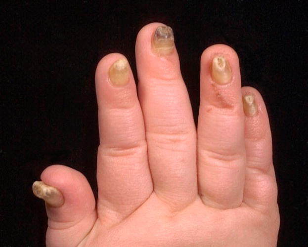
Nails, thick, right hand, F1-5. Note that this image also shows Hyperconvex nails.
Comment: No objective standard for nail thickness could be identified although an unsupported claim suggests that nails are 0.5 mm in females and 0.6 mm in males (http://www.emedicine.com/orthoped/topic421.htm). There is a build up of keratin causing the nail plate to lift away from the nail bed. The thickened nail plate is usually very hard.
Replaces: Onychogryposis; Pachyonychia
Nail, thin
Definition: Nail that appears thin when viewed on end (Fig. 103). subjective
Fig. 103.
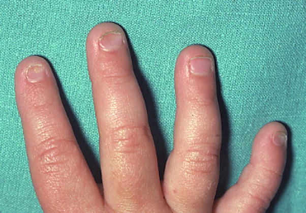
Nails, thin, F2,5, right hand. This image also shows Concave nail, F2, left hand.
Comment: No objective standard for nail thickness could be identified. An unsupported claim suggests that nails are 0.5 mm in females and 0.6 mm in males (http://www.emedicine.com/orthoped/topic421.htm). Thin nails are usually brittle, may easily fray, or break at the free edge. Thin nails usually grow slowly but this definition does not require slow growth of the nail. Note that the term koilonychia is an abnormal shape of the fingernail where the nail has raised ridges and is thin and concave. Since it indicates also other characteristics than thin nails, it should not be used to indicate this. The affected digits should be specified.
Onychogryposis: See Nails, thick
Pachyonychia: See Nails, thick
Acknowledgments
This paper was reviewed and edited by the co-Chairs of the International Dysmorphologic Terminology working group (Judith Allanson, Leslie G. Biesecker, John Carey, and Raoul C. M. Hennekam). This review and editing was necessary to increase the consistency of formatting and content among the six manuscripts [Allanson et al., 2009b; Carey et al., 2009; Hall et al., 2009; Hennekam et al., 2009; and Hunter et al., 2009] and this paper. While the authors of the papers are responsible for the original definitions and drafting of the papers, final responsibility for the content of each paper is shared by the authors and the four co-Chairs. Images were provided by several of the authors, Raoul C. M. Hennekam, Helen Hughes, and Julia Fekecs. The efforts of LGB were supported by the Intramural Research Program of the National Human Genome Research Institute of the National Institutes of Health.
References
- Aase JM. Diagnostic dysmorphology. New York: Plenum Medical Book Co; 1990. p. 299. [Google Scholar]
- Allanson JE, Biesecker LG, Carey JC, Hennekam RC. Defining morphology: Introduction. Am J Med Genet …. 2008a doi: 10.1002/ajmg.a.32601. [DOI] [PMC free article] [PubMed] [Google Scholar]
- Allanson JE, Cunniff C, Hoyme HE, McGaughran J, Muenke M, Neri G. Defining morphology: Head and face. Am J Med Genet …. 2008 doi: 10.1002/ajmg.a.32612. [DOI] [PMC free article] [PubMed] [Google Scholar]
- Anderson M, Blais MM, Green WT. Lengths of the growing foot. J Bone Joint Surg Am. 1956;38-A:998–1000. [PubMed] [Google Scholar]
- Biesecker LG. A maneuver to assess the presence of metacarpal or metatarsal osseous syndactyly: A physical finding useful for the differential diagnosis of polydactyly. Am J Med Genet A. 2007;143:1788–1789. doi: 10.1002/ajmg.a.31829. [DOI] [PubMed] [Google Scholar]
- Carey JC, Cohen MM, Jr, Curry C, Devriendt K, Holmes L, Verloes A. Defining morphology: Oral region. Am J Med Genet …. 2008 doi: 10.1002/ajmg.a.32602. [DOI] [PubMed] [Google Scholar]
- Feingold M, Bossert WH. Normal values for selected physical parameters: an aid to syndrome delineation. Birth Defects Orig Artic Ser. 1974;10:1–16. [PubMed] [Google Scholar]
- Frantz CH, O’Rahilly R. Congenital skeletal limb deficiencies. J Bone Joint Surg Am. 1961;43A:1202–1224. [Google Scholar]
- Hall BD, Graham JM, Jr, Cassidy SB, Opitz JM. Defining morphology: periorbital area. Am J Med Genet …. 2008 [Google Scholar]
- Hall JG, Allanson J, Gripp KW, Slavotinek A. Handbook of Physical Measurements. Oxford: Oxford University Press; 2007. p. 507. [Google Scholar]
- Hennekam RC, Cormier-Daire V, Hall J, Méhes K, Patton M, Stevenson R. Defining morphology: nose and philtrum. Am J Med Genet …. 2008 doi: 10.1002/ajmg.a.32600. [DOI] [PubMed] [Google Scholar]
- Hook EB, Bonenfant R, Powers ML, Greenberg M, Shapiro LR. The human simian crease and its variants. A model for investigation of serious congenital malformation. Birth Defects Orig Artic Ser. 1974;10:7–16. [PubMed] [Google Scholar]
- Hunter AGW, Frias F, Gillessen-Kaesbach G, Hughes HE, Jones KL, Wilson L. Defining morphology: ear. Am J Med Genet …. 2008 doi: 10.1002/ajmg.a.32599. [DOI] [PubMed] [Google Scholar]
- Malina RM, Hamill PVV, Lemeshow S. In: Manual of physical status and performance in childhood. Roche AF, Malina RM, editors. New York: Plenum; 1973. pp. 1048–1056. [Google Scholar]
- Merlob P, Sivan Y, Reisner SH. Lower limb standards in newborns. Am J Dis Child. 1984;138:140–142. doi: 10.1001/archpedi.1984.02140400026006. [DOI] [PubMed] [Google Scholar]
- Ozonoff MB. Pediatric orthopedic radiology. xii. Philadelphia: Saunders; 1992. p. 803. [Google Scholar]
- Poznanski AK. The hand in radiologic diagnosis. 2. Philadelphia: Saunders; 1984. pp. 266–278. [Google Scholar]
- Saul RA, Seaver LH, Sweet KM, Geer JS, Phelan MC, Mills CM. Growth References: Third Trimester to Adulthood. Greenville: Keys Printing; 1998. [Google Scholar]
- Seaborg B, Bodurtha J. Nail size in normal infants. Establishing standards for healthy term infants. Clin Pediatr (Phila) 1989;28:142–145. doi: 10.1177/000992288902800309. [DOI] [PubMed] [Google Scholar]
- Swanson AB. A classification for congenital limb malformations. J Hand Surg [Am] 1976;1:8–22. doi: 10.1016/s0363-5023(76)80021-4. [DOI] [PubMed] [Google Scholar]
- Swanson AB, Barsky AJ, Entin MA. Classification of limb malformations on the basis of embryological failures. Surg Clin North Am. 1968;48:1169–1179. doi: 10.1016/s0039-6109(16)38644-3. [DOI] [PubMed] [Google Scholar]
- Temtamy S, McKusick VA. Synopsis of hand malformations with particular emphasis on genetic factors. Birth Defects Orig Artic Ser. 1969;5:125–184. [Google Scholar]
- Young CC, Niedfeldt MW, Morris GA, Eerkes KJ. Clinical examination of the foot and ankle. Prim Care. 2005;32:105–132. doi: 10.1016/j.pop.2004.11.002. [DOI] [PubMed] [Google Scholar]


