Abstract
Gastric adenocarcinoma of fundic gland type is extremely rare. A 78-year-old Japanese man with carcinomatosis of unknown primary origin underwent upper gastrointestinal endoscopy. The endoscopy revealed an elevated lesion with central ulcer (1.3 × 2 × 1.5 cm) in the gastric corpus of fundic gland area. One biopsy was taken. The biopsy showed well differentiated adenocarcinoma. The adenocarcinoma was composed of basophilic chief cell-like cells and acidophilic parietal cells. The biopsy diagnosis was well differentiated adenocarcinoma of the stomach composed of chief cell-like cells and parietal cells (gastric adenocarcinoma of fundic gland type).
Keywords: Stomach, adenocarcinoma, chief cells, parietal cells, fundic gland type
Introduction
Gastric adenocarcinoma with features of chief cell differentiation is extremely rare [1-3]. It is not described in WHO blue book [4]. Herein reported a case of gastric well differentiated adenocarcinoma composed of chief cell-like cells and parietal cells (gastric adenocarcinoma of fundic gland type).
Case report
A 78-year-old Japanese man was admitted to our hospital because of abdominal pain and weakness. Imaging modalities including US and CT showed multiple tumors of the liver and a vague tumor-like lesion in the colon. The bile ducts were mildly dilated. Because of weakness of the patient, further examination could not be done. Clinically, carcinomatosis of uncertain origin was diagnosed. However, colon cancer or bile duct carcinoma was suspected.
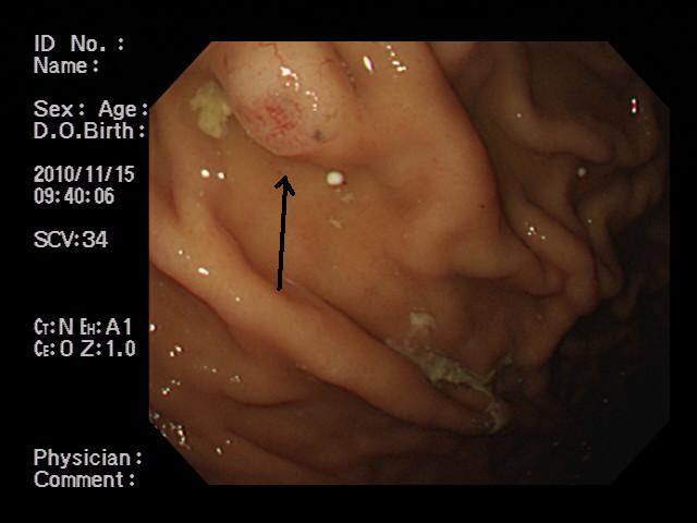
Upper gastrointestinal endoscopy revealed an elevated lesion with central erosion (1.3 × 2 × 1.5 cm) (Figure 1) in the gastric corpus of fundic gland area. One biopsy was taken. The biopsy showed monotonous proliferation of complex atypical glands with hyperchromatic nuclei in the gastric mucosa (Figures 2A, and 2B). Nuclear atypia is mild. Maturation was not seen. The lesion was diagnosed as well differentiated adenocarcinoma. The atypical carcinomatous glands were composed of chief cell-like basophilic cells and acidophilic parietal cells (Figures 2B and 2C). The nuclei of both cell types were located in the basal part. The chief cell-like cells accounted for 80%, and parietal cell for 20%.
Figure 1.

Gastric endoscopy. An elevated lesion with central erosion (arrow) is seen.
Figure 2.

The biopsy of the gastric lesion. A: Low power view. The biopsy shows monotonous proliferation of atypical glands with glandular complexity and hyperchromatic nuclei. The atypical glands do not show maturation. HE, × 50. B: Middle power view. The biopsy shows chief cell-like atypical cells and carcinomatous acidophilic parietal cells. HE, × 200. C: High power view. The nature of chief cells and parietal cells of adenocarcinoma are obvious. Some of parietal cells shows triangular shape (arrow). HE, ×400.
Occasionally, the parietal cells took the form of triangular shape (Figure 2B and 2C). The pathological diagnosis was primary gastric well differentiated adenocarcinoma composed of chief cell-like cells and parietal cells (gastric adenocarcinoma of fundic glands type). The patient died of carcinomatosis three months after the admission. Autopsy was not performed.
Discussion
The gastric adenocarcinoma in the present case appears primary gastric carcinoma, because the carcinoma resided in the gastric mucosa and the carcinoma consisted of chief cell-like cells and parietal cells.
The present gastric adenocarcinoma resembles gastric fundic gland. Very recently, gastric adenocarcinoma of fundic glands type was proposed [3]. Tsukamoto et al [1] recently reported a case of gastric well differentiated adenocarcinoma with chief cell differentiation. They found that the chief cell-like cells were immunohisto-chemically positive for pepsinogens I and II and Runt-related transcription factor gene 3 (RUNX3), markers of chief cells. Ogasawara et al [2] showed that RUNX3 expression correlated with chief cell differentiation in gastric adeno-carcinomas. Ueyama et al [3] very recently reported gastric adenocarcinoma of fundic glands type. They showed the carcinoma cells were positive for pepsinogen I and MUC6, markers of chief cells. Focal areas showed H+/K+-ATPase immunoreactivity, suggesting parietal cell differentiation [3]. In the current case, the immuno-histochemical study of pepsinogens, RUNX3, and H+/K+-ATPase, because of the lack of these antibodies in our laboratory. However, the current case showed apparent HE findings of chief cells and parietal cells. The present gastric adenocarcinoma occurred in the fundic gland area of gastric corpus, supporting the diagnosis.
In conclusion, the author reported an extremely rare case of gastric well differentiated adenocarcinoma histologically composed of chief cell-like cells and parietal cells (gastric adenocarcinoma of fundic gland type) occurring in the fundic gland area of the gastric corpus.
References
- 1.Tsukamoto T, Yokoi T, Maruta S, Kitamura M, Yamamoto T, Ban H, Tatematsu M. Gastric adenocarcinoma with chief cell differentiation. Pathol Int. 2007;57:517–522. doi: 10.1111/j.1440-1827.2007.02134.x. [DOI] [PubMed] [Google Scholar]
- 2.Ogasawara N, Tsukamoto T, Mozoshita T, Inada KI, Ban H, Kondo S, Takasu S, Ushijima T, Ito K, Ito Y, Ichinose M, Ogawa T, Joh T, Tatemastu M. RUNX3 expression correlates with chief cell differentiation in human gastric cancers. Histol Histopathol. 2009;24:31–40. doi: 10.14670/HH-24.31. [DOI] [PubMed] [Google Scholar]
- 3.Ueyama H, Yao T, Nakashima Y, Hirakawa K, Oshiro Y, Hirahashi M, Iwashita A, Watanabe S. Gastric adenocarcinoma of fundic gland type (chief cell predominant type): proposal for a new entity of gastric adenocarcinoma. Am J Surg Pathol. 2010;34:609–619. doi: 10.1097/PAS.0b013e3181d94d53. [DOI] [PubMed] [Google Scholar]


