CASE DESCRIPTION
Over the last year, a 40-year-old man had complained of weekly episodes of facial flushing, diaphoresis, irritability,and diarrhea, which were triggered by emotional stress or exercise. He also presented with chest discomfort and had lost 6 kg. He had poorly controlled essential hypertension that had lasted over the last six years. On examination, his systolic blood pressure was 160 mm Hg, his diastolic blood pressure was 110 mm Hg, his heart rate was 110 beats/min, and his body mass index was 32 kg/m2; he did not present with postural hypotension. A small goiter without nodules and a slight lid retraction were noted. He had no facial flushing or hand tremor. During the chest discomfort investigation, the chest X-ray revealed an anterior mediastinal enlargement.
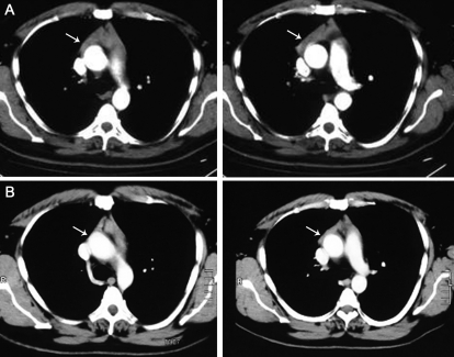
A thoracic computed tomography (CT) scan revealed a homogeneous mass with no adjacent structure invasion or calcification at the topography of the thymus enlargement (Figure 1A). Goiter was also detected but did not extend to the intrathoracic compartment. A carcinoid syndrome caused by a primary neuroendocrine tumor was suspected.
Figure 1.
CT-scan images showing a thymus enlargement before (A) and after (B) treatment for hyperthyroidism due to Graveś′ disease, with remarkable shrinkage of the thymus (arrows).
The patient´s exams depicted normal values of urinary 5-hydroxyindoleacetic acid (2.8 and 3.1 mg/24 h, normal values-NV 2-6), urinary metanephrines (1.0 and 0.09 mcg/mg creatinine, NV <1.2), serum calcitonin (7.3 pg/mL, NV <11.5), negative tumoral markers β-hCG and alpha-fetoprotein. Thyrotoxicosis was diagnosed from the following fluoroimmunoassay measurements: total T3, 371 ng/dL (NV 40–180 ng/dL); total T4, 20.9 mcg/dL (NV 4.5–12 mcg/dL); free T4, 4.9 ng/dL (NV 0.7–1.5 ng/dL); and a suppressed TSH<0.03 mUI/L (NV 0.4–4.5 mUI/L). Serum antiperoxidase and antithyroglobulin antibodies measured by fluoroimmunoassay were positive (859 and 72 U/mL, NV<35 U/mL), with levels of TSH-receptor antibody (TRab) of 49% (radioimmunometric assay NV<8%). Ultrasound thyroid imaging revealed a 43.2 g homogeneous hypoechogenic goiter, which did not have nodules and was limited to the neck.
Therefore, Graves' disease was diagnosed, and a thymus growth was then attributed to hyperplasia caused by Graves' disease.
Methimazol (20 mg/day) was initiated, and after four months of treatment, symptoms had improved, and thyroid hormones had normalized. Goiter and TRab values had reduced. After six months of thyroid hormonal control, a new CT revealed marked thymus shrinkage (Figure 1B).
After 24 months, the patient was in remission of Graveś disease, with negative TRab values and a normal thoracic CT scan. Graveś ophthalmopathy was never presented.
DISCUSSION
We described a patient with systemic arterial hypertension and thoracic discomfort that presented an anterior mediastinal mass and Graveś disease. In adults, thymoma is a common neoplasm of the anterior mediastinal compartment and occurs between 40 and 60 years of age with a slightly male predominance. Chest discomfort or pain is the most common symptom.
However, our patient also presented episodic flushing, diaphoresis, and diarrhea, which are common symptoms for carcinoid syndrome. Together with the anterior mediastinal enlargement, carcinoid syndrome that had resulted from a neuroendocrine tumor was initially suspected. Primary neuroendocrine tumors of the thymus account for less than 5% of all anterior mediastinal neoplasms. They are highly aggressive and more prevalent in men during the fourth and fifth decades of life. The symptoms are related to structure compression, distant metastasis, or endocrinopathies, including carcinoid syndrome. A CT-scan usually shows a lobulated thymic mass with heterogeneous enhancement and central areas of low attenuation, before necrosis and hemorrhage, and eventually calcifications.1 The presence of normal 24-hour urinary vanillylmandelic acid and metanephrine levels ruled out the possibility of neuroendocrine tumors. Normal testing physical examination and undetectable alpha-fetoprotein and beta human chorionic gonadotropin levels also ruled out the possibility of nonseminomatous germ-cell tumors.1
Combining the hormonal thyroid abnormalities with the thoracic CT characteristics, thymic hyperplasia due to Graveś disease was diagnosed. Thymic hyperplasia is commonly associated with Graves' disease, but it is not emphasized in major endocrinology texts and must be recognized by all physicians.
There are two morphologic forms of thymus hyperplasia associated with Graves' disease: lymphoid hyperplasia and true hyperplasia. The first is characterized by a thymus medullary lymphoid-follicle formation that is not visualized as an enlargement of the anterior mediastinal compartment.2 In contrast, the second form, true hyperplasia, presents as an increase in thymic tissue. The mechanism of both hyperplasias is not well established and could be caused by excessive stimulation by the thyroid hormone or TRab itself.3,4
Few cases of detectable massive enlargements of the thymus have been reported; most of these cases involved thymectomy or biopsies to exclude the thymomas and treatment of the remaining tissue with hyperthyroidism.5,6 Because the association between Graveś disease and thymic hyperplasia is unclear, unnecessary approaches such as sternotomy and transthoracic biopsy may be used.7
In conclusion, thymic enlargement associated with Graves' disease, especially when a homogeneous mass without a surrounding invasion, calcifications or a cystic image is revealed from a thoracic CT-scan, allows for judicious clinical and radiologic follow-up during the hyperthyroidism treatment.
Footnotes
No potential conflict of interest was reported.
REFERENCES
- 1.Chaer R, Massad MG, Evans A, Snow NJ, Geha AS. Primary neuroendocrine tumors of the thymus. Ann Thorac Surg. 2002;74:1733–40. doi: 10.1016/s0003-4975(02)03547-6. 10.1016/S0003-4975(02)03547-6 [DOI] [PubMed] [Google Scholar]
- 2.Gunn A, Michie W. Biopsy of the thymus. British Journal of Surgery. 1965;52:957–63. doi: 10.1002/bjs.1800521212. 10.1002/bjs.1800521212 [DOI] [PubMed] [Google Scholar]
- 3.Murakami M, Hosoi Y, Negishi T, Kamiya Y, Miyashita K, Yamada M, et al. Thymic hyperplasia in patients with Graves' disease. Identification of thyrotropin receptors in human thymus. J Clin Invest. 1996;98:2228–34. doi: 10.1172/JCI119032. [DOI] [PMC free article] [PubMed] [Google Scholar]
- 4.Nakamura T, Murakami M, Horiguchi H, Nagasaka S, Ishibashi S, Mori M, et al. A case of thymic enlargement in hyperthyroidism in a young woman. Thyroid. 2004;14:307–10. doi: 10.1089/105072504323030979. 10.1089/105072504323030979 [DOI] [PubMed] [Google Scholar]
- 5.Kirkeby KM, Pont A. Image in endocrinology: thymic hyperplasia in a patient with Graves' disease. J Clin Endocrinol Metab. 2006;91:1. doi: 10.1210/jc.2005-1811. 10.1210/jc.2005-1811 [DOI] [PubMed] [Google Scholar]
- 6.Yamanaka K, Nakayama H, Watanabe K, Kameda Y. Anterior mediastinal mass in a patient with Graves' disease. Ann Thorac Surg. 2006;81:1904–6. doi: 10.1016/j.athoracsur.2005.07.081. 10.1016/j.athoracsur.2005.07.081 [DOI] [PubMed] [Google Scholar]
- 7.Popoveniuc G, Sharma M, Devdhar M, Wexler JA, Carroll NM, Wartofsky L, et al. Graves' disease and thymic hyperplasia: the relationship of thymic volume to thyroid function. Thyroid. 2010;20:1015–8. doi: 10.1089/thy.2009.0383. 10.1089/thy.2009.0383 [DOI] [PubMed] [Google Scholar]


