Abstract
With the increasing number of reports of aberrant root canal morphology, the clinician needs to be aware of the variable anatomy. Various case reports have been published with the finding of middle mesial canal in mandibular first molar, however finding of middle distal canal in distal root of mandibular first molar is rare. This case report describes root canal treatment of two rooted mandibular first molar with five root canals (three in distal and two in mesial root), and Sert and Bayirli Type XVIII canal configuration in distal root.
Keywords: Distal root, mandibular first molar, middle distal canal, root canals
INTRODUCTION
The main objective of root canal treatment is thorough mechanical and chemical cleansing of the entire pulp space followed by complete obturation with an inert filling material.[1] Therefore, it is imperative that aberrant anatomy is identified prior to, and during the root canal treatment of such teeth. In 1974, Vertucci and William[2] described the presence of an independent middle mesial canal in a mandibular first molar. Since then unusual canal anatomy associated with mandibular first molar has been reported in several clinical studies / case reports. The present case report describes root canal treatment in a mandibular first molar with three separate root canal orifices and a single exiting foramen.
CASE REPORT
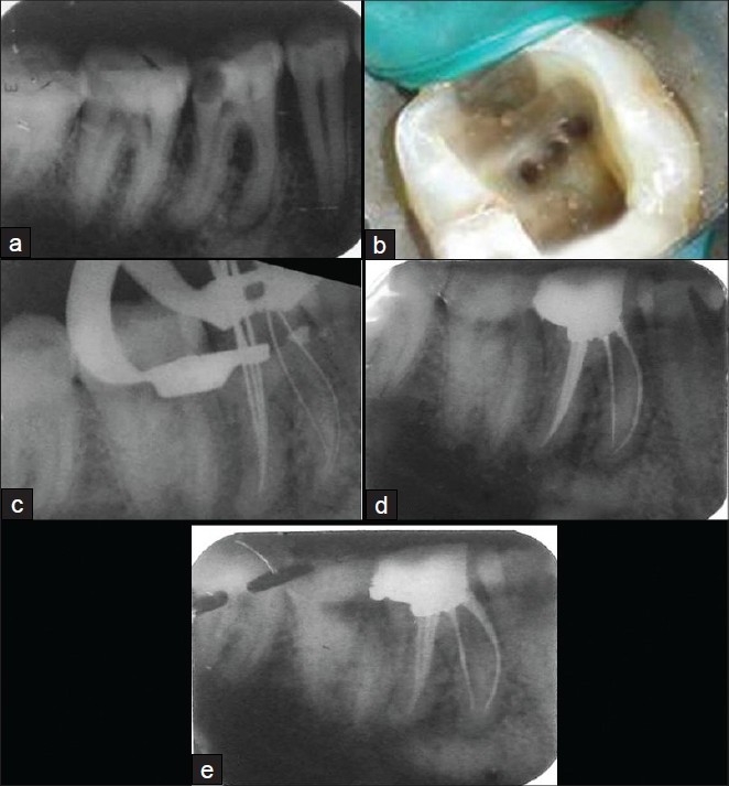
A 20-year old female patient was referred to our department of Conservative Dentistry and Endodontics with the chief complaint of pain in the lower right back tooth since the past 3 days. Her medical history was non-contributory. Clinical examination revealed a deep disto-occlusal carious lesion in relation to right mandibular first molar (tooth # 46). The tooth was tender to vertical percussion. There was no tenderness on palpation in buccal and lingual vestibule. Tooth mobility and periodontal probing around the tooth was within physiologic limits. Thermal and electrical pulp testing elicited a negative response. The preoperative radiograph demonstrated a disto-occlusal radiolucency approaching the pulp space and widening of periodontal ligament space in relation to the mesial and distal root apices [Figure 1a]. A diagnosis of necrotic pulp with symptomatic apical periodontitis was established and endodontic therapy was scheduled.
Figure 1.

(a) Preoperative radiograph of 46, (b) Intraoral photograph showing three distal canal orifices in access cavity preparation, (c) Working length radiograph, (d) Postobturation radiograph, (e) Postobturation radiograph with 30 degree mesial angulation
Following local anaesthesia, an endodontic access cavity was prepared under rubber dam isolation on tooth # 46. Examination of pulp chamber floor revealed five distinct root canal orifices: two were detected mesially (mesiobuccal and mesiolingual); and, three distally (distobuccal, middle distal and distolingual) [Figure 1b]. Three canals in the distal root were confirmed on radiograph [Figure 1c]. Cleaning and shaping of canals was done, and the canals were dried with absorbent points. The obturation was done by cold lateral compaction of gutta-percha [Figure 1d,e]. The patient was asymptomatic during the follow-up period.
DISCUSSION
The majority of mandibular first molars have two roots, one mesial and one distal, and their usual root canal distribution is two canals in the mesial root and one or two canals in the distal root.[1] The major variant of root canal system of mandibular first molar is the presence of a middle mesial canal with 1-15 % incidence.[3] Incidence of three canals in distal root of mandibular first molar in an Indian population is 1.7%; 0.2% in Senegalese population; 1.7% in Turkish population; 0.7% in Burmese population; 1.6% in Thai population; and, in Sudanese population 3% incidence has been reported.[4]
This case demonstrates a rare anatomical configuration and supports previous reports of the existence of such configuration in mandibular first molars. Detailed review of case reports with three canals in the distal root/roots is summarized in Table 1. In this case report, distal root has three distinct root canal orifices with single apical termination, that could be described as Type XVIII canal configuration according to Sert and Bayirli supplemental canal configurations of root canal morphology.[6] Type XVIII canal pattern in distal root of mandibular first molar is previously reported in only one case.[4] Diagnostic measures are important aids in location of root canal orifices including multiple pretreatment radiographs, examination of pulp chamber floor with a sharp explorer, troughing grooves with ultrasonic tips, staining of chamber floor with 1% methylene blue dye, performing sodium hypochlorite “Champagne bubble test”, visualizing canal bleeding points, and use of dental operating microscope and magnification loupes.[1]
Table 1.
Review of the case reports with the finding of three or more canals in distal root/roots of mandibular first molar

CONCLUSIONS
Usually, a prudent inspection of the pulp chamber floor by proper visualization allows the clinician to search for additional canals. Proper and thorough instrumentation is one of the key factors in the success of endodontic therapy; therefore, the clinician should be aware of the incidence of these extra canals in the mandibular first molar.
Footnotes
Source of Support: Nil
Conflict of Interest: None declared.
REFERENCES
- 1.Vertucci FJ, Haddix JE, Britto LR. Tooth morphology and access cavity preparation. In: Cohen S, Hargreaves KM, editors. Pathways of the pulp. 9th ed. Louis MO USA: Mosby; 2006. pp. 148–232. [Google Scholar]
- 2.Vertucci F, Williams R. Root canal anatomy of the mandibular first molar. JNJ Dent Assoc. 1974;48:27–8. [PubMed] [Google Scholar]
- 3.Baugh D, Wallace J. Middle mesial canal of the mandibular first molar: A case report and literature review. J Endod. 2004;30:185–6. doi: 10.1097/00004770-200403000-00015. [DOI] [PubMed] [Google Scholar]
- 4.Kottoor J, Sudha R, Velmurugan N. Middle distal canal of the mandibular first molar: A case report and literature review. Int Endod J. 2010;43:714–22. doi: 10.1111/j.1365-2591.2010.01737.x. [DOI] [PubMed] [Google Scholar]
- 5.Mushtaq M, Farooq R, Rashid A, Robbani I. Spiral computed tomographic evaluation and endodontic management of mandibular first molar with three distal canals. J Conserv Dent. 2011;14:196–8. doi: 10.4103/0972-0707.82602. [DOI] [PMC free article] [PubMed] [Google Scholar]
- 6.Sert S, Bayirli GS. Evaluation of the root canal configurations of the mandibular and maxillary permanent teeth by gender in the turkish population. J Endod. 2004;30:391–8. doi: 10.1097/00004770-200406000-00004. [DOI] [PubMed] [Google Scholar]


