Abstract
Bonfil's rigid fibroscope is an instrument used to perform tracheal intubation, proven to be effective both in patients with normal and in those with difficult airways. We use this device in awake intubation in a patient presenting with a large right neck mass and a tonsillar tumor which limited the mouth opening. Also, we describe our technique of insertion of Bonfil's retromolar fibroscope from the right side of the mouth across the tongue.
Keywords: Aged, anesthesia, awake intubation, Bonfil's fibroscope, difficult intubation, tonsillar tumor
INTRODUCTION
According to the American Society of Anesthesiologists practice guidelines for management of a difficult airway, an awake intubation is considered the primary method to secure a suspected difficult airway.[1] Bonfil's rigid fibroscope (Karl storzGasbH, Tutlingham, Germany) is an instrument used to perform tracheal intubation, proven to be effective both in patients with normal and in those with difficult airways.[2–7] Bonfil's fiberscope is a rigid 40 cm long endoscope with 40 curved ends, on which a tracheal tube with an internal diameter of at least 5.5 mm can be loaded, and a 5.5 mm tracheal tube adaptor that allows the position of the distal end of the tube to be adjusted a few millimeters beyond the tip of fiberscope.[2–7]
Techniques of insertion of Bonfil's retromolar fiberscope
Both midline and retromolar approaches were used in adults; we used a third technique, lateral entry into the hypopharynx at a right angle across the tongue.
CASE REPORT
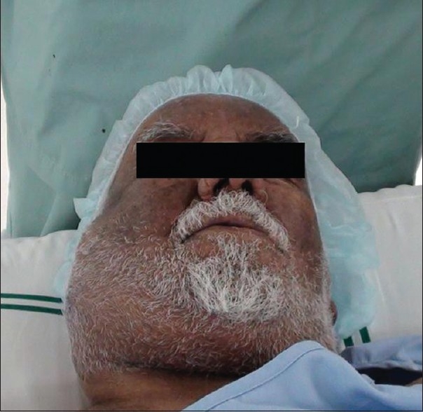
A 79-year-old male presented with history of neck swelling since 2 months. Airway examination revealed malampatti class III, short and thick neck swelling of right side covering the angle of the mandible interfering and limiting the mouth opening [Figure 1].
Figure 1.

Right hard and fixed neck mass
The patient received 1 g paracetamol infusion, and 50 mcg dexmedotomidine over 30 minutes, 0.6 mg atropine intramuscular (IM) injection plus nebulization with 5 ml of 4% lignocaine hydrochloride topical solutions via nebulizer and face mask over 15 minutes, 30 minutes before intubation.
The oropharynx topically anesthetized with two puffs of 10% lignocaine.
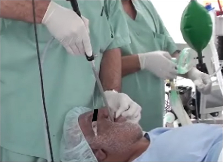
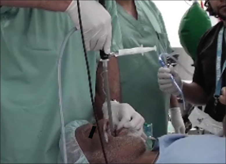
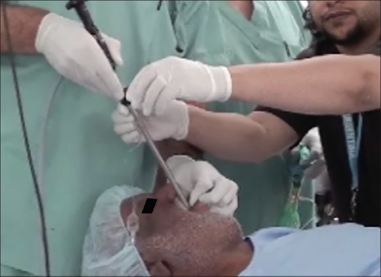
Bonfil's fibroscope (preloaded with an 7.5 mm cuffed endotracheal tube) was advanced via the lateral side of mouth to the hypopharynx and advanced toward the posterior pharynx; when Bonfil's fibroscope was positioned immediately in front of the vocal cords an additional 2 ml of 2% lignocaine was injected via the tube adaptor [Figures [Figures 2 and 3]. After 30 seconds Bonfil's fibroscope was advanced further until the tip off the scope passed the glottis opening; an additional 2 ml of 2% lignocaine was injected onto the trachea. The endotracheal tube was advanced over the scope [Figure 4]. Bonfil's fibroscope was then removed leaving the endotracheal tube in place.
Figure 2.

Bonfil's fibroscope advanced via the lateral side of mouth
Figure 3.

2 ml of 2% lignocaine was injected via the tube adaptor
Figure 4.

Endotracheal tube advanced over the scope
DISCUSSION
Traditionally, an awake intubation is performed by flexible fiber optic scope; however many new devices have been developed to assist anesthesiologists with both routine and difficult airway management, one of which is Bonfil's fiberscope.[8] It has been used in patients who have limited or no neck mobility.[8,9] Its design allows easy navigation through the oral cavity of patients with a large tongue or large tonsils; it allows for a faster intubation than other difficult airway devices.[10] We used it in a patient with a compromised and difficult airway; this device may be more beneficial than a flexible fiberscope since it can be readily navigated through soft tissues and physically lift the airway structure, is more affordable, durable, and easy to clean,[9] and should be considered when intubating the patient with a difficult and or compromised airway.
Footnotes
Source of Support: Nil
Conflict of Interest: None declared.
REFERENCES
- 1.Practice guidelines for management of the difficult airway, an updated report by the American society of anesthesiologist's task force on management of the difficult airway. Anesthesiology. 2003;98:1269–77. doi: 10.1097/00000542-200305000-00032. [DOI] [PubMed] [Google Scholar]
- 2.Halligan M, Charters P. A clinical evaluation of the Bonfils intubation fibroscope. Anaesthesia. 2003;58:1087–91. doi: 10.1046/j.1365-2044.2003.03407.x. [DOI] [PubMed] [Google Scholar]
- 3.Wong P. Intubation times for using the Bonfils intubation fibroscpe. Br J Anaesth. 2003;91:757. [PubMed] [Google Scholar]
- 4.Rudolph C, Schlender M. Clinical experiences with fiber optic intubationwith the Bonfils intubation fibroscope. Anaesthesiol Reanim. 1996;21:127–30. [PubMed] [Google Scholar]
- 5.Wahlen BM, Gercek E. Three-dimensional cervicalspine movement durinhg intubation using the Macitosh and Bullard laryngoscopes, the Bonfils fibroscope and the intubating laryngeal mask airway. Eur J Anaesthesiol. 2004;21:907–13. doi: 10.1017/s0265021504000274. [DOI] [PubMed] [Google Scholar]
- 6.Rudolph C, Schneider JP, Wallenborn J, Schaffranietz L. Movement of the upper cervical spine during laryngoscopy, a comparison of the Bonfils intubation fibroscope and the Macintosh laryngoscope. Anaesthesia. 2005;60:668–72. doi: 10.1111/j.1365-2044.2005.04224.x. [DOI] [PubMed] [Google Scholar]
- 7.Bein B, Yan M, Tonner PH, Scholz J, Steinfath M, Dörges V. Tracheal intubation using the Bonfils intubation fibroscope after failed direct laryngoscopy. Anaesthesia. 2004;59:1207–09. doi: 10.1111/j.1365-2044.2004.03967.x. [DOI] [PubMed] [Google Scholar]
- 8.Corbanese U, Possamai C. Awake intubation with Bonfils Fiberscope in patients with difficult Airways. Eur J Anaesthesiol. 2009;26:837–41. doi: 10.1097/EJA.0b013e32832c6076. [DOI] [PubMed] [Google Scholar]
- 9.Wahlen BM, Gercek E. Three-dimensional cervical spine movement during intubation using the Macintosh and Bullard laryngoscopes, the Bonfils fiberscope and the intubating laryngeal mask airway. Eur J Anaesthesiol. 2004;21:907–13. doi: 10.1017/s0265021504000274. [DOI] [PubMed] [Google Scholar]
- 10.Bein B, Worthmann F, Scholz J, Brinkmann F, Tonner PH, Steinfath M, et al. A comparison of the intubating laryngeal mask airway and Bonfils intubating fiberscope in patients with predicted difficult airways. Anaesthesia. 2004;59:68–74. doi: 10.1111/j.1365-2044.2004.03778.x. [DOI] [PubMed] [Google Scholar]


