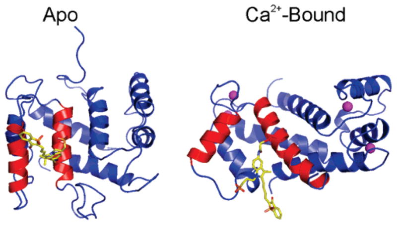
Figure 2.

Model of Mero-S100A4. Model of the apo and Ca2+-bound Mero-S100A4. Helices 3 and 4 of one monomer are shown in red, and the merocyanine dye attached to Cys81 is shown in yellow. Merocyanine attached to only one monomer is shown for simplicity. The bound calcium ions are depicted as pink spheres.
