Abstract
Here we describe a model of medial temporal lobe organization in which parallel “what” and “where” processing streams converge within the hippocampus to represent events in the spatio-temporal context in which they occurred; this circuitry also mediates the retrieval of context from event cues and vice versa, which are prototypes of episodic recall. Evidence from studies in animals are reviewed in support of this model, including experiments that distinguish characteristics of episodic recollection from familiarity, neuropsychological and recording studies that have identified a key role for the hippocampus in recollection and in associating events with the context in which they occurred, and distinct roles for parahippocampal region areas in separate “what” and “where” information processing that contributes to recollective and episodic memory.
Keywords: episodic memory, hippocampus, recollection, familiarity, context, place cells
For over 50 years we have known that the medial temporal lobe (MTL) plays a critical role in memory, and since the 1980s it has become clear that this role is especially important for episodic memory, our ability for recollection of specific events in their spatiotemporal context. A fundamental question, then, is how does the MTL support episodic memory? Recent studies have suggested a functional organization of the MTL that could support encoding and retrieval of events in context (Davachi, 2006; Eichenbaum et al., 2007; Diana et al., 2007). The model is based on a combination of evidence from amnesia and functional imaging in humans, as well as lesion and single neuron recording studies in animals, several of which will be described below. Work in our laboratory is focused on the development of an animal model of episodic memory, because a complete and detailed understanding about the distinct roles of specific MTL areas and how they represent information relevant to episodic memory will depend on experiments where interventions can be localized within particular brain areas and where neural activity throughout the MTL system can be systematically characterized.
Here we will first consider whether animals, and in particular rats, have memory capacities that parallel episodic memory in humans, and we will consider the role of the hippocampus in this capacity. Then we will provide an overview of the MTL model and describe experiments that are beginning to identify the functional roles of specific components of the MTL, consistent with this model.
Do rats have episodic memory – is the hippocampus involved?
A major challenge in the development of an animal model of episodic memory concerns the question of whether animals have this capacity and how to measure it. In humans, episodic memory is readily observed in the verbal recall of specific experiences, but this approach is obviously not possible in animals. Our own work towards addressing this question has adopted a different, quantitative methodology that has been used extensively in humans to investigate distinctions between recall of episodic memories and a sense of familiarity for previous experienced materials. This method uses the Receiver Operating Characteristic (ROC) function to generate separate indices of episodic recollection and familiarity in accordance with theories that consider these two processes as distinct and independent (Yonelinas, 2002).
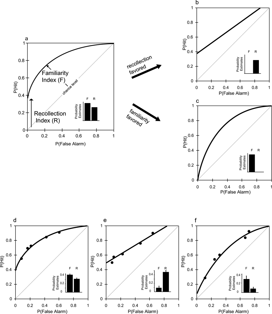
In a typical ROC experiment on recognition of single items (e.g., words, faces), human subjects are initially given a study list then later tested with a longer list that includes both the items that were studied (old items) and an equal number of not studied items, and the subjects must declare each test item as “old” or “new.” The proportion of correct identifications of old items (“hits”) is compared to the proportion of incorrect identifications of new items as “old” (“false alarms”) across a large range of response criteria, either manipulated by varying the payoff ratio of rewards for hits and correct rejections or by using subjects’ confidence judgments on each response (Macmillan & Creelman, 1991). The ROC function is typically characterized by two prominent features, an above-zero Y-intercept that makes the curve asymmetrical, and a bowing of the curve away from the chance line, making the function curvilinear (Figure 1a). According to the dual process model, the magnitude of the asymmetry reflects the contribution of recollection, whereas the degree of curvilinearity indexes the contribution of familiarity (Yonelinas, 2001). Thus, when episodic recollection is favored, the ROC function becomes linear and asymmetrical (Figure 1b). Conversely, when familiarity is favored, the ROC becomes curvilinear and symmetrical (Figure 1c).
Figure 1.
Ideal ROC functions for human recognition memory predicted by Dual Process Signal Detection (DPSD) theory (see Yonelinas, 2001) and observed ROC functions for recognition memory in rats. a–c. Humans. a. Item recognition. The ROC curve is typically asymmetrical and curvilinear. Quantitiative measurements of the contributions of recollection (R) and familiarity (F) are calculated as probability estimates shown in the inset of this and other figures (see Yonelinas et al., 2002). b. ROC function observed when performance is based only on recollection. c. ROC function observed when performance is based only on familiarity. d–f. Rats. d. Item recognition (data from Fortin et al., 2004). Recollection and familiarity components are both robust, similar to the ideal item recognition ROC in humans (Panel a.). e. Associative recognition (data from Sauvage et al., 2008). The ROC becomes linear, similar to the ideal ROC of humans when performance is based on recollection only (panel b). f. Item recognition with a speeded response deadline demand (data from Sauvage et al., 2010a). The ROC becomes symmetrical and curvilinear, similar to the ideal ROC of humans when performance is based on familiarity only (Panel c).
A striking aspect of ROC analysis is that this method is equally applicable to studying memory in animals as it is in humans, because subjects are not required to explicitly recall studied items; instead they simply have to respond differentially to new and old items under a range of response biases. To perform an ROC analysis of recognition memory in rats, the memory cues were taken from a large pool of ordinary household odors (e.g., lemon, thyme, cumin) mixed in sand within small plastic cups (Fortin et al., 2004). Initially, rats sampled a series of 10 stimuli, each baited with a bit of sweetened cereal buried in the sand of each stimulus cup; digging for the reward in each cup ensured that the animal had sampled all of the odors. After a 30-min memory retention period, recognition memory was tested using a series of 20 test stimuli, which involved a random ordering of the 10 odors presented in the sample phase (old odors) plus 10 new odors taken from the pool. Rewards were based on a non-match response contingency, such that rats could obtain rewards by digging only in test cups containing new odors. In addition, when old test odors were presented, rats were required to refrain from digging in the test cup in order to obtain a reward in an alternate cup located in the back of the cage. To manipulate the animal’s response bias towards “old” or “new” responses, both the height of the test cup and the ratio of reward magnitude in the test cup versus that in the alternate cup were varied. If the effort to dig in the test cup was high (tall cup) and the reward amount was small, then rats were biased to refrain from digging, which is their signal that the test cup was “old” (i.e., was experienced in the study phase). This bias condition is equivalent to a liberal criterion for the “old” response in humans and results in high hit and high false alarm rates. Conversely, if the effort was low (short cup) and the reward amount relatively large, then rats were biased to dig in the target cup, that is, to emit a “new” response. This bias condition is equivalent to a conservative criterion for the “old” response in humans and results in lower hit and false alarm rates.
Results across several studies showed that the ROC curve of rats for odor recognition contained both an asymmetrical component (above-zero Y-intercept) and a strong curvilinear component (Figure 1d; Fortin et al., 2004; for review see Eichenbaum et al., 2010). This pattern is strikingly similar to the ROC function in humans on verbal recognition performance (Figure 1a) and is consistent with the view that, as in humans, in rats both recollection and familiarity contribute to recognition memory.
In further studies, we modified our behavioral protocol to examine whether the recollection and familiarity components of the ROC function of rats were independent and influenced by memory demands that, in humans, favor the use of recollection and familiarity consistent with the dual process model. In humans, a requirement to remember stimulus associations, called associative recognition, has been shown to favor the use of recollection and reduce the contribution of familiarity (Yonelinas 1997, Yonelinas 1999). We developed a version of the associative recognition paradigm for rats using stimulus pairs composed of combinations of an odor mixed into one of several digging media (e.g., wood chips, beads, sand) contained in a cup (Sauvage et al., 2008). Rats can readily learn to separately attend to odors and media as distinct stimulus dimensions (Birrell & Brown, 2000), so we expected the rats to distinguish these elements and to rely on recollection of their associations (e.g., lemon is associated with wood chips). Each day the animals would initially sample a series of 10 odor-medium pairings; then, following a 30-min delay, the rats had to distinguish re-presentations of the 10 original (old) pairings from 10 rearranged (new) pairings of the same odors and media. We employed the same non-matching rule and manipulations of bias that were used in our study on odor recognition described above. Consistent with the findings on associative recognition in humans, the resulting associative recognition memory ROC function was highly asymmetrical, indicating the presence of a strong recollection component (Figure 1e; Sauvage et al., 2008). Furthermore, the shape of the ROC was linear, indicating the absence of a significant familiarity component. This finding indicates that the contribution of recollection can be isolated from that of familiarity by manipulating the memory demands of the recognition task.
A full dissociation of ROC parameters of recollection and familiarity requires that we are also able to manipulate the task in a way that will produce the opposite pattern, reduction in the contribution of recollection while sparing familiarity, thereby generating results complementary to those observed in the associative recognition experiment. Several theoretical views have suggested that familiarity is characterized as a perceptually driven, pattern matching process which is completed rapidly, whereas recollection is characterized as a conceptually driven, organizational process which requires more time (for reviews see Yonelinas, 2001; Mandler, 2008). Therefore we tested the prediction from dual process theory that the addition of an early response deadline at the memory test phase would reduce the contribution of the slower recollection process while sparing that of the more rapid familiarity process (Sauvage et al., 2010a). We initially trained and tested animals using the odor recognition task (Fortin et al., 2004) with no response deadline and observed an ROC function that was both asymmetrical and curvilinear, replicating our earlier results. In subsequent ‘deadline’ testing, we invoked response deadlines that were half that of the unrestricted response latencies, by covering the test cup when the deadline arrived. The deadline ROC function was curvilinear, suggesting that performance was supported by familiarity (Figure 1f: Sauvage et al., 20010a). However, in striking contrast to the ROC function in the no-deadline condition, the ROC function in the deadline condition became almost perfectly symmetrical, suggesting that recollection did not contribute significantly to recognition performance. These observations support the view that familiarity-based responses are made more rapidly than recollection-based responses, consistent with the dual process model. Furthermore these observations show that the contribution of familiarity can be isolated from that of recollection by manipulating memory demands of the recognition task. Moreover, combined with the findings on associative recognition (Sauvage et al., 2008), these results doubly dissociate the contributions of recollection and familiarity, consistent with the dual process model and inconsistent with single process models.
Is the hippocampus essential for episodic recollection?
These analyses of memory performance in intact rats provide a strong foundation for examining the role of the hippocampus in episodic recollection. In humans, damage to the hippocampus results in a severe deficit in episodic memory. Conversely, in functional imaging studies, a common observation is that the hippocampus is selectively activated during episodic recollection (reviewed in Eichenbaum et al., 2007). We asked whether the hippocampus is also critical to the recollection component of the ROC function in rats performing the item recognition task (Fortin et al., 2004). Thus, animals initially trained in the recognition task and assessed by the ROC method were given bilateral localized hippocampal lesions and re-tested using the same procedures. The results showed that selective damage to the hippocampus did not affect animals’ ability to perform the procedures of the non-matching task, and did not alter response biases. Furthermore, the ROC function of animals with hippocampal damage remained curvilinear, indicating intact familiarity. However, the ROC function became symmetrical, indicating loss of the recollection component. Thus hippocampal lesions selectively eliminated the contribution of recollection while sparing the contribution of familiarity.
Importantly, an alternative interpretation of these data, consistent with the single process model, is that hippocampal damage simply weakened memory and that the recollection (asymmetry) component of the ROC function was more sensitive to this weakening than the familiarity (curvilinearity) component. To address this possibility, we examined the ROC function of normal rats with memory weakened by increasing the delay between study and test from 30 min to 75 min (Fortin et al., 2004). The single process model predicts that the ROC should become symmetrical, similar to the effects of hippocampal damage. However, the opposite result was observed. Thus, under the long delay condition, while both the recollection and familiarity components of the ROC function were decreased, the curvilinearity (familiarity) component was virtually eliminated, whereas a substantial asymmetry (contribution of recollection) persisted. Furthermore, a direct comparison of the ROC functions of rats with hippocampal damage and normal rats with weakened memory was possible because the overall recognition performance (measured by percent correct, which combines the contribution of recollection and familiarity) was equivalent under these conditions (64% in normal rats at long delay; 66% in rats with hippocampal lesions at short delay). This comparison revealed that the recognition performance of the two groups was supported by distinct performance strategies, such that normal rats exclusively used recollection, whereas rats with hippocampal damage relied exclusively on familiarity, thus providing a double dissociation of strategies that is entirely consistent with the dual process model, and inconsistent with single process models, and unequivocally shows that the hippocampus supports recollection, but not familiarity.
Yet further evidence for the distinction between episodic recollection and familiarity and for a selective role of the hippocampus in recollection came from our study on associative recognition. Rats were initially trained on the associative recognition task described above, then retested after selective damage to the hippocampus (Sauvage et al., 2008). In this task, because the rats had been repeatedly exposed to the same odors and digging media with many different pairings, we expected that they would distinguish these elements and rely on recollection of their associations (e.g., lemon is associated with wood chips). Alternatively, however, odors and media could readily be unitized into scented medium configurations (lemon smelling wood chips), allowing the use of familiarity to make recognition judgments. As described above, the ROC function of normal rats in associative recognition was strongly asymmetrical and linear, consistent with strong recollection and absence of familiarity, respectively (Figure 1e). This pattern is congruent with the interpretation that animals recalled the associations between items and their paired media and did not use the familiarity of the item-medium combinations to recognize the test stimuli. Consistent with our earlier observation of recollection impairment in animals with hippocampal damage performing the item recognition task, animals with hippocampal damage also suffered a significant decrease in the asymmetry of the associative recognition ROC, indicating impairment in recollection. However, the shape of the ROC function became curvilinear in animals with hippocampal damage, consistent with a compensatory enhancement and reliance on familiarity to recognize old test pairs. This observation is consistent with studies on humans showing that, when recollection and familiarity were put into competition, memory based on these processes can be affected in opposite directions by hippocampal damage (Diana et al, 2008; Giovanello et al, 2006; Quamme et al, 2007). Thus, when the item pairs are processed as two distinct stimulus elements, memory performance depends largely on recollection of the acquired associations, as described above. Alternatively, when the elements of a pair are readily “unitized” into a single odor-medium configuration, familiarity can support memory for stimulus pairings just as it does for single stimuli. Thus, our results are consistent with the hypothesis that, unlike normal rats but like patients with amnesia, rats with hippocampal damage unitize the odor-medium combinations, allowing them to employ their intact familiarity capacity to support recognition. Thus, in the absence of hippocampal function, the contribution of familiarity is exaggerated, again dissociating recollection and familiarity, and supporting a key role for the hippocampus in recollection.
These studies using ROC analyses of recognition memory provide strong evidence of distinct processes of episodic recollection and familiarity in rats, very similar to the results in humans, and indicate that localized damage to the hippocampus results in a selective deficit in recollection. These findings validate an investigation of the functional organization of the MTL in support of episodic memory in rodents, which we will describe next.
What is the functional organization of the MTL that supports the features of episodic memory?
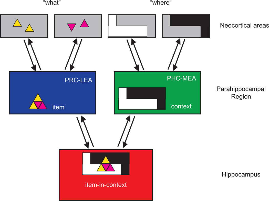
The evidence described above indicates that rats have a memory capacity that shares features with episodic memory in humans, and the hippocampus plays an essential role. However, these studies do not provide insights into how other components of the MTL contribute, nor do these studies inform us about the nature of the neural representations supporting this capacity. Over the last decade cognitive neuroscience studies have revealed important roles in memory for specific areas within the MTL, including prominently, subdivisions of the parahippocampal region and hippocampus (Davachi, 2006; Eichenbaum et al., 2007; Diana et al., 2007). These findings can be summarized in a simple model of the functional organization of this system (Figure 2). The major cortical inputs to the MTL originate in two streams. In one stream (the “what” stream), projections from unimodal and polymodal sensory areas send inputs about perceptual objects and behavioral events principally to the perirhinal cortex (PRC) and thence to lateral entorhinal cortex (LEC). In the other stream (the “where” stream), visuospatial information processed in parietal and retrosplenial cortex and conveyed to the parahippocampal cortex (PHC) and medial entorhinal cortex (MEC; Suzuki & Amaral, 1994; Furtak et al., 2007; Kerr et al., 2007; van Strien et al., 2009; Wang et al., 2011). LEC and MEC then project to each major subdivision of the hippocampus in different patterns of convergence, supporting hippocampal representations of objects and events mapped within spatial context. The outputs of hippocampal processing return to the LEC and MEC, thence to PRC and MEC, respectively, then back to widespread neocortical areas. On a functional level, the encoding of episodic memories involves the convergence of information about events and their context within the hippocampus. Then, during the retrieval of episodic memories, cueing by a previously experienced object can drive the circuit to reactivate the convergent representation in the hippocampus which, via the feedback pathway, then reactivates the “where” stream to retrieve the context representation (or vice versa, cuing with context can reactivate event information in the “what” stream). The ability to retrieve context from item information, that is, to remember where an event occurred (or vice versa), is a classic prototype of episodic memory.
Figure 2.
Schematic diagram of the medial temporal lobe memory system in mammals. The “where” stream of the neocortex projects differentially to the perirhinal cortex (PRC) and lateral entorhinal area (LEA), whereas the “what” stream of the neocortex projects differentially to the parahippocampal cortex (PHC) and medial entorhinal area (MEA). In those parahippocampal regions, the PRC-LEA represents individual objects (items), whereas the PHC-MEA represents contextual information. Those streams converge in the hippocampus where items are represented in the context in which they were experienced. Outputs of the hippocampus are directed back to the parahippocampal areas, and then the neocortical areas, that were the origins of the “what” and “where” stream inputs.
Key evidence in support of the model is derived from studies that distinguish roles of MTL structures in episodic recollection and memory for associations of events and context versus familiarity with events alone (reviewed in Davachi, 2006; Eichenbaum et al., 2007; Diana et al., 2007). These reviews describe a large number of studies in which damage to the hippocampus results in deficits in episodic recollection and memory for associations and context, as we observed in rats in the studies described above. Correspondingly, these reviews describe a large number of functional imaging studies have reported that activation the hippocampus occurs with episodic recollection and memory for associations and context, whereas activation of the perirhinal cortex is linked with familiarity and activation of the parahippocampal cortex is linked to context retrieval. Most striking are double dissociations between deficits in familiarity or item memory following perirhinal cortex damage versus deficits in episodic recollection or associative memory following hippocampal damage (Bowles et al., 2010). Parallel to these observations, there are striking double dissociations in functional imaging of hippocampal activation in recollection versus perirhinal cortex activation in familiarity (Ranaganath et al., 2003; Daselaar et al., 2006). These studies strongly support the dual process model of recognition, in which episodic recollection and familiarity reflect distinct memory processes that are supported by different neural substrates.
A prominent alternative view is that the functional distinctions in MTL structures described above can be explained by a single process model in which differences in stronger memory during recollection of associations versus weaker memory in familiarity for single items (Squire et al., 2004, 2007; Wixted & Squire, 2010). The single process model cannot account for the double dissociations in amnesia and fMRI studies cited above, nor can single process theories account for the double dissociations between the role of the hippocampus in recollection and familiarity in our studies on rats described above. Nevertheless, studies on human amnesia and fMRI studies continue to build evidence that favors one view or the other. There is agreement that identifying the key brain areas and underlying neuronal representations in cortical and MTL structures can provide a deeper understanding of the functional organization of this system (e.g., Wixted et al., 2010; Montaldi & Mayes, 2010) and now we will focus on this approach, first comparing findings on damage to different MTL areas in animals performing recognition memory tests, and second, considering how information is encoded by single neurons within different MTL areas.
Animal models of recognition memory
Animal models offer a substantial improvement in the resolution with which we can examine the effects of selective damage to particular MTL areas and in characterizing neural activation at the level of the units of information processing. These methods have been applied in models of recognition memory in which monkeys or rats are trained to respond differentially to new and previously experienced stimuli or in which we observe their natural tendency to explore novel stimuli. Interpretations of the findings on the role of the hippocampus and other MTL areas have been contentious, so we will begin with an overview of early work then describe more recent studies that have clarified the issues. Early on in research on the roles of MTL areas in recognition memory, there was a strong focus on the delayed non-match to sample (DNMS) test. In the DNMS paradigm (Gaffan 1974; Mishkin & Delacour, 1975) an initially novel three-dimensional “junk” object is presented, then after a variable temporal delay, the animal is rewarded for selecting another novel (i.e. non-matching) object over the sample. Following ablation of the entire medial temporal lobe area, monkeys perform well if the delay is a few seconds, but memory deteriorates rapidly (Mishkin, 1978, Zola-Morgan & Squire, 1985) modeling the rapid memory decline observed in amnesic patients (Squire et al., 1988; Aggleton et al., 1988). A similar severe and rapid decline in recognition memory is also observed after damage limited to the perirhinal cortex alone or in combination with parahippocampal or entorhinal cortex in monkeys (Zola-Morgan et al., 1989; Meunier et al., 1993) and in rats (Otto & Eichenbaum, 1992a; Mumby & Pinel, 1994; Wiig & Bilkey, 1995; reviewed in Steckler et al., 1998; Brown & Aggleton, 2000; although see Winters et al., 2004).
In contrast, monkeys with selective damage to the hippocampus (Murray & Mishkin, 1998) or entorhinal cortex (Buckmaster et al., 2004) perform normally in DNMS at substantial memory delays. In some studies, a small, but statistically significant deficit was observed at a very long delay (Zola et al., 2000), but in other studies no deficit was observed even at long memory delays or when animals are required to remember a long list of stimuli (Murray & Mishkin, 1998). In rats, performance on DNMS is also intact following selective hippocampal damage (see Steckler et al., 1998 and Mumby, 2001 for detailed reviews), although some studies report partial impairment at long delay intervals or when the list of sample objects was numerous (Mumby et al., 1992, 1995; Steele & Rawlins, 1993; Wiig & Bilkey, 1995; Dudchencko et al., 2000; Clark et al., 2001).
Succeeding studies have examined recognition memory by monitoring spontaneous exploration of familiar and novel stimuli, wherein an object or picture is first presented, then re-presented after a delay along with another novel stimulus. Animals typically spend approximately twice as much time investigating the novel stimulus over the previously experienced stimulus. Superficially, these paradigms would seem to present the same memory requirement for recognition of a novel stimulus as the DNMS task. However, there are differences in the nature of the stimuli and behavioral responses, as well as in the motivational basis for performance, that could influence the use of different memory strategies in the expression of recognition memory (Nemaniac et al., 2004). Indeed, in monkeys, even the same animals with selective hippocampal or parahippocampal cortex damage that show little or no deficit in DNMS have severe and rapidly apparent deficits on the spontaneous novelty exploration task (Zola et al., 2000; Nemanic et al., 2004). In rats, some studies report no deficit (Save et al., 1992; Mumby et al., 2002; Lee et al., 2005; Winters et al., 2004; Langston & Wood, 2010) whereas other studies have observed an impairment at long delays (Clark et al., 2000; Hammond et al, 2004; Rampon et al., 2000), and observation of the deficit may depend on the amount of damage to the hippocampus (Moses et al., 2002; Broadbent et al., 2004). In contrast to the variability of findings on the hippocampus, damage to the perirhinal cortex consistently results in a severe and rapidly developing deficit in monkeys (Nemaniac et al., 2004) and in rats (Ennaceur et al., 1996; Mumby et al., 2002; Norman & Eacott, 2004, 2005; Winters et al., 2004; Winters & Bussey, 2005a,b; Young et al., 1997).
Importantly, in variants of recognition tasks where rats must remember places, the hippocampus consistently plays an important role. Hippocampal damage causes severe and immediate deficits on DNMS tasks in which animals must remember cues that are composed as visually elaborated arms of a maze (Yee & Rawlins, 1994; Prusky et al., 2004). In a variant of the spontaneous novelty exploration task where an initially presented object is moved to a new location or to a new environment during the test phase, selective hippocampal lesions consistently result in deficits even following relatively small lesions of the hippocampus that have no effect on exploration of novel objects (Mumby et al., 2002; Eacott & Norman, 2004; Lee et al., 2005). Similarly, damage to the parahippocampal cortex does not affect exploration of novel objects but results in severe impairment in recognizing objects after a change in position or context (Eacott et al., 2004; Norman and Eacott, 2005) or sometimes both (Langston & Wood, 2010). Strikingly, in a double dissociation with perirhinal cortex, Normal and Eacott (2005) showed that whereas parahippocampal damage produced a deficit in object-location memory, perirhinal damage resulted in a deficit in object-object memory. These results suggest that the hippocampus and parahippocampal cortex may be particularly involved in memory for spatial scenes or context whereas the perirhinal cortex processes object information without context.
Why do hippocampal lesions result in partial and variable impairments in recognition memory across tasks and across species? Based on the findings from human memory, one possibility is that the hippocampus supports only one of two processes that contributes to recognition performance (e.g., recollection) and the demand for this component varies across behavioral paradigms. As described above, this hypothesis was addressed in our study in which rats were trained on a variant of DNMS in which they initially sampled a series of odors and then judged old and new test stimuli across a range of response criteria to derive ROC functions (Fortin et al., 2004). The results of that study revealed that recognition in rats was supported by two processes akin to recollection and familiarity, and that selective hippocampal damage eliminated the contribution of recollection and spared that of familiarity. These findings offer an explanation of the mixture of findings on hippocampal damage in previous studies on recognition memory, suggesting that the presence and magnitude of the deficit is dependent on the relative contributions of recollection and familiarity processes in the performance of particular tasks.
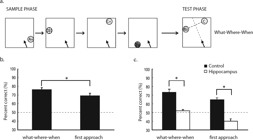
An additional study provides further evidence that the hippocampus is critical in memory for a combination of information about what happened, where it happened, and when it occurred, three key features of episodic memory. In this study we trained rats on a task which assesses memory for events from single episodes involving a combination of odors (“what”) presented in unique places (“where”) and in a specific order (“when”; Figure 3a; Ergorul & Eichenbaum, 2004). On each trial, rats sequentially sampled a unique series of four rewarded odor stimulus cups, each in a different place along the periphery of a large open field. Then, memory for the order of those events was tested by presenting a choice between an arbitrarily selected pair of the odor cups in their original locations, and selection of the earlier presented item was reinforced with a buried reward. Because rats could employ memory for the locations of the cups (“where”) without using odor information (“what”), we also measured responses based purely on location information in two ways: First, we recorded the initial stimulus the animal approached; we separately determined that rats cannot tell which odor is inside until they approach the odor cup. Second, we presented probe memory tests in which the odors were omitted and the rats had to use the locations only to identify which odor was presented earlier.
Figure 3.
The hippocampus and “what,” “where,” “when” memory. a. Behavioral protocol. b. Control rats make an initial good guess about which item occurred first (“when”) based on location information (“where”) and then they confirmed or disconfirmed their choice based on the odor in the cup (“what”). c. Rats with hippocampal damage perform at chance level on the final choice and below chance in their first visit.
Normal animals performed well in the standard what-where-when tests. Furthermore, in the measure of first cup visited on the test trials, the animals performed less well, albeit still above chance (Figure 3b). Therefore, it appears that normal rats make an initial good ‘guess’ about which item occurred first (“when”) based on location information (“where”) and then they confirmed or disconfirmed their choice based on the odor in the cup (“what”). Furthermore, normal rats fall to chance performance in the probe tests that omitted the odors, indicating that they considered items that lacked the correct “what” component distinct from either correct item. This combination of findings provides compelling evidence that normal rats form strongly integrated representations of what happened when and where. Rats with hippocampal damage were severely impaired on the standard what-where-when memory judgments, performing no better than chance (Figure 3c). Interestingly, animals with hippocampal damage tended to first approach the more recently reinforced cup, in opposition to their training to approach the earlier presented cup, suggesting their performance was driven by an intact system guided by recent reinforcement. These observations indicate that normal rats can remember single episodes of what happened, where, and when, and that this ability is based on highly integrated what-where-when representations that are supported by the hippocampus.
The combined findings on recognition memory and our what-where-when test strongly support the model illustrated in Figure 2. Whereas the perirhinal and lateral entorhinal cortex are essential to memory for specific objects, the parahippocampal region and medial entorhinal cortex are critical to memory for the context in which events occur. These results are consistent with the idea that the hippocampus is involved in the convergence of object and context information and confirm the critical role of the hippocampus in memory for integrating the what-where-when features of episodic memory. In the next section, we will consider parallel studies on the nature of memory representations contained in neuronal activity in MTL areas.
Activity patterns of single neurons in the MTL
Observations on the responses of neurons in the medial temporal areas in animals performing recognition tasks provides evidence about the functions of MTL areas that complement the findings from lesion studies, and offer insights into the nature of information represented within each of the relevant brain areas. There are three prominent responses of neurons in the perirhinal and lateral entorhinal areas in both monkeys and rats performing delayed matching and non-matching to sample tasks (reviewed in Brown & Xiang, 1998; Desimone et al., 1995; Fuster, 1995; Suzuki & Eichenbaum, 2000). First, many cells encode specific stimulus representations. Second, some cells maintain stimulus-specific activity during the memory delay when the sample is no longer present, indicating a persistent a representation of the sample. Third, many cells show enhanced or suppressed responses to the familiar stimuli when they re-appear in the memory test. Some of these neurons show diminished responses to stimulus repetition within a recognition memory trial, but then recover their maximal responses; these cells could signal the recency of stimulus presentations. Other cells exhibit long lasting decrements in responsiveness to stimuli, which could support recognition over extended periods (Brown et al., 1987; Miller et al., 1991). These findings are consistent with the critical role of the perirhinal cortex in item recognition, and indicate that the perirhinal cortex supports familiarity by modulation of responses to stimulus re-presentation.
Unlike perirhinal and lateral entorhinal cortex neurons, neurons in the parahippocampal cortex lack repetition-related changes in responses to specific stimuli (Riches et al., 1988, 1991). Instead, neurons in the parahippocampal and medial entorhinal cortex demonstrate strong spatial coding (Quirk et al., 1992; Burwell et al., 1998; Burwell & Hafeman, 2003; Fyhn et al., 2004; Hargreaves et al., 2005). These findings are consistent with the lesion studies showing the importance of the parahippocampal cortex in recognition that relies on spatial context.
Hippocampal neurons do not show stimulus selective activations or repetition related firing patterns in monkeys (Riches et al., 1991), rats (Otto & Eichenbaum, 1992; Young et al., 1997; Sakurai, 1994), or humans (Rutishauser et al., 2006) performing delayed matching and non-matching tasks where they recognize individual stimuli. Instead, these studies reveal that hippocampal neurons show only generic responses to novelty or familiarity that are the same across a broad range of stimuli. This finding indicates that hippocampal neurons do not encode memories for specific stimuli, but rather suggest a role in encoding the outcome of recognition experiences. On the other hand, hippocampal neurons do encode specific sensory stimuli when they are associated with a location or behavioral context in which they occurred in rats (Wible et al, 1986; Young et al., 1994; Hampson et al., 1993; Deadwyler et al., 1996; Wood et al., 1999, 2000; Wiebe & Staubli, 1999; Moita et al., 2003, 2004), monkeys (Rolls et al.,1989; Feigenbaum & Rolls, 1991; Wirth et al., 2003; Cahusac et al., 1993; Yanike et al., 2004), and humans (Ekstrom et al., 2003; Kreiman et al., 2000). These results indicate that hippocampal firing patterns reflect unique conjunctions of stimuli with their significance, the animal’s specific behaviors, and the places and contexts in which the stimuli occur (Eichenbaum, 2004). Also, the observations from single neuron recordings have been confirmed by differential activation of the immediate early gene fos in neurons in the MTL (Zhu et al., 1995;Wan et al, 1999). In these studies rats are trained to view visual stimuli that are novel, familiar, or familiar but spatially rearranged. Fos is activated by novel stimuli in the perirhinal and lateral entorhinal cortex, but not in the hippocampus or postrhinal cortex. Conversely, fos is expressed in response to novel spatial arrangements of familiar stimuli, as well as in spatial learning (Jenkins et al., 2004), selectively in the hippocampus and postrhinal cortex, but not in perirhinal cortex.
The development of hippocampal neuronal representations of events in their spatial and temporal context predicts successful learning
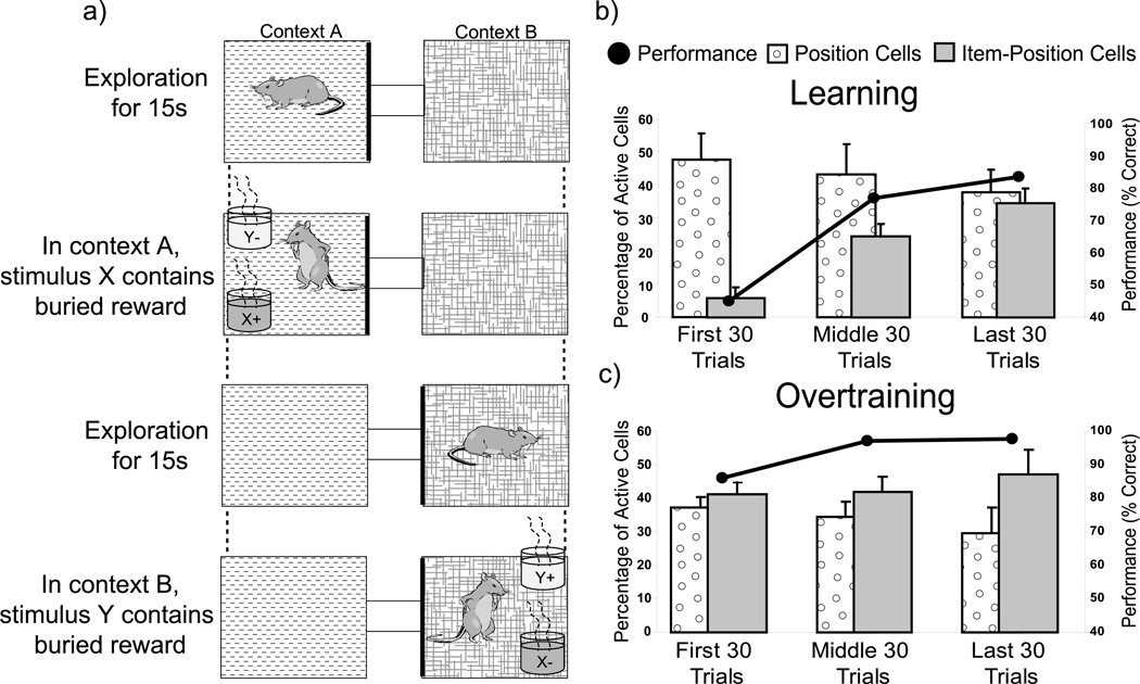
We have also obtained parallel electrophysiological data showing that hippocampal neurons develop representations of stimulus elements (“what”) in the context in which they occur (“where”) in rats while performing a task which requires them to remember what happened where (Komorowski et al., 2009). In this experiment rats moved between environmental contexts that differed in visual, textural, and olfactory cues. On each trial, rats were initially allowed time to orient to the environment; then, they were presented with two cups that were distinguished by both their odors and their digging media. In one environmental context (A), one of the stimuli (X) had a buried reward and the other stimulus (Y) did not, whereas in the other environmental context, the contingency was reversed (Y was baited and X was not; Figure 4a). Therefore the rat had to learn which of the two stimuli had been rewarded within each environment.
Figure 4.
Hippocampal neurons develop item-place representations in parallel with learning what happens where. a. Object-context association task. The two contexts (represented by different shadings) differed in their flooring and wallpaper. The stimulus items (X or Y) differed in odor and in the medium that filled the pots. Items with a plus contained reward, whereas those with a minus did not, each depending upon the spatial context. b. Changes in proportions of Item-Position and Position cells in learning vs. c. overtraining sessions.
We found that rats required several training sessions to acquire an initial problem of this type, but a subsequent second problem with new stimuli and new environmental contexts was typically acquired in the middle of a single 100-trial training session. This rapid learning allowed us to track the firing patterns of single neurons during the course of training on the second problem. We could therefore examine how neuronal firing patterns in the hippocampus might encode the relevant object-context associations.
We focused on the firing rates of hippocampal principal cells in areas CA1 and CA3 for a 1-s period when rats sampled the stimuli during each trial. Early in training, we found that a large percentage of neurons fired when animals sampled either stimulus in a particular location in one of the two environments (Figure 4b; first 30 trials). These likely correspond to so-called “place cells” which fire when rats occupy a location in their environment. Some of these cells maintained the same place-specific firing patterns throughout training. At this stage, the firing patterns of virtually none of the cells distinguished the stimuli. However, as the animals acquired the conditional discrimination, some neurons began to fire selectively during the sampling of one of the objects in one of the contexts and these cells continued to exhibit item-context specificity after learning (Figure 4b; middle 30 trials). The magnitude of item-context representation was robust in that, by the end of the training session, the percentage of hippocampal neurons that fired selectively during the sampling of one of the objects in a particular context equaled that of the percentage of place cells (Figure 4b; last 30 trials). This item-context representation remained strong throughout recording sessions in which animals were highly overtrained on the task (Figure 4c). Thus, a large percentage of hippocampal neurons developed representations of task-relevant item-context associations, and their evolution was closely correlated with learning those associations. Furthermore, subsequent analyses showed that the item-context representations developed from pre-existing spatial representations into enhanced activations when particular items were sampled in specific locations. Conversely, the representation of the items alone was minimal throughout learning and the representation of places where any object was sampled, although strong, remained unchanged throughout training. These findings strongly suggest that the development of conjunctive item-context representations within the hippocampus underlies memories for items in the places where they occur.
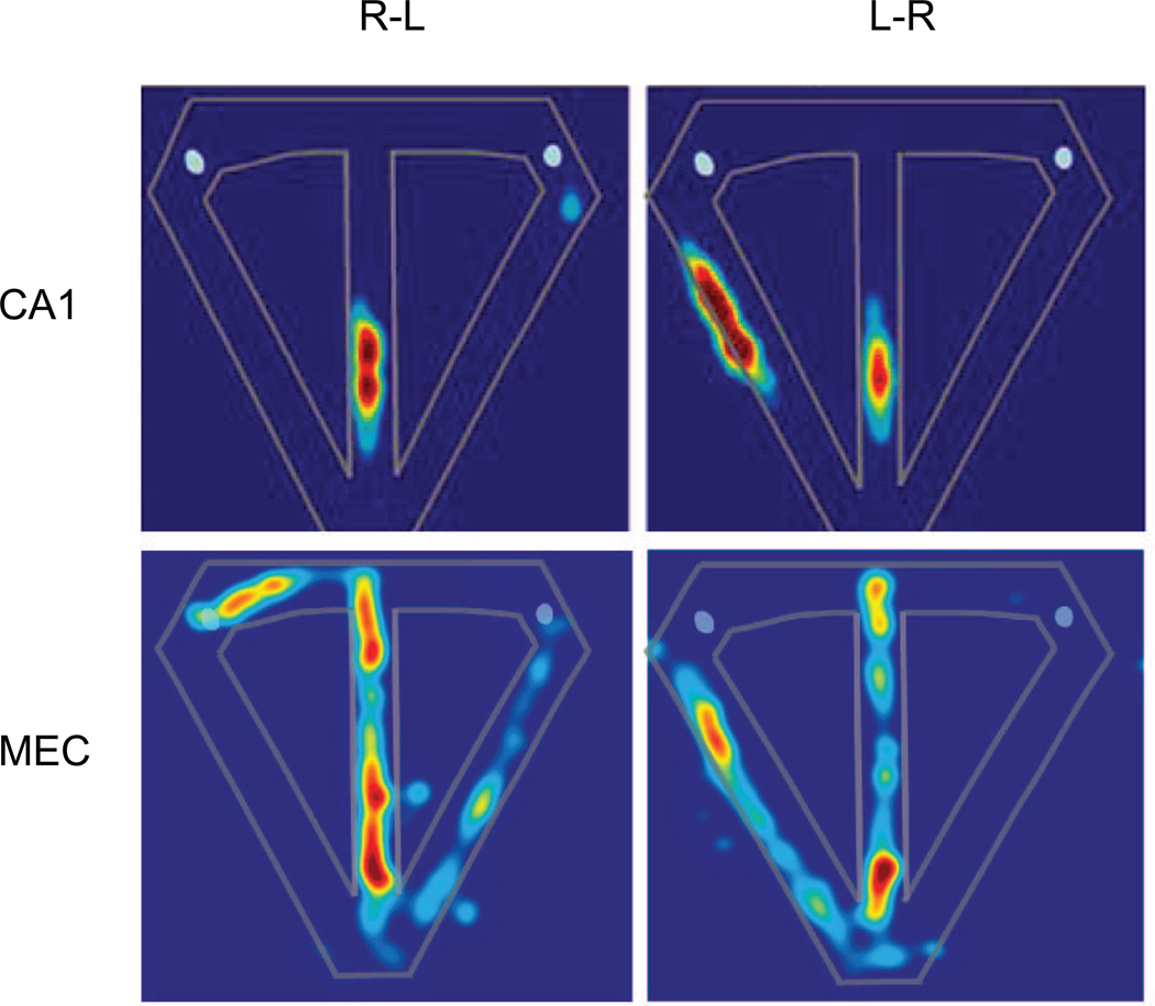
We have also explored the organization of hippocampal neuronal representations in spatial memory, focusing on how hippocampal and medial entorhinal neurons encode sequences of places that compose navigational episodes in a maze. In one study, rats were trained on the classic spatial T-maze alternation task in which successful performance depends on distinguishing left- and right-turn episodes to guide each subsequent choice (Wood et al., 2000). If hippocampal neurons encode each sequential behavioral event within one type of episode, then neuronal activity at locations that overlap in left-to-right and right-to-left turn trials should vary according to the route currently under way. Indeed, virtually all cells that were active as the rat traversed these common locations were differentially active on left-to-right versus right-to-left trials. For example, the CA1 cell shown in Figure 5 fired more strongly as the rat traversed the stem when rats performed a right-to-left trial that when it performed a left-to-right trial. Although most cells exhibited similar quantitative differentiation of trial types, other cells fired exclusively on one type of trial.
Figure 5.
Spatial firing rate plots for example neurons in CA1 and MEC in rats performing a T-maze alternation task. Left panels show firing rate plots for right-to-left trials and right panels show firing rates plots for left-to-right trials. Each trial begins when the rat leaves the reward site (small light-blue circles) on the left or right side of the T-maze (outlined in white), then the rat runs down the start arm, turns into and traverses the central stem, then turns in the direction opposite to the start arm to receive a reward. Note the animal occupies only the central stem on all trials, such that comparisons of firing patterns between left-to-right and right-to-left trials focus on that area alone. Red = highest normalize firing rate; blue = zero firing rate.
Similar results have subsequently been observed in several versions of this task (Bower et al., 2005; Ferbinteanu and Shapiro, 2003; Frank et al., 2000; Griffin et al., 2007; Lee et al., 2006; Ainge et al., 2007; Pastalkova et al., 2008; for review, see Shapiro et al., 2006; but not all versions of the task Lenck-Santini et al., 2001; Bower et al., 2005). Furthermore, these observations are consistent with recent results in animals and humans showing that hippocampal neuronal activity captures sequential events that compose distinct memories (Ginther et al., 2011; Paz et al., 2010). These findings suggest a reconciliation of the current controversy about spatial navigation and episodic memory views of hippocampal function: Place cells represent the series of places where events occur in sequences that compose distinct, “episodic” memories.
We have also begun to examine how the cortical areas of the MTL contribute to spatial episodic memory, and have begun with the medial entorhinal cortex (MEC), the site of the so-called “grid cells” that provide a mapping of the environment (Fyhn et al., 2004). When animals forage in random directions within an open field the grid-like spatial representation is composed from increased firing rates of MEC neurons that form a hexagonal pattern in the environment. It is notable, however, that the grid structure breaks down when animals are constrained to make hairpin turns within the previously unconstrained open field (Derdikman et al., 2009). This observation is important because in the standard, random foraging experimental protocol, spatial cues provide the only regularities and constraints. In contrast, what differed between the hairpin turn maze and the open field condition was the imposition of behavioral constraints while spatial cues were held constant. Importantly, it is only in the unconstrained open field condition that hippocampal cells display purely allocentric spatial firing patterns. It appears that when stimulus or behavioral regularities are imposed, the activity of neurons in medial entorhinal cortex, like neurons in the hippocampus, might reflect the corresponding regularities embedded in the task protocol.
We have also adopted the same spatial memory task used previously (Wood et al., 2000) to compare the activity of hippocampal and medial entorhinal neurons in animals performing a continuous spatial alternation on a T-maze in which hippocampal neurons encode sequences of locations traversed and disambiguate overlapping routes (Lipton et al., 2007). Consistent with previous reports (Fyhn et al., 2004; Hargreaves et al., 2005), activity of neurons in medial entorhinal cortex also signaled an animal’s position along the maze. However, we did not observe the grid-like firing pattern of MEC activity on the T-maze, although some MEC neurons did exhibit a high degree of spatial specificity as the animal traversed multiple loci on the maze (Figure 5). Many of these MEC neurons also exhibited differential firing patterns along the central stem of the maze during the two types of trials, similar to hippocampal neurons. For example, the cell shown in Figure 5 had different multi-peaked patterns of activity for right-to-left and left-to-right trials. This pattern of activity was an exclusive feature of MEC neurons such that, unlike MEC neurons, no hippocampal units had poorly localized, trial-type specific firing that extended the length of the central stem. Consequently, MEC neurons performed better than hippocampal neurons at distinguishing left-to-right and right-to-left trials.
Conversely, hippocampal neurons had smaller place fields, higher spatial tuning, and higher spatial information content than MEC neurons. Therefore, hippocampal neurons showed considerably greater spatial specificity than MEC neurons. The combined results of this study suggest that disambiguation of overlapping experiences occurs prior to the hippocampus, and that hippocampal and medial entorhinal circuits play distinct and complementary roles in the continuous spatial alternation. MEC neurons more successfully distinguished what kind of trial the animal was performing, i.e., stronger coding of temporal or meaningful context, whereas hippocampal neurons more successfully signaled specific events with the trial the animal was performing. Together both regions supply requisite elements of a neural code for particular events as they occur within unique episodes.
These observations suggest that the hippocampus and medial entorhinal cortex play distinct roles in episodic memory. Consistent with the model presented in Figure 2, the MEC may support a representation of the spatiotemporal context that distinguishes the two routes through the maze that distinguish trial episodes. The hippocampus may represent specific sequential events, each signaled by the animal’s location, that compose each type of episode. Broadening this interpretation of MEC as representing the context of each memory, we recently found that damage to the dorsocaudal MEC, the site of the grid cells, results in a selective deficit in the contribution of recollection to the ROC function in recognition memory (Sauvage et al., 2010b), consistent with a role in contextual representations supporting episodic memory (Eichenbaum et al., 2007).
Conclusions
The convergence of findings from behavioral, lesion, and recording approaches in animals strongly supports the idea that different components of the MTL make distinct contributions to the memory capacities that share features with episodic memory in humans. The combined findings suggest that perirhinal cortex plays an essential role in object recognition memory, and conversely, the perirhinal cortex can support relatively intact recognition memory even when the hippocampus is eliminated. Correspondingly, neurons in the perirhinal cortex encode and maintain representations of individual stimuli and signal their familiarity. By contrast, the parahippocampal and medial entorhinal cortex are essential to spatial recognition, and correspondingly, neurons in these areas convey information about spatial contextual features of distinct experiences and not individual stimuli or locations; the role of MEC may also extend to non-spatial context that contributes to recollective memory. The hippocampus makes a selective essential contribution to recognition memory, specifically to the recollection component of item recognition, associative recognition, and memory for spatial context, all of which have in common a demand for representing stimuli in context. Correspondingly, hippocampal neurons encode configurations items and events in the spatial and temporal context in which they were experienced, a central feature of episodic recollection. These findings support a model in which object and event information processed by the perirhinal and lateral entorhinal cortex and information about spatial and temporal context processed by the parahippocampal and medial entorhinal cortex converge on the hippocampus (Figure 2). The hippocampus maps events within a spatio-temporal contextual framework, supporting a “memory space” that binds events and their context and links related memories (Eichenbaum et al., 1999; 2007; Eichenbaum, 2004).
Footnotes
Publisher's Disclaimer: This is a PDF file of an unedited manuscript that has been accepted for publication. As a service to our customers we are providing this early version of the manuscript. The manuscript will undergo copyediting, typesetting, and review of the resulting proof before it is published in its final citable form. Please note that during the production process errors may be discovered which could affect the content, and all legal disclaimers that apply to the journal pertain.
References
- Aggleton JP, Nicol RM, Huston AE, Fairbairn AF. The performance of amnesic subjects on tests of experimental amnesia in animals, delayed matching-to-sample and concurrent learning. Neuropsychologia. 1988;26:265–272. doi: 10.1016/0028-3932(88)90079-6. [DOI] [PubMed] [Google Scholar]
- Ainge JA, Tamosiunaite M, Woergoetter F, Dudchencko PA. Hippocampal CA1 place cells encode intended destination on a maze with multiple choice points. J. Neurosci. 2007;27:9769–9779. doi: 10.1523/JNEUROSCI.2011-07.2007. [DOI] [PMC free article] [PubMed] [Google Scholar]
- Birrell JM, Brown VJ. Medial frontal cortex mediates perceptual attentional set shifting in the rat. J. Neurosci. 2000;20:4320–4324. doi: 10.1523/JNEUROSCI.20-11-04320.2000. [DOI] [PMC free article] [PubMed] [Google Scholar]
- Bower MR, Euston DR, McNaughton BL. Sequential-context-dependent hippocampal activity is not necessary to learn sequences with repeated elements. J. Neurosci. 2005;25:1313–1323. doi: 10.1523/JNEUROSCI.2901-04.2005. [DOI] [PMC free article] [PubMed] [Google Scholar]
- Bowles B, Crupi C, Pigott S, Parrent A, Wiebe S, Janzen L, Köhler S. Double dissociation of selective recollection and familiarity impairments following two different surgical treatments for temporal-lobe epilepsy. Neuropsychologia. 2010;48:2640–2647. doi: 10.1016/j.neuropsychologia.2010.05.010. [DOI] [PubMed] [Google Scholar]
- Broadbent NJ, Squire LR, Clark RE. Spatial memory, recognition memory, and the hippocampus. Proc Natl Acad Sci U S A. 2004;101:14515–14520. doi: 10.1073/pnas.0406344101. [DOI] [PMC free article] [PubMed] [Google Scholar]
- Brown MW, Aggleton JP. Recognition memory: what are the roles of the perirhinal cortex and hippocampus? Nat Rev Neurosci. 2001;2:51–61. doi: 10.1038/35049064. [DOI] [PubMed] [Google Scholar]
- Brown MW, Xiang JZ. Recognition memory, Neuronal substrates of the judgment of prior occurrence. Prog. in Neurobiol. 1998;55:149–189. doi: 10.1016/s0301-0082(98)00002-1. [DOI] [PubMed] [Google Scholar]
- Brown MW, Wilson FA, Riches IP. Neuronal evidence that inferomedial temporal cortex is more important than hippocampus in certain processes underlying recognition memory. Brain Res. 1987;409:158–162. doi: 10.1016/0006-8993(87)90753-0. [DOI] [PubMed] [Google Scholar]
- Buckmaster CA, Eichenbaum H, Amaral DG, Suzuki WA, Rapp P. Entorhinal cortex lesions disrupt the relational organization of memory in monkeys. J. Neurosci. 2004;24:9811–9825. doi: 10.1523/JNEUROSCI.1532-04.2004. [DOI] [PMC free article] [PubMed] [Google Scholar]
- Burwell RD, Hafeman DM. Positional firing properties of postrhinal cortex neurons. Neuroscience. 2003;119:577–588. doi: 10.1016/s0306-4522(03)00160-x. [DOI] [PubMed] [Google Scholar]
- Burwell RD, Shapiro ML, O'Malley MT, Eichenbaum H. Positional firing properties of perirhinal cortex neurons. Neuroreport. 1998;9:3013–3018. doi: 10.1097/00001756-199809140-00017. [DOI] [PubMed] [Google Scholar]
- Cahusac PMB, Rolls ET, Miyashita Y, Niki H. Modification of the responses of hippocampal neurons in the monkey during the learning of a conditional spatial response task. Hippocampus. 1993;3:29–42. doi: 10.1002/hipo.450030104. [DOI] [PubMed] [Google Scholar]
- Clark RE, Zola SM, Squire LR. Impaired recognition memory in rats after damage to the hippocampus. J. Neurosci. 2000;20:8853–8860. doi: 10.1523/JNEUROSCI.20-23-08853.2000. [DOI] [PMC free article] [PubMed] [Google Scholar]
- Clark RE, West AN, Zola SM, Squire LR. Rats with lesions of the hippocampus are impaired on the delayed nonmatching-to-sample task. Hippocampus. 2001;11:176–186. doi: 10.1002/hipo.1035. [DOI] [PubMed] [Google Scholar]
- Daselaar SM, Fleck MS, Cabeza RE. Triple Dissociation in the Medial Temporal Lobes, Recollection, Familiarity, and Novelty. J. Neurophysiol. 2006;96:1902–1911. doi: 10.1152/jn.01029.2005. [DOI] [PubMed] [Google Scholar]
- Davachi L. Item, context and relational episodic encoding in humans. Curr. Opin. Neurobiol. 2006;16:693–700. doi: 10.1016/j.conb.2006.10.012. [DOI] [PubMed] [Google Scholar]
- Diana RA, Yonelinas AP, Ranganath C. Imaging recollection and familiarity in the medial temporal lobe: a three-component model. Trends Cogn Sci. 2007;11:379–386. doi: 10.1016/j.tics.2007.08.001. [DOI] [PubMed] [Google Scholar]
- Diana RA, Yonelinas AP, Ranganath C. The effects of unitization on familiarity-based source memory: testing a behavioral prediction derived from neuroimaging data. J. Exper Psychol: Learning, memory, and cognition. 2008;34:730–740. doi: 10.1037/0278-7393.34.4.730. [DOI] [PMC free article] [PubMed] [Google Scholar]
- Deadwyler SE, Bunn T, Hampson RE. Hippocampal ensemble activity during spatial delayed nonmatch to sample performance in rats. J. Neurosci. 1996;16:354–372. doi: 10.1523/JNEUROSCI.16-01-00354.1996. [DOI] [PMC free article] [PubMed] [Google Scholar]
- Derdikman D, Whitlock JR, Tsao A, Fyhn M, Hafting T, Moser MB, Moser EI. Fragmentation of grid cell maps in a multicompartment environment. Nat. Neurosci. 2009;12:1325–1332. doi: 10.1038/nn.2396. [DOI] [PubMed] [Google Scholar]
- Desimone R, Miller EK, Chelazzi L, Lueschow A. Multiple memory systems in the visual cortex. In: Gazzaniga MS, editor. The Cognitive neurosciences. Cambridge, MA: MIT Press; 1995. pp. 475–486. [Google Scholar]
- Dudchencko P, Wood E, Eichenbaum H. Neurotoxic hippocampal lesions have no effect on odor span and little effect on odor recognition memory, but produce significant impairments on spatial span, recognition, and alternation. J. Neurosci. 2000;20:2964–2977. doi: 10.1523/JNEUROSCI.20-08-02964.2000. [DOI] [PMC free article] [PubMed] [Google Scholar]
- Eacott MJ, Norman G. Integrated memory for object, place, and context in rats, a possible model of episodic-like memory? J Neurosci. 2004;24:1948–1953. doi: 10.1523/JNEUROSCI.2975-03.2004. [DOI] [PMC free article] [PubMed] [Google Scholar]
- Eichenbaum H, Dudchencko P, Wood E, Shapiro M, Tanila H. The hippocampus, memory, and place cells, Is it spatial memory or a memory space? Neuron. 1999;23:209–226. doi: 10.1016/s0896-6273(00)80773-4. [DOI] [PubMed] [Google Scholar]
- Eichenbaum H. Hippocampus, Cognitive processes and neural representations that underlie declarative memory. Neuron. 2004;44:109–120. doi: 10.1016/j.neuron.2004.08.028. [DOI] [PubMed] [Google Scholar]
- Eichenbaum H, Yonelinas AP, Ranganath C. The medial temporal lobe and recognition memory. Annual Review of Neuroscience. 2007;30:123–152. doi: 10.1146/annurev.neuro.30.051606.094328. [DOI] [PMC free article] [PubMed] [Google Scholar]
- Eichenbaum H. Neuroscience. Dedicated to Memory? Science. 2010;330:1331–1332. doi: 10.1126/science.1199462. [DOI] [PubMed] [Google Scholar]
- Ekstrom AD, Kahana MJ, Caplan JB, Fields TA, Isham EA, Newman EL, Fried I. Cellular networks underlying human spatial navigation. Nature. 2003;425:184–187. doi: 10.1038/nature01964. [DOI] [PubMed] [Google Scholar]
- Ennaceur A, Neave N, Aggleton JP. Neurotoxic lesions of the perirhinal cortex do not mimic the behavioural effects of fornix transection in the rat. Behav Brain Res. 1996;80:9–25. doi: 10.1016/0166-4328(96)00006-x. [DOI] [PubMed] [Google Scholar]
- Ergorul C, Eichenbaum H. The hippocampus and memory for "what," "where," and "when". Learning and Memory. 2004;11:397–405. doi: 10.1101/lm.73304. [DOI] [PMC free article] [PubMed] [Google Scholar]
- Feigenbaum JD, Rolls ET. Allocentric and egocentric information processing in the hippocampal formation of the behaving primate. Psychobiology. 1991;19:21–40. [Google Scholar]
- Ferbinteanu J, Shapiro ML. Prospective and retrospective memory coding in the hippocampus. Neuron. 2003;40:1227–1239. doi: 10.1016/s0896-6273(03)00752-9. [DOI] [PubMed] [Google Scholar]
- Fortin NJ, Wright SP, Eichenbaum H. Recollection-like memory retrieval in rats is dependent on the hippocampus. Nature. 2004;431:188–191. doi: 10.1038/nature02853. [DOI] [PMC free article] [PubMed] [Google Scholar]
- Frank L, Brown EN, Wilson M. Trajectory encoding in the hippocampus and entorhinal cortex. Neuron. 2000;27:169–178. doi: 10.1016/s0896-6273(00)00018-0. [DOI] [PubMed] [Google Scholar]
- Furtak SC, Wei S-M, Agster KL, Burwell RD. Functional neuroanatomy of the parahippocampal region in the rat. The perirhinal and postrhinal cortices. Hippocampus. 2007;17:709–722. doi: 10.1002/hipo.20314. [DOI] [PubMed] [Google Scholar]
- Fuster JM. Memory in the cerebral cortex: An empirical approach to neural networks in the human and nonhuman primate. Cambridge: MIT Press; 1995. [Google Scholar]
- Fyhn M, Molden S, Witter MP, Moser EI, Moser MB. Spatial representation in the entorhinal cortex. Science. 2004;305:1258–1264. doi: 10.1126/science.1099901. [DOI] [PubMed] [Google Scholar]
- Fyhn M, Molden S, Witter MP, Moser EI, Moser M. Spatial representation in the entorhinal cortex. Science. 2004;305:1258–1264. doi: 10.1126/science.1099901. [DOI] [PubMed] [Google Scholar]
- Gaffan D. Recognition impaired and association intact in the memory of monkeys after transection of the fornix. J Comp Physiol Psychol. 1974;86:1100–1109. doi: 10.1037/h0037649. [DOI] [PubMed] [Google Scholar]
- Ginther MR, Walsh DF, Ramus SJ. Hippocampal neurons encode different episodes in an overlapping sequence of odors task. J. Neurosci. 2011;31:2706–2711. doi: 10.1523/JNEUROSCI.3413-10.2011. [DOI] [PMC free article] [PubMed] [Google Scholar]
- Giovanello KS, Keane MM, Verfaellie M. The contribution of familiarity to associative memory in amnesia. Neuropsychologia. 2006;44:1859–1865. doi: 10.1016/j.neuropsychologia.2006.03.004. [DOI] [PMC free article] [PubMed] [Google Scholar]
- Griffin AL, Eichenbaum H, Hasselmo ME. Spatial representations of hippocampal CA1 neurons are modulated by behavioral context in a hippocampus-dependent memory task. J. Neurosci. 2007;27:2416–2423. doi: 10.1523/JNEUROSCI.4083-06.2007. [DOI] [PMC free article] [PubMed] [Google Scholar]
- Hammond RS, Tull LE, Stackman RW. On the delay-dependent involvement of the hippocampus in object recognition memory. Neurobiol Learn Mem. 2004;82:26–34. doi: 10.1016/j.nlm.2004.03.005. [DOI] [PubMed] [Google Scholar]
- Hampson RE, Heyser CJ, Deadwyler SA. Hippocampal cell firing correlates of delayed-match-to-sample performance in the rat. Behav. Neurosci. 1993;107:715–739. doi: 10.1037//0735-7044.107.5.715. [DOI] [PubMed] [Google Scholar]
- Hargreaves EL, Rao G, Lee I, Knierim JJ. Major dissociation between medial and lateral entorhinal input to dorsal hippocampus. Science. 2005;308:1792–1794. doi: 10.1126/science.1110449. [DOI] [PubMed] [Google Scholar]
- Jenkins TA, Amin E, Pearce JM, Brown MW, Aggleton JP. Novel spatial arrangements of familiar visual stimuli promote activity in the rat hippocampal formation but not the parahippocampal cortices: a c-fos expression study. Neuroscience. 2004;124:43–52. doi: 10.1016/j.neuroscience.2003.11.024. [DOI] [PubMed] [Google Scholar]
- Kerr KM, Agster KL, Furtak SC, Burwell RD. Functional neuroanatomy of the parahippocampal region, The lateral and medial entorhinal areas. Hippocampus. 2007;17:697–708. doi: 10.1002/hipo.20315. [DOI] [PubMed] [Google Scholar]
- Komorowski RW, Manns JR, Eichenbaum H. Robust conjunctive item-place coding by hippocampal neurons parallels learning what happens where. J. Neurosci. 2009;29:9918–9929. doi: 10.1523/JNEUROSCI.1378-09.2009. [DOI] [PMC free article] [PubMed] [Google Scholar]
- Kreiman G, Koch C, Fried I. Category specific visual responses of single neurons in the human medial temporal lobe. Nat. Neurosci. 2000;3:946–953. doi: 10.1038/78868. [DOI] [PubMed] [Google Scholar]
- Langston RF, Wood ER. Associative recognition and the hippocampus: Differential effects of hippocampal lesions on object-place, object-context and object-place-context memory. Hippocampus. 2010;20:1139–1153. doi: 10.1002/hipo.20714. [DOI] [PubMed] [Google Scholar]
- Lee I, Hunsaker MR, Kesner RP. The role of hippocampal subregions in detecting spatial novelty. Beh. Neurosci. 2005;119:145–153. doi: 10.1037/0735-7044.119.1.145. [DOI] [PubMed] [Google Scholar]
- Lee I, Griffin AL, Zilli EA, Eichenbaum H, Hasselmo ME. Gradual translocation of spatial correlates of neuronal firing in the hippocampus toward prospective locations. Neuron. 2006;51:639–650. doi: 10.1016/j.neuron.2006.06.033. [DOI] [PubMed] [Google Scholar]
- Lench-Santini PP, Save E, Poucet B. Place-cell firing does not depend on the direction of turn in a Y-maze alternation task. Eur, J. Neurosci. 2001;13:1055–1058. doi: 10.1046/j.0953-816x.2001.01481.x. [DOI] [PubMed] [Google Scholar]
- Lipton PA, White JA, Eichenbaum H. Disambiguation of overlapping experiences by neurons in medial entorhinal cortex. J. Neurosci. 2007;27:5787–5795. doi: 10.1523/JNEUROSCI.1063-07.2007. [DOI] [PMC free article] [PubMed] [Google Scholar]
- Macmillan NA, Creelman CD. Detection theory, A user's guide. New York, NY: Cambridge University Press; 1991. [Google Scholar]
- Mandler G. Familiarity breeds attempts, A critical review of dual process theories of recognition. Perspect. Psychol. Sci. 2008;3:392–401. doi: 10.1111/j.1745-6924.2008.00087.x. [DOI] [PubMed] [Google Scholar]
- Meunier M, Bachevalier J, Mishkin M, Murray EA. Effects on visual recognition of combined and separate ablations of the entorhinal and perirhinal cortex in rhesus monkeys. J. Neurosci. 1993;13:5418–6532. doi: 10.1523/JNEUROSCI.13-12-05418.1993. [DOI] [PMC free article] [PubMed] [Google Scholar]
- Miller EK, Li L, Desimone R. A neural mechanism for working and recognition memory in inferior temporal cortex. Science. 1991;254:1377–1379. doi: 10.1126/science.1962197. [DOI] [PubMed] [Google Scholar]
- Mishkin M, Delacour J. An analysis of short-term visual memory in the monkey. J. Exp. Psychol. Anim. Behav. Process. 1975;1:326–334. doi: 10.1037//0097-7403.1.4.326. [DOI] [PubMed] [Google Scholar]
- Mishkin M. Memory in monkeys severely impaired by combined but not by separate removal of amygdala and hippocampus. Nature. 1978;273:297–298. doi: 10.1038/273297a0. [DOI] [PubMed] [Google Scholar]
- Moita MAP, Moisis S, Zhou Y, LeDoux JE, Blair HT. Hippocampal place cells acquire location specific location specific responses to the conditioned stimulus during auditory fear conditioning. Neuron. 2003;37:485–497. doi: 10.1016/s0896-6273(03)00033-3. [DOI] [PubMed] [Google Scholar]
- Moita MAP, Rosis S, Zhou Y, LeDoux JE, Blair HT. Putting fear in its place, Remapping of hippocampal place cells during fear conditioning. J. Neurosci. 2004;24:7015–7023. doi: 10.1523/JNEUROSCI.5492-03.2004. [DOI] [PMC free article] [PubMed] [Google Scholar]
- Montaldi D, Mayes AR. The role of recollection and familiarity in the functional differentiation of the medial temporal lobes. Hippocampus. 2010;20:1291–1314. doi: 10.1002/hipo.20853. [DOI] [PubMed] [Google Scholar]
- Moses SN, Sutherland RJ, McDonald RJ. Differential involvement of amygdala and hippocampus in responding to novel objects and contexts. Brain Res. Bull. 2002;58:517–527. doi: 10.1016/s0361-9230(02)00820-1. [DOI] [PubMed] [Google Scholar]
- Mumby DG, Pinel PJ. Rhinal cortex lesions and object recognition in rats. Behav. Neurosci. 1994;108:11–18. doi: 10.1037//0735-7044.108.1.11. [DOI] [PubMed] [Google Scholar]
- Mumby DG, Wood ER, Pinel JP. Object recognition memory is only mildly impaired in rats with lesions of the hippocampus and amygdala. Psychobiology. 1992;20:18–27. [Google Scholar]
- Mumby DG, Mana MJ, Pinel JP, David E, Banks K. Pyrithiamine-induced thiamine deficiency impairs object recognition in rats. Behav Neurosci. 1995;109:1209–1214. [PubMed] [Google Scholar]
- Mumby DG, Glenn MJ, Nesbitt C, Kyriazis DA. Dissociation in retrograde memory for object discriminations and object recognition in rats with perirhinal cortex damage. Behav Brain Res. 2002;132:215–226. doi: 10.1016/s0166-4328(01)00444-2. [DOI] [PubMed] [Google Scholar]
- Mumby DG. Perspectives on object-recognition memory following hippocampal damage, lessons from studies in rats. Behav Brain Res. 2001;127:159–181. doi: 10.1016/s0166-4328(01)00367-9. [DOI] [PubMed] [Google Scholar]
- Murray EA, Mishkin M. Object recognition and location memory in monkeys with excitotoxic lesions of the amygdala and hippocampus. J. Neurosci. 1998;18:6568–6582. doi: 10.1523/JNEUROSCI.18-16-06568.1998. [DOI] [PMC free article] [PubMed] [Google Scholar]
- Nemanic S, Alvarado MC, Bachevalier J. The hippocampal/parahippocampal regions and recognition memory, insights from visual paired comparison versus object-delayed nonmatching in monkeys. J. Neurosci. 2004;24:2013–2026. doi: 10.1523/JNEUROSCI.3763-03.2004. [DOI] [PMC free article] [PubMed] [Google Scholar]
- Norman G, Eacott MJ. Impaired object recognition with increasing levels of feature ambiguity in rats with perirhinal cortex lesions. J. Neurosci. 2004;148:79–91. doi: 10.1016/s0166-4328(03)00176-1. [DOI] [PubMed] [Google Scholar]
- Norman G, Eacott MJ. Dissociable effects of lesions to the perirhinal cortex and the postrhinal cortex on memory for context and objects in rats. Behav. Neurosci. 2005;119:557–566. doi: 10.1037/0735-7044.119.2.557. [DOI] [PubMed] [Google Scholar]
- Otto T, Eichenbaum H. Neuronal activity in the hippocampus during delayed non-match to sample performance in rats: Evidence for hippocampal processing in recognition memory. Hippocampus. 1992a;2:323–334. doi: 10.1002/hipo.450020310. [DOI] [PubMed] [Google Scholar]
- Otto T, Eichenbaum H. Complementary roles of the orbital prefrontal cortex and the perirhinal-entorhinal cortices in an odor-guided delayed-nonmatching-to-sample task. Behav. Neurosci. 1992b;106:762–775. doi: 10.1037//0735-7044.106.5.762. [DOI] [PubMed] [Google Scholar]
- Pastalkova E, Itskov V, Amarasingham A, Buzsáki G. Internally generated cell assembly sequences in the rat hippocampus. Science. 2008;321:322–327. doi: 10.1126/science.1159775. [DOI] [PMC free article] [PubMed] [Google Scholar]
- Paz R, Gelbard-Sagiv H, Mukamel R, Harel M, Malach R, Fried I. A neural substrate in the human hippocampus for linking successive events. Proc. Natl. Acad. Sci. U S A. 2010;107:6046–6051. doi: 10.1073/pnas.0910834107. [DOI] [PMC free article] [PubMed] [Google Scholar]
- Prusky GT, Douglas RM, Nelson L, Shabanpoor A, Sutherland RJ. Visual memory task for rats reveals an essential role for hippocampus and perirhinal cortex. Proc. Natl. Acad. Sci. USA. 2004;101:5064–5068. doi: 10.1073/pnas.0308528101. [DOI] [PMC free article] [PubMed] [Google Scholar]
- Quamme JR, Yonelinas AP, Norman KA. Effect of unitization on associative recognition in amnesia. Hippocampus. 2007;17:192–200. doi: 10.1002/hipo.20257. [DOI] [PubMed] [Google Scholar]
- Quirk GJ, Muller RU, Kubie JL, Ranck JB., Jr The positional firing properties of medial entorhinal neurons, Description and comparison with hippocampal place cells. J. Neurosci. 1992;12:1945–1963. doi: 10.1523/JNEUROSCI.12-05-01945.1992. [DOI] [PMC free article] [PubMed] [Google Scholar]
- Rampon C, Tang Y-P, Goodhouse J, Shimizu E, Kyin M, Tsien J. Enrichment induces structural changes and recovery from non-spatial memory deficits in CA1 NMDAR1-knockout mice. Nat. Neurosci. 2000;3:238–244. doi: 10.1038/72945. [DOI] [PubMed] [Google Scholar]
- Ranganath C, Yonelinas AP, Cohen MX, Dy CJ, Tom SM, D'Esposito M. Dissociable correlates of recollection and familiarity within the medial temporal lobes. Neuropsychologia. 2003;42:2–13. doi: 10.1016/j.neuropsychologia.2003.07.006. [DOI] [PubMed] [Google Scholar]
- Riches IP, Wilson FAW, Brown MW. The effects of visual stimulation and memory on neurons of the hippocampal formation and the neighboring parahippocampal gyrus and inferior temporal cortex of the primate. J. Neurosci. 1991;11:1763–1779. doi: 10.1523/JNEUROSCI.11-06-01763.1991. [DOI] [PMC free article] [PubMed] [Google Scholar]
- Riches IP, Wilson FAW, Brown MW. Neurones of the medial temporal lobes and recognition memory. In: Haas HL, Buzsaki G, editors. Synaptic Plasticity in the Hippocampus. Berlin: Springer-Verlag; 1988. pp. 193–197. [Google Scholar]
- Rolls ET, Baylis GC, Hasselmo ME, Nalwa V. The effect of learning on the face selective responses of neurons in the cortex in the superior temporal sulcus of the monkey. Exp. Brain Res. 1989;76:153–164. doi: 10.1007/BF00253632. [DOI] [PubMed] [Google Scholar]
- Rutishauser U, Mamelak AN, Schuman EM. Single-trial learning of novel stimuli by individual neurons of the human hippocampus-amygdala complex. Neuron. 2006;49:805–813. doi: 10.1016/j.neuron.2006.02.015. [DOI] [PubMed] [Google Scholar]
- Sakurai Y. Involvement of auditory cortical and hippocampal neurons in auditory working memory and reference memory in the rat. J. Neurosci. 1994;14:2606–2623. doi: 10.1523/JNEUROSCI.14-05-02606.1994. [DOI] [PMC free article] [PubMed] [Google Scholar]
- Sauvage MM, Fortin NJ, Owens CB, Yonelinas AP, Eichenbaum H. Recognition memory: Opposite effects of hippocampal damage on recollection and familiarity. Nature Neuroscience. 2008;11:16–18. doi: 10.1038/nn2016. [DOI] [PMC free article] [PubMed] [Google Scholar]
- Sauvage MM, Beer Z, Eichenbaum H. Recognition memory: Adding a response deadline eliminates recollection but spares familiarity. Learning and Memory. 2010a;17:104–108. doi: 10.1101/lm.1647710. [DOI] [PMC free article] [PubMed] [Google Scholar]
- Sauvage MM, Beer Z, Ekovich M, Ho L, Eichenbaum H. The caudal medial entorhinal cortex: a selective role in recollection-based recognition memory. J. Neurosci. 2010b;30:15695–15699. doi: 10.1523/JNEUROSCI.4301-10.2010. [DOI] [PMC free article] [PubMed] [Google Scholar]
- Save E, Poucet B, Foreman N, Buhot MC. Object exploration and reactions to spatial and nonspatial changes in hooded rats following damage to parietal cortex or hippocampal formation. Behav. Neurosci. 1992;106:447–456. [PubMed] [Google Scholar]
- Shapiro ML, Kennedy PJ, Ferbinteanu J. Representing episodes in the mammalian brain. Curr. Opin, Neurobiol. 2006;16:701–709. doi: 10.1016/j.conb.2006.08.017. [DOI] [PubMed] [Google Scholar]
- Squire LR, Zola-Morgan S, Chen KS. Human amnesia and animal models of amnesia, performance of amnesic patients on tests designed for the monkey. Behav. Neurosci. 1988;102:210–221. doi: 10.1037//0735-7044.102.2.210. [DOI] [PubMed] [Google Scholar]
- Squire LR, Stark CEL, Clark RE. The medial temporal lobe. Ann. Rev. Neurosci. 2004;27:279–306. doi: 10.1146/annurev.neuro.27.070203.144130. [DOI] [PubMed] [Google Scholar]
- Squire LR, Wixted JT, Clark RE. Recognition memory and the medial temporal lobe, A new perspective. Nat. Rev. Neurosci. 2007;8:872–883. doi: 10.1038/nrn2154. [DOI] [PMC free article] [PubMed] [Google Scholar]
- Steckler T, Drinkenburg WH, Saghal A, Aggleton JP. Recognition memory in rats II. Neuroanatomical substrates. Prog. Neurobiol. 1998;54:313–332. doi: 10.1016/s0301-0082(97)00061-0. [DOI] [PubMed] [Google Scholar]
- Steele K, Rawlins JN. The effects of hippocampectomy on performance by rats of a running recognition task using long lists of non-spatial items. Behav. Brain Res. 1993;54:1–10. doi: 10.1016/0166-4328(93)90043-p. [DOI] [PubMed] [Google Scholar]
- Suzuki WA, Amaral DG. Perirhinal and parahippocampal cortices of the macaque monkey. Cortical afferents. J. Comp. Neurol. 1994;350:497–533. doi: 10.1002/cne.903500402. [DOI] [PubMed] [Google Scholar]
- Suzuki W, Eichenbaum H. The neurophysiology of memory. Ann NY Acad Sci. 2000;911:175–191. doi: 10.1111/j.1749-6632.2000.tb06726.x. [DOI] [PubMed] [Google Scholar]
- Van Strien NM, Cappaert NL, Witter MP. The anatomy of memory: an interactive overview of the parahippocampal-hippocampal network. Nat. Rev. Neurosci. 2009;10:272–282. doi: 10.1038/nrn2614. [DOI] [PubMed] [Google Scholar]
- Vann SD, Brown MW, Erichsen JT, Aggleton JP. Fos imaging reveals differential patterns of hippocampal and parahippocampal subfield activation in rats in response to different spatial memory tests. J. Neurosci. 2000;20:2711–2718. doi: 10.1523/JNEUROSCI.20-07-02711.2000. [DOI] [PMC free article] [PubMed] [Google Scholar]
- Wan H, Aggleton JP, Brown MW. Different contributions of the hippocampus and perirhinal cortex to recognition memory. J. Neurosci. 1999;19:1142–1148. doi: 10.1523/JNEUROSCI.19-03-01142.1999. [DOI] [PMC free article] [PubMed] [Google Scholar]
- Wang Q, Enquan G, Burkhalter A. Gateways of ventral and dorsal streams in mouse visual cortex. J. Neurosci. 2011;31:1905–1918. doi: 10.1523/JNEUROSCI.3488-10.2011. [DOI] [PMC free article] [PubMed] [Google Scholar]
- Wible CG, Findling RL, Shapiro M, Lang EJ, Crane S, Olton DS. Mnemonic correlates of unit activity in the hippocampus. Brain Res. 1986;399:97–110. doi: 10.1016/0006-8993(86)90604-9. [DOI] [PubMed] [Google Scholar]
- Wiebe SP, Staubli UV. Dynamic filtering of recognition memory codes in the hippocampus. J. Neurosci. 1999;19:10562–10574. doi: 10.1523/JNEUROSCI.19-23-10562.1999. [DOI] [PMC free article] [PubMed] [Google Scholar]
- Wiig KA, Bilkey DK. Lesions of rat perirhinal cortex exacerbate the memory deficit observed following damage to the fimbria-fornix. Behav. Neurosci. 1995;109:620–630. doi: 10.1037//0735-7044.109.4.620. [DOI] [PubMed] [Google Scholar]
- Winters BD, Bussey TJ. Transient inactivation of perirhinal cortex disrupts encoding, retrieval, and consolidation of object recognition memory. J. Neurosci. 2005a;25:52–61. doi: 10.1523/JNEUROSCI.3827-04.2005. [DOI] [PMC free article] [PubMed] [Google Scholar]
- Winters BD, Bussey TJ. Glutamate receptors in perirhinal cortex mediate encoding, retrieval and consolidation of object recognition memory. J. Neurosci. 2005b;25:4243–4251. doi: 10.1523/JNEUROSCI.0480-05.2005. [DOI] [PMC free article] [PubMed] [Google Scholar]
- Winters BD, Forwood SE, Cowell RA, Saksida LM, Bussey TJ. Double dissociation between the effects of peri-postrhinal cortex and hippocampal lesions on tests of object recognition and spatial memory, Heterogeneity of function within the temporal lobe. J. Neurosci. 2004;24:5901–5908. doi: 10.1523/JNEUROSCI.1346-04.2004. [DOI] [PMC free article] [PubMed] [Google Scholar]
- Wirth S, Yanike M, Frank LM, Smith AC, Brown EN, Suzuki WA. Single neurons in the monkey hippocampus and learning of new associations. Science. 2003;300:1578–1581. doi: 10.1126/science.1084324. [DOI] [PubMed] [Google Scholar]
- Wixted JT, Squire LR. The role of the human hippocampus in familiarity-based and recollection-based recognition memory. Behav. Brain. Res. 2010;215:197–208. doi: 10.1016/j.bbr.2010.04.020. [DOI] [PMC free article] [PubMed] [Google Scholar]
- Wixted JT, Mickes L, Squire LR. Measuring recollection and familiarity in the medial temporal lobe. Hippocampus. 2010;20:1195–1205. doi: 10.1002/hipo.20854. [DOI] [PMC free article] [PubMed] [Google Scholar]
- Wood E, Dudchenko PA, Eichenbaum H. The global record of memory in hippocampal neuronal activity. Nature. 1999;397:613–616. doi: 10.1038/17605. [DOI] [PubMed] [Google Scholar]
- Wood E, Dudchenko P, Robitsek JR, Eichenbaum H. Hippocampal neurons encode information about different types of memory episodes occurring in the same location. Neuron. 2000;27:623–633. doi: 10.1016/s0896-6273(00)00071-4. [DOI] [PubMed] [Google Scholar]
- Wood ER, Dudchenko PA, Robitsek RJ, Eichenbaum H. Hippocampal neurons encode information about different types of memory episodes occurring in the same location. Neuron. 2000;27:623–633. doi: 10.1016/s0896-6273(00)00071-4. [DOI] [PubMed] [Google Scholar]
- Yanike M, Wirth S, Suzuki WA. Representations of well-learned information in the monkey hippocampus. Neuron. 2004;42:477–487. doi: 10.1016/s0896-6273(04)00193-x. [DOI] [PubMed] [Google Scholar]
- Yee BK, Rawlins JN. The effects of hippocampal formation ablation or fimbria-fornix section on performance of a nonspatial radial arm maze task by rats. J. Neurosci. 1994;14(6):3766–3774. doi: 10.1523/JNEUROSCI.14-06-03766.1994. [DOI] [PMC free article] [PubMed] [Google Scholar]
- Yonelinas AP. Recognition memory ROCs for item and associative information, the contribution of recollection and familiarity. Memory and Cognition. 1997;25:747–763. doi: 10.3758/bf03211318. [DOI] [PubMed] [Google Scholar]
- Yonelinas AP. The contribution of recollection and familiarity to recognition and source-memory judgments, A formal dual-process model and an analysis of receiver operating characteristics. J. Exp. Psychol. Learn Mem. Cogn. 1999;25:1415–1434. doi: 10.1037//0278-7393.25.6.1415. [DOI] [PubMed] [Google Scholar]
- Yonelinas AP. Components of episodic memory, the contribution of recollection and familiarity. Phil. Trans. Roy. Soc. Lond. Biol. Sci. 2001;356:1363–1374. doi: 10.1098/rstb.2001.0939. [DOI] [PMC free article] [PubMed] [Google Scholar]
- Yonelinas AP. The nature of recollection and familiarity, A review of 30 years of research. J. Mem. Lang. 2002;46:441–517. [Google Scholar]
- Young BJ, Fox GD, Eichenbaum H. Correlates of hippocampal complex-spike cell activity in rats performing a nonspatial radial maze task. J. Neurosci. 1994;14:6553–6563. doi: 10.1523/JNEUROSCI.14-11-06553.1994. [DOI] [PMC free article] [PubMed] [Google Scholar]
- Young BJ, Otto T, Fox GD, Eichenbaum H. Memory representation within the parahippocampal region. J. Neurosci. 1997;17:5183–5195. doi: 10.1523/JNEUROSCI.17-13-05183.1997. [DOI] [PMC free article] [PubMed] [Google Scholar]
- Zhu XO, Brown MW, Aggleton JP. Neuronal signaling of information important to visual recognition memory in rat rhinal and neighbouring cortices. Eur. J. Neurosci. 1995;7:753–765. doi: 10.1111/j.1460-9568.1995.tb00679.x. [DOI] [PubMed] [Google Scholar]
- Zola SM, Squire LR, Teng E, Stefanacci L, Buffalo EA, Clark RE. Impaired recognition memory in monkeys after damage limited to the hippocampal region. J. Neurosci. 2000;20:451–643. doi: 10.1523/JNEUROSCI.20-01-00451.2000. [DOI] [PMC free article] [PubMed] [Google Scholar]
- Zola-Morgan S, Squire LR. Medial temporal lesions in monkeys impair memory on a variety of tasks sensitive to human amnesia. Behav Neurosci. 1985;99:22–34. doi: 10.1037//0735-7044.99.1.22. [DOI] [PubMed] [Google Scholar]
- Zola-Morgan S, Squire LR, Amaral DC, Suzuki WA. Lesions of perirhinal and parahippocampal cortex that spare the amygdala and hippocampal formation produce sever memory impairment. J. Neurosci. 1989;9:4355–4370. doi: 10.1523/JNEUROSCI.09-12-04355.1989. [DOI] [PMC free article] [PubMed] [Google Scholar]


