

Abstract
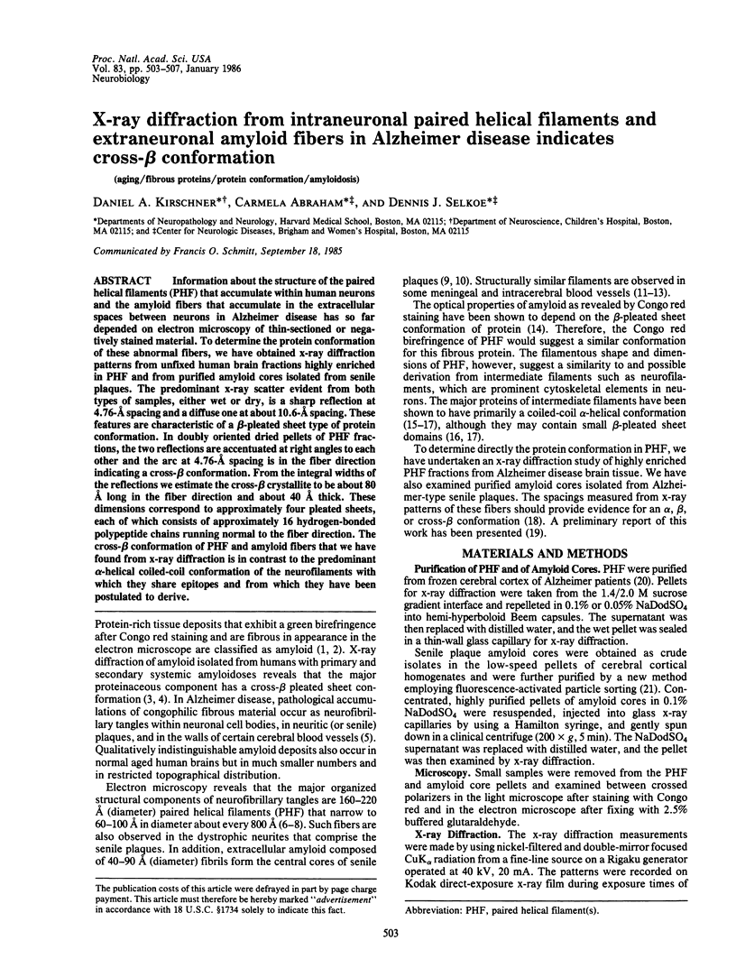
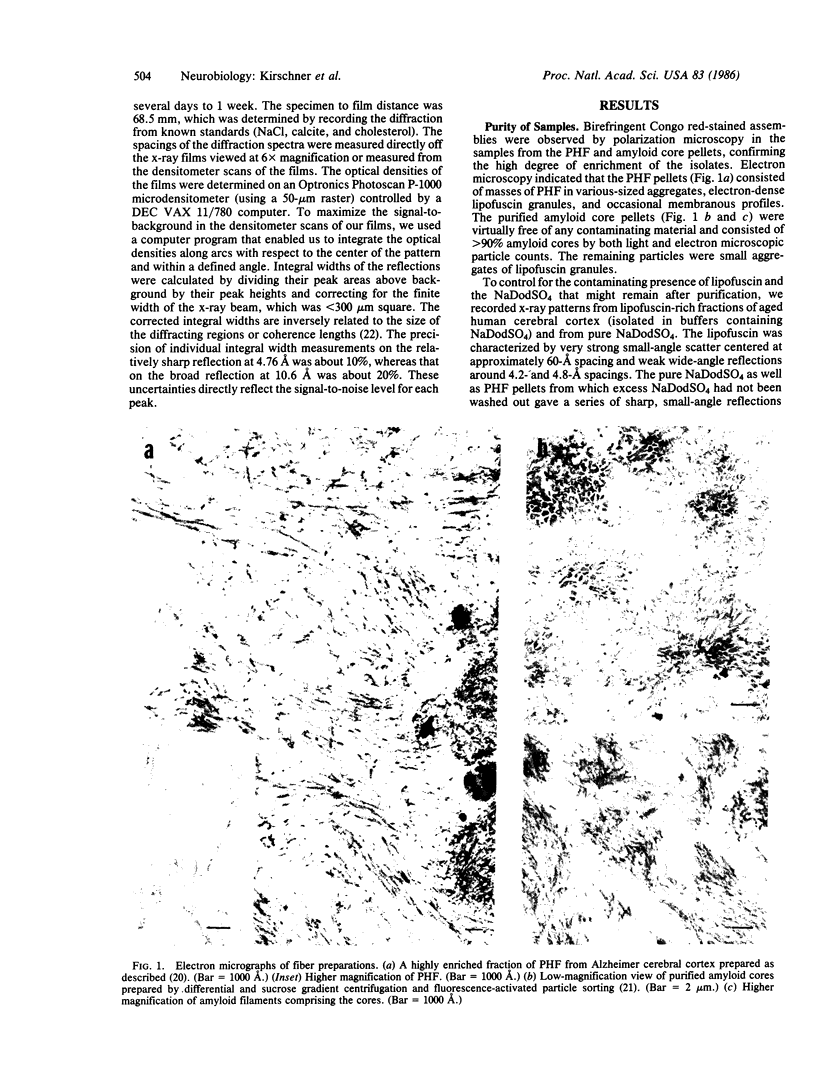
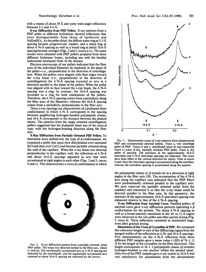
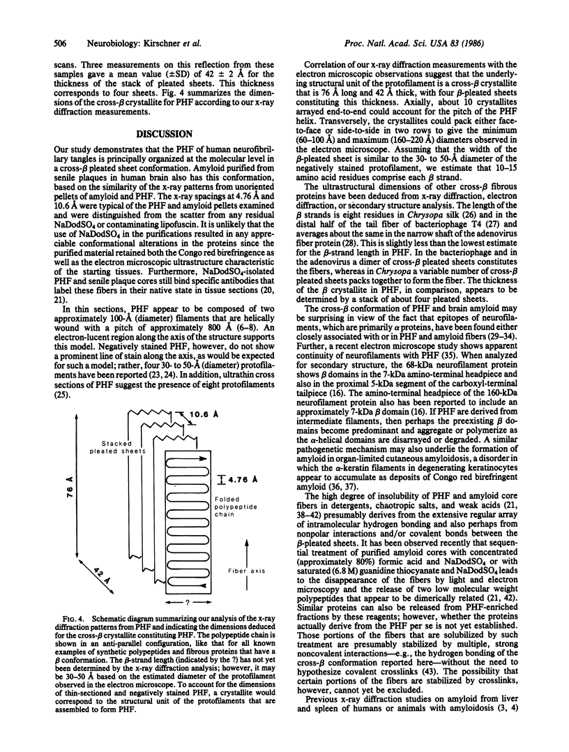
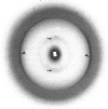
Information about the structure of the paired helical filaments (PHF) that accumulate within human neurons and the amyloid fibers that accumulate in the extracellular spaces between neurons in Alzheimer disease has so far depended on electron microscopy of thin-sectioned or negatively stained material. To determine the protein conformation of these abnormal fibers, we have obtained x-ray diffraction patterns from unfixed human brain fractions highly enriched in PHF and from purified amyloid cores isolated from senile plaques. The predominant x-ray scatter evident from both types of samples, either wet or dry, is a sharp reflection at 4.76-A spacing and a diffuse one at about 10.6-A spacing. These features are characteristic of a beta-pleated sheet type of protein conformation. In doubly oriented dried pellets of PHF fractions, the two reflections are accentuated at right angles to each other and the arc at 4.76-A spacing is in the fiber direction indicating a cross-beta conformation. From the integral widths of the reflections we estimate the cross-beta crystallite to be about 80 A long in the fiber direction and about 40 A thick. These dimensions correspond to approximately four pleated sheets, each of which consists of approximately 16 hydrogen-bonded polypeptide chains running normal to the fiber direction. The cross-beta conformation of PHF and amyloid fibers that we have found from x-ray diffraction is in contrast to the predominant alpha-helical coiled-coil conformation of the neurofilaments with which they share epitopes and from which they have been postulated to derive.
Full text
PDF
Images in this article
Selected References
These references are in PubMed. This may not be the complete list of references from this article.
- Anderton B. H., Breinburg D., Downes M. J., Green P. J., Tomlinson B. E., Ulrich J., Wood J. N., Kahn J. Monoclonal antibodies show that neurofibrillary tangles and neurofilaments share antigenic determinants. Nature. 1982 Jul 1;298(5869):84–86. doi: 10.1038/298084a0. [DOI] [PubMed] [Google Scholar]
- Bonar L., Cohen A. S., Skinner M. M. Characterization of the amyloid fibril as a cross-beta protein. Proc Soc Exp Biol Med. 1969 Sep;131(4):1373–1375. doi: 10.3181/00379727-131-34110. [DOI] [PubMed] [Google Scholar]
- Eanes E. D., Glenner G. G. X-ray diffraction studies on amyloid filaments. J Histochem Cytochem. 1968 Nov;16(11):673–677. doi: 10.1177/16.11.673. [DOI] [PubMed] [Google Scholar]
- Earnshaw W. C., Goldberg E. B., Crowther R. A. The distal half of the tail fibre of bacteriophage T4. Rigidly linked domains and cross-beta structure. J Mol Biol. 1979 Jul 25;132(1):101–113. doi: 10.1016/0022-2836(79)90498-4. [DOI] [PubMed] [Google Scholar]
- Geddes A. J., Parker K. D., Atkins E. D., Beighton E. "Cross-beta" conformation in proteins. J Mol Biol. 1968 Mar 14;32(2):343–358. doi: 10.1016/0022-2836(68)90014-4. [DOI] [PubMed] [Google Scholar]
- Geisler N., Kaufmann E., Fischer S., Plessmann U., Weber K. Neurofilament architecture combines structural principles of intermediate filaments with carboxy-terminal extensions increasing in size between triplet proteins. EMBO J. 1983;2(8):1295–1302. doi: 10.1002/j.1460-2075.1983.tb01584.x. [DOI] [PMC free article] [PubMed] [Google Scholar]
- Glenner G. G. Amyloid deposits and amyloidosis. The beta-fibrilloses (first of two parts). N Engl J Med. 1980 Jun 5;302(23):1283–1292. doi: 10.1056/NEJM198006053022305. [DOI] [PubMed] [Google Scholar]
- Glenner G. G., Eanes E. D., Page D. L. The relation of the properties of Congo red-stained amyloid fibrils to the -conformation. J Histochem Cytochem. 1972 Oct;20(10):821–826. doi: 10.1177/20.10.821. [DOI] [PubMed] [Google Scholar]
- Glenner G. G., Wong C. W. Alzheimer's disease: initial report of the purification and characterization of a novel cerebrovascular amyloid protein. Biochem Biophys Res Commun. 1984 May 16;120(3):885–890. doi: 10.1016/s0006-291x(84)80190-4. [DOI] [PubMed] [Google Scholar]
- Green N. M., Wrigley N. G., Russell W. C., Martin S. R., McLachlan A. D. Evidence for a repeating cross-beta sheet structure in the adenovirus fibre. EMBO J. 1983;2(8):1357–1365. doi: 10.1002/j.1460-2075.1983.tb01592.x. [DOI] [PMC free article] [PubMed] [Google Scholar]
- Hashimoto K., Kobayashi H. Histogenesis of amyloid in the skin. Am J Dermatopathol. 1980 Summer;2(2):165–171. doi: 10.1097/00000372-198000220-00014. [DOI] [PubMed] [Google Scholar]
- Ihara Y., Abraham C., Selkoe D. J. Antibodies to paired helical filaments in Alzheimer's disease do not recognize normal brain proteins. Nature. 1983 Aug 25;304(5928):727–730. doi: 10.1038/304727a0. [DOI] [PubMed] [Google Scholar]
- KIDD M. Paired helical filaments in electron microscopy of Alzheimer's disease. Nature. 1963 Jan 12;197:192–193. doi: 10.1038/197192b0. [DOI] [PubMed] [Google Scholar]
- Kumakiri M., Hashimoto K. Histogenesis of primary localized cutaneous amyloidosis: sequential change of epidermal keratinocytes to amyloid via filamentous degeneration. J Invest Dermatol. 1979 Aug;73(2):150–162. doi: 10.1111/1523-1747.ep12581609. [DOI] [PubMed] [Google Scholar]
- Masters C. L., Simms G., Weinman N. A., Multhaup G., McDonald B. L., Beyreuther K. Amyloid plaque core protein in Alzheimer disease and Down syndrome. Proc Natl Acad Sci U S A. 1985 Jun;82(12):4245–4249. doi: 10.1073/pnas.82.12.4245. [DOI] [PMC free article] [PubMed] [Google Scholar]
- Merz P. A., Wisniewski H. M., Somerville R. A., Bobin S. A., Masters C. L., Iqbal K. Ultrastructural morphology of amyloid fibrils from neuritic and amyloid plaques. Acta Neuropathol. 1983;60(1-2):113–124. doi: 10.1007/BF00685355. [DOI] [PubMed] [Google Scholar]
- Narang H. K. High-resolution electron microscopic analysis of the amyloid fibril in Alzheimer's disease. J Neuropathol Exp Neurol. 1980 Nov;39(6):621–631. doi: 10.1097/00005072-198011000-00001. [DOI] [PubMed] [Google Scholar]
- Powers J. M., Schlaepfer W. W., Willingham M. C., Hall B. J. An immunoperoxidase study of senile cerebral amyloidosis with pathogenetic considerations. J Neuropathol Exp Neurol. 1981 Nov;40(6):592–612. doi: 10.1097/00005072-198111000-00002. [DOI] [PubMed] [Google Scholar]
- Rasool C. G., Abraham C., Anderton B. H., Haugh M., Kahn J., Selkoe D. J. Alzheimer's disease: immunoreactivity of neurofibrillary tangles with anti-neurofilament and anti-paired helical filament antibodies. Brain Res. 1984 Sep 24;310(2):249–260. doi: 10.1016/0006-8993(84)90148-3. [DOI] [PubMed] [Google Scholar]
- Selkoe D. J., Abraham C., Ihara Y. Brain transglutaminase: in vitro crosslinking of human neurofilament proteins into insoluble polymers. Proc Natl Acad Sci U S A. 1982 Oct;79(19):6070–6074. doi: 10.1073/pnas.79.19.6070. [DOI] [PMC free article] [PubMed] [Google Scholar]
- Selkoe D. J., Ihara Y., Salazar F. J. Alzheimer's disease: insolubility of partially purified paired helical filaments in sodium dodecyl sulfate and urea. Science. 1982 Mar 5;215(4537):1243–1245. doi: 10.1126/science.6120571. [DOI] [PubMed] [Google Scholar]
- TERRY R. D. THE FINE STRUCTURE OF NEUROFIBRILLARY TANGLES IN ALZHEIMER'S DISEASE. J Neuropathol Exp Neurol. 1963 Oct;22:629–642. doi: 10.1097/00005072-196310000-00005. [DOI] [PubMed] [Google Scholar]
- Vanley C. T., Aguilar M. J., Kleinhenz R. J., Lagios M. D. Cerebral amyloid angiopathy. Hum Pathol. 1981 Jul;12(7):609–616. doi: 10.1016/s0046-8177(81)80044-5. [DOI] [PubMed] [Google Scholar]
- Wais-Steider C., Eagles P. A., Gilbert D. S., Hopkins J. M. Structural similarities and differences amongst neurofilaments. J Mol Biol. 1983 Apr 5;165(2):393–400. doi: 10.1016/s0022-2836(83)80263-0. [DOI] [PubMed] [Google Scholar]
- Wisniewski H. M., Merz P. A., Iqbal K. Ultrastructure of paired helical filaments of Alzheimer's neurofibrillary tangle. J Neuropathol Exp Neurol. 1984 Nov;43(6):643–656. doi: 10.1097/00005072-198411000-00008. [DOI] [PubMed] [Google Scholar]
- Wiśniewski H. M., Narang H. K., Terry R. D. Neurofibrillary tangles of paired helical filaments. J Neurol Sci. 1976 Feb;27(2):173–181. doi: 10.1016/0022-510x(76)90059-9. [DOI] [PubMed] [Google Scholar]
- Yoshimura N. Evidence that paired helical filaments originate from neurofilaments--electron microscope observations of neurites in senile plaques in the brain in Alzheimer's disease. Clin Neuropathol. 1984 Jan-Feb;3(1):22–27. [PubMed] [Google Scholar]