Abstract
Wooden objects are often used as nonhuman primate enrichment to provide variety and novelty, promote exploratory behavior, and supply an outlet for curiosity. However, concerns have been raised regarding the ability to sanitize wood by using conventional cage-wash procedures. To address this concern, we examined sanitation outcomes between soiled plastic toys and manzanita wooden manipulanda immediately after a cage-wash cycle. Both an ATP luminometer device, which is capable of providing an immediate assessment of sanitation levels, and traditional bacterial culture were used, with the secondary goal of comparing these methods for sanitation monitoring. Results showed that the wooden objects did not differ from plastic toys with respect to the overall efficacy of cage-wash sanitization. Therefore, manzanita wood can be used as nonhuman primate enrichment without risking pathogen transmission when items are rotated among animals.
Abbreviation: RLU, relative light units
Environmental enrichment of nonhuman primates is implemented in the biomedical research setting as a way to provide variety and novelty, promote exploratory behavior, and supply an outlet for curiosity. Among the many available forms of enrichment, simple inanimate objects are often used as they are generally durable, safe, inexpensive, and easily maintained.24,36 Wooden objects can be used as either perches or manipulanda in an attempt to simulate aspects of the natural habitat.37 Wood encourages noninjurious, species-typical behaviors such as gnawing, perching, climbing, manipulating, and playing.18,33,36 Previous reports have shown that nonhuman primates prefer wooden objects to other toys such as nylon balls, rubber toys, and swing toys19,24 with minimal loss of interest over time.4,31 This is likely due to the novelty of the wooden items since the configuration and texture change with wear.38 Types of wood used as enrichment include box elder,5,31,33 cherry,5,18,19 oak,32 almond,24 maple,36 and manzanita wood.36 Manzanita, the common name for the genus Arctostaphylos, includes evergreen shrubs and small trees found in northwestern North America.27
Environmental enrichment devices offer multiple benefits; however, the risk of injury to nonhuman primates invariably exists. Since the implementation of inanimate enrichment in the biomedical research setting, isolated reports have been published regarding harm due to a ring toy,30 rope,17 wire from an automobile tire,12 and wooden objects such as perches,22 foraging substrates,26,40 and manipulanda.6 These incidents must be viewed in light of the number of nonhuman primates that manipulate these environmental items on a daily basis. The potential benefits of the wooden enrichment items outweigh the possible risks and therefore, enrichment programs were not revised after these noted circumstances.6,26,40 Moreover, one study showed improved dental health in animals that used cedar wood as manipulanda.4
Environmental enrichment items have the potential to interfere with facility operations. Shreds of wood from maple and red oak manipulanda created drain obstructions at one facility.13 However, similar reports do not exist for all types of wood used, as various wood species wear differently. For example, in contrast to the study just cited,13 another reported that nonhuman primates do not shred red oak. Instead, red oak wears into small flakes that are unlikely to obstruct drains.34 This report34 discouraged the use of white oak, black locust, box elder, black cherry, weeping willow, and silver maple because they wear into large strips. Therefore, the species of wood must be considered prior to its use for enrichment in a biomedical research facility.
The ability to sanitize wooden objects is a concern in the animal research setting; therefore, some facilities discard wooden enrichment items at regular intervals or forgo their use altogether.36 When facilities opt to sanitize wood, different approaches are implemented. Wood can be autoclaved, cleaned with disinfectants, water-blasted, or put through a cage-wash cycle.36 However, there is disagreement on whether these procedures adequately sanitize the wooden objects to avoid transmission of pathogens when manipulanda are rotated among animals.3,21,36,37 In fact, a previous report using bacterial culture demonstrated that sanitizing rubber toys in a commercial cage washer was inadequate in eliminating all bacterial organisms.3 However, the organisms were presumably environmental contaminants that did not pose a significant disease threat to nonhuman primates.
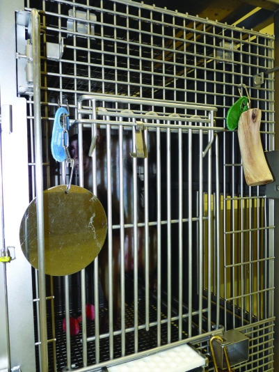
At our institution, manzanita is the only type of wood used as enrichment for caged rhesus macaques. It is denser and less porous than other wood types and therefore less absorbent and easier to clean. Manzanita is used as a device attached to the outside of the cage (Figure 1) or as manipulanda placed inside of cages and runs. Enrichment items remain with the cage during the cage-wash cycle and rotate among animals to enhance the novelty of the objects. Consistent with many previous reports,13,24,35,36 no health issues due to the wooden segments have emerged at our institution since their implementation 8 y ago. Moreover, there are no reports of drain malfunctions due to the manzanita wood, and neither shreds nor flakes have been noted in the drains. However, because of concerns raised over the ability to sanitize wood sufficiently by using standard cage-wash procedures, we examined sanitation outcomes after a traditional cage-wash cycle between nonhuman primate enrichment items made of 2 different materials. We hypothesized that cage-wash procedures were equally effective at sanitizing wooden items and other enrichment objects. We used 2 methods, an ATP luminometer device and traditional bacterial culture, to test this hypothesis, with the secondary goal of comparing these methods of sanitation monitoring.
Figure 1.
Manzanita wood used as an enrichment device on the outside of a nonhuman primate cage.
Materials and Methods
Test objects.
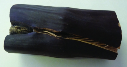
This study used 8 nonhuman primate cages (Allentown, Allentown, NJ), each containing one moderately worn plastic toy (Hercules Dental Device, Bio-Serv, Frenchtown, NJ) and one moderately worn section of manzanita wood (Manzanita Burlworks, Borrego Springs, CA). Swabs were obtained from the toy, wood segment, and cage surface after 2 wk of occupation by rhesus macaques (Macaca mulatta); these were the ‘soiled’ samples. These items were then cleaned using an automated cage wash cycle and resampled (‘sanitized’ samples). All objects were swabbed with culturettes for aerobic bacterial culture and with surface sampler swabs supplied by the manufacturer of the ATP detection device (Accupoint 2, Neogen, Lansing, MI). Each swabbed surface measured roughly 4 × 4 cm, and the swab areas for the 2 different testing methods were adjacent but not overlapping. For the plastic and wooden toys, swabs for bacterial culture were taken from one half of the object and for the ATP detection device from the other half; the deepest crevices available were sampled (Figure 2). At approximately the same location in each cage, samples were taken from an interior corner and wall surface. The cage surface location was implemented as a control because it is a smooth, impervious surface that is easily sanitized. In addition, known soiled surfaces were swabbed to evaluate the efficacy of the ATP luminometer and bacterial culture as sanitation assessment methods.
Figure 2.
Representative manzanita wood crevice swabbed for bacterial culture and ATP luminometer sanitation assessment.
This study was performed at Tulane National Primate Research Center, which is an AAALAC-accredited facility. The housing and care provided were in accordance with the regulations of the Animal Welfare Act1 and recommendations of The Guide for the Care and Use of Laboratory Animals.20 Animals were housed singly indoors in stainless steel cages equipped with perches and multiple enrichment devices. Cage floor, cage pan, and enrichment items were rinsed daily with water. Animals were maintained on a 12:12-h light:dark cycle with ambient temperature of 64 to 72° F (17.8 to 22.2° C) and a relative humidity of 30% to 70%. The subjects were fed commercial diet formulated for nonhuman primates (Purina Diet 5037, PMI Feeds, St Louis, MO) twice daily, provided water ad libitum, and given supplemental fruit and forage throughout the week. This study was performed independently of weekly quality assurance monitoring at our facility.
Bacterial culture.
Aerobic cultures were inoculated into thioglycollate broth and incubated at 37 °C for 24 h; if no growth was observed, incubation was extended to 48 h. Broth was gram-stained and examined at 100× magnification; results were categorized as presence (‘fail’) or absence (‘pass’) of growth. Growth was identified as gram-positive cocci or rods, gram-negative rods, and yeast.
ATP detection device.
The results of the ATP detection device were recorded in RLU (relative light units). Because no institutional benchmark value existed at our facility, the pass–fail cut-off value was set at 250 RLU by using published results,15,23,41,43 to provide stringent control measures.
Statistical analysis.
Student t tests (Statistica, StatSoft, Tulsa, OK) were used to compare ATP luminometer results of the various test objects. Results are expressed in mean ± SE, and differences were considered statistically significant at a P value of less than 0.05.
Results
Bacterial culture.
Organisms were cultured from all soiled test objects. After sanitization, growth was recorded from a lower percentage of wooden manipulanda (13%) than from toys (50%) or cages (32%). Before sanitation, gram-negative rods and gram-positive cocci and rods were present on all toys, wooden pieces, and cages; cages also tested positive for yeast. After sanitation, all items sampled carried gram-positive rods; cages also carried gram-positive cocci. Gram-negative rods were cultured from 2 of the 16 sanitized cage surfaces but not from the sanitized wooden pieces or toys.
ATP detection device.
Soiled wood showed the highest mean luminosity (2093.5 ± 1070.9 RLU). Mean luminosity was significantly (t = 2.7; P < 0.05) higher for soiled wooden pieces than soiled cages (119 ± 34.8 RLU); the mean luminosity reading of soiled toys was 310 ± 291.4 RLU. After sanitization, all test objects had a mean RLU result of 0.
Comparison of ATP detection device and bacterial culture.
When evaluating pass–fail results for the ATP luminometer and bacterial culture swabs, 68.8% of the results showed agreement between the 2 evaluation methods for the sanitized trial and 28.1% for the soiled trial.
Discussion
The current study addressed the concerns regarding the ability to sanitize manzanita wood segments used as environmental enrichment for nonhuman primates. After 2 wk of use by rhesus macaques, all materials harbored bacteria; manzanita wood had the highest level of organic debris, according to the ATP luminometer. This result is not surprising, because the ability of an enrichment item to harbor microorganisms is related to its surface complexity.3 Although wood accumulated the greatest amount of organic debris, bacterial culture and ATP luminometer results showed that this wood was as sanitizable as were toys and cage surfaces. This finding was further confirmed by using the absence of gram-negative rods as an indicator of adequate sanitization, in agreement with a previous report investigating rubber toys.3 Therefore, manzanita wood was not found to be a potential fomite, in comparison to the other objects we evaluated.
The growth of gram-negative rods on the cage surface is concerning, but the validity of the results must be questioned. Although we swabbed cages promptly after cage-wash procedures, bacterial contamination may have occurred before the results were collected. In addition, environmental contamination from the microbiologic laboratory cannot be ruled out. Because the current study was performed independently of weekly quality assurance monitoring at our facility, these results were not investigated further.
The secondary goal of the study was to compare two methods of sanitation monitoring used in the biomedical research setting, traditional bacterial culture and novel ATP luminometery. The monitoring of various morphologic bacterial groups by culture is a practice that has been consistent over the last century.14 The growth of gram-negative organisms is considered the most important indicator of ineffective sanitization.7,39,44 In addition, these organisms can be quantified by replicate organism detection process and counting (RODAC) plating. This method of quality assurance has considerable limitations because it detects only easily grown aerobic bacteria and fungi and cannot detect parasites or organic debris.43 Moreover, the recovery of pathogenic microorganisms can be limited when swabbing dry surfaces.8 In addition, bacterial culture takes several days to complete, thus limiting the value of these results.29 Finally, culture is relatively expensive and may have to be performed off-site.
ATP-based monitoring devices are hand-held instruments originally developed for instantaneous sanitation monitoring in food production facilities.11 These devices are now used widely by drug companies, healthcare laboratories, and hospitals, and recent publications have demonstrated their effectiveness in the laboratory animal setting.11,43 The use of ATP luminometers is advocated as an acceptable replacement for or adjunct to bacterial culture for quality assurance.43 These detection devices extract ATP from organic material on surfaces sampled by using a swab. The ATP reacts with the firefly reagent luciferin luciferase to produce light, which then is quantified and expressed in RLU. ATP is present in microorganisms and, perhaps more importantly, organic matter, which provides a conditioning film to which microorganisms attach and acts as a source of nutrients for growth.28 Therefore, the absence of organic debris, rather than bacterial organisms, is considered a superior indicator of cleanliness.16,45
ATP detection devices offer multiple advantages over traditional bacterial culture because they are cost-effective and time-efficient and show superior reproducibility and sensitivity. In addition, these devices offer real-time results, after which immediate corrective action may be taken.8,9,14,28 However, this technology has limitations, in that both gram-negative bacteria43 and protein-only soiling are poorly detected,25 and bleach,10 temperature, and pH can affect the accuracy of the results.25 In addition, the sensitivity of the instruments available varies by manufacturer and depends on the ability of the machine to extract cellular ATP from surface debris and to produce and quantify light.16,23 The optimal sensitivity required by a facility depends on the surface swabbed and the intended use of the resultant information.16 Therefore, no standard RLU baseline values exist, and they are instead established at the institutional level.43 This process involves establishing a pass–fail ATP RLU cut-off value by producing a calibration curve from repeated assays of known clean and dirty surfaces.14,45 Once a mean RLU is set for known clean surfaces, a cut-off value can be defined as 2 standard deviations above the mean RLU value or by adding 20% to the mean RLU.14 Benchmark value are not standardized and vary from 250 to 1000 RLU in published reports.15,23,41,43 Setting these levels has proven to be difficult at various institutions.42
Previous reports have shown correspondence between the use of an ATP detection device and bacterial culture for sanitation assessment when examining floors and rodent caging in an animal research facility11 and worktops and equipment in a hospital kitchen.2 However, because ATP detection devices and bacterial culture measure different parameters, the value of comparing the results is questionable.23,41 In addition, the majority of ATP on human-hand–contacted surfaces (approximately 77%) is the result of nonmicrobial organisms, which are not detected by bacterial culture.15 Therefore, the lack of agreement in the current study and previous reports examining surfaces in hospital medical and surgical wards is not surprising.23,41
The current study includes several limitations. The bacterial culture results were not quantified by replicate organism detection process and counting (RODAC) plating, a lack that prevented the establishment of a quantitative cut-off pass–fail value of the bacterial culture results. Instead, we determined this cut-off by using the presence or absence of organism growth. In addition, no institutional benchmark value was used for the ATP luminometer, given that a calibration curve had not yet been performed at our facility. Lastly, the results of the current study must be interpreted in light of the limited sample size.
Traditional bacterial culture and ATP luminometry are common methods for monitoring surface hygiene in the biomedical research setting. These methods demonstrated that traditional cage-wash procedures are adequate for cleaning manzanita wood segments. Therefore, manzanita wood can be used as nonhuman primate enrichment without risking pathogen transmission if items are rotated among animals.
Acknowledgments
We thank Brooke Cardenas, Kathrine Phillippi-Falkenstein, Dr Lara Doyle, and the clinical pathology laboratory at Tulane National Primate Research Center. This work was supported by the training grant R25 RR024231 and Tulane National Primate Research Center base grant P51 RR000164 from NIH.
References
- 1.Animal Welfare Act as Amended 2007. 7 USC §2131-2159.
- 2.Aycicek H, Pguz U, Karci K. 2006. Comparison of results of ATP bioluminescence and traditional hygiene swabbing methods for the determination of surface cleanliness at a hospital kitchen. Int J Hyg Environ Health 209:203–206 [DOI] [PubMed] [Google Scholar]
- 3.Bayne KA, Dexter SL, Hurst JK, Strange GM, Hill EE. 1993. Kong toys for laboratory primates: are they really an enrichment or just fomites? Lab Anim Sci 43:78–85 [PubMed] [Google Scholar]
- 4.Brinkman C. 1996. Toys for the boys: environmental enrichment for singly housed adult male macaques (Macaca fascicularis). Lab Prim News 35:5–9 [Google Scholar]
- 5.Champoux M, Hempel M, Reinhardt V. 1987. Environmental enrichment with sticks for singly caged adult rhesus monkeys. Lab Prim News 26:5–7 [Google Scholar]
- 6.Cliett ML, Stewart LA. 2010. Epistaxis and nasal swelling in a cynomolgus macaque. Lab Anim (NY) 39:300–303 [DOI] [PubMed] [Google Scholar]
- 7.Committee on Long-term Holding of Laboratory Rodents 1976. Long-term holding of laboratory rodents. Ilar News 19:L1–L25 [Google Scholar]
- 8.Davidson CA, Griffith CJ, Peters AC, Fielding LM. 1999. Evaluation of 2 methods for monitoring surface cleanliness—ATP bioluminescence and traditional hygiene swabbing. Luminescence 14:33–38 [DOI] [PubMed] [Google Scholar]
- 9.Day D. 2007. Validation of quality assurance program changes. Tech Talk 12:2 [Google Scholar]
- 10.Dumigan DG, Boyce JM, Golebiewski M, Balogun O, Rizvani R. 2010. Who is really caring for your environment of care? Developing standardized cleaning procedures and effective monitoring techniques. Am J Infect Control 38:387–392 [DOI] [PubMed] [Google Scholar]
- 11.Ednie DL, Wilson RP, Lang CM. 1998. Comparison of 2 sanitation monitoring methods in an animal research facility. Contemp Top Lab Anim Sci 37:71–7412456174 [Google Scholar]
- 12.Etheridge ME, O'Malley J. 1996. Diarrhea and peritonitis due to traumatic perforation of the stomach in a rhesus macaque (hardware disease). Contemp Top Lab Anim Sci 35:57–59 [PubMed] [Google Scholar]
- 13.Gallucci P, Cliett ML, Stewart LA. 2009. Wood as an enrichment device for primates. Tech Talk 14:1 [Google Scholar]
- 14.Griffith CJ, Blucher A, Fleri J, Fielding L. 1994. An evaluation of luminometry as a technique in food microbiology and a comparison of 6 commercially available luminometers. Food Sci Technol Today 8:209–216 [Google Scholar]
- 15.Griffith CJ, Cooper RA, Gilmore J, Davies C, Lewis M. 2000. An evaluation of hospital cleaning regimes and standards. J Hosp Infect 45:19–28 [DOI] [PubMed] [Google Scholar]
- 16.Griffith CJ, Davidson CA, Peters AC, Fielding LM. 1997. Towards a strategic cleaning assessment programme: hygiene monitoring and ATP luminometry, an options appraisal. Food Sci Technol Today 11:15–24 [Google Scholar]
- 17.Hahn NE, Lau D, Eckert K, Markowitz H. 2000. Environmental enrichment-related injury in a macaque (Macaca fascicularis): intestinal linear foreign body. Comp Med 50:556–558 [PubMed] [Google Scholar]
- 18.Hienz RD, Pyle DA, Frey JJ, Zarcone TJ, Adams RJ, Turkkan JS. 2000. Enrichment device use by baboons during long-term vs intermittent availability. Lab Prim News 39:1–3 [Google Scholar]
- 19.Hienz RD, Zarcone TJ, Turkkan JS, Pyle DA, Adams RJ. 1998. Measurement of enrichment device use and preference in singly caged baboons. Lab Prim News 37:6–10 [Google Scholar]
- 20.Institute for Laboratory Animal Research 2010. Guide for the care and use of laboratory animals, 8th ed. Washington (DC): National Academies Press [Google Scholar]
- 21.Kelley ST, Hall AS. 1995. Housing, p 193–209. In: Bennett BT, Abee CR, Henrickson R. Nonhuman primates in biomedical research: biology and management. San Diego (CA): Academic Press [Google Scholar]
- 22.Kessler MJ, Kupper JL. 1976. Obstructive gastric foreign body in rhesus monkey (Macaca mulatta). Lab Anim Sci 26:619–621 [PubMed] [Google Scholar]
- 23.Lewis T, Griffith C, Gallo M, Weinbren M. 2008. A modified ATP benchmark for evaluating the cleaning of some hospital environmental surfaces. J Hosp Infect 69:156–163 [DOI] [PubMed] [Google Scholar]
- 24.Line SW, Morgan KN. 1991. The effects of 2 novel objects on the behavior of singly caged adult rhesus macaques. Lab Anim Sci 41:365–369 [PubMed] [Google Scholar]
- 25.Lipscomb IP, Sihota AK, Botham M, Harris KL, Keevil CW. 2006. Rapid method for the sensitive detection of protein contamination on surgical instruments. J Hosp Infect 62:141–148 [DOI] [PubMed] [Google Scholar]
- 26.Mätz-Rensing K, Floto A, Kaup FJ. 2004. Intraperitoneal foreign body disease in a baboon (Papio hamadryas). J Med Primatol 33:113–116 [DOI] [PubMed] [Google Scholar]
- 27.Meyer S. 2008. Arctostaphylos Adans: manzanita, p 266–270. In: Bonner FT, Karrfalt RP. The woody plant seed manual. Agriculture handbook, no 727. Washington (DC): US Department of Agriculture, Forest Service [Google Scholar]
- 28.Moore G, Griffith C. 2002. A comparison of traditional and recently developed methods for monitoring surface hygiene within the food industry: an industry trial. Int J Environ Health Res 12:317–329 [DOI] [PubMed] [Google Scholar]
- 29.Moore G, Griffith C, Fielding L. 2001. A comparison of traditional and recently developed methods for monitoring hygiene within the food industry: a laboratory study. Dairy, Food, and Environmental Sanitation 21:478–488 [Google Scholar]
- 30.Murchinson MA. 1993. Potential animal hazard with ring toys. Lab Prim News 32:1–2 [Google Scholar]
- 31.Reinhardt V. 1989. Evaluation of the long-term effectiveness of 2 environmental enrichment objects for singly caged rhesus macaques. Lab Anim 18:31–33 [Google Scholar]
- 32.Reinhardt V. 1990. Comparing the effectiveness of PVC perches versus wooden perches as environmental enrichment objects for singly caged rhesus monkeys. Lab Prim News 29:13–14 [Google Scholar]
- 33.Reinhardt V. 1990. Time budget of caged rhesus monkeys exposed to a companion, a PVC perch, and a piece of wood for an extended time. Am J Primatol 20:51–56 [DOI] [PubMed] [Google Scholar]
- 34.Reinhardt V. 1992. Environmental enrichment branches that do not clog drains. Lab Prim News 31:8 [Google Scholar]
- 35.Reinhardt V. 1997. The Wisconsin gnawing stick. AWIC Newslett 7:11–12 [Google Scholar]
- 36.Reinhardt V. 2000. Wooden objects for enrichment: a discussion. Lab Prim News 39:1–4 [Google Scholar]
- 37.Reinhardt V, Houser WD, Cowley D, Champoux M. 1987. Preliminary comments on environmental enrichment with branches for individually caged rhesus monkeys. Lab Prim News 26:1–3 [Google Scholar]
- 38.Reinhardt V, Reinhardt A. 2008. Environmental enrichment and refinement for nonhuman primates kept in research laboratories: a photographic documentation and literature review. Washington (DC): Animal Welfare Institute [Google Scholar]
- 39.Scholndelmeyer CW, Dillehay DL, Webb SK, Huerkamp MJ, Mook DM, Pullium JK. 2006. Investigation of appropriate sanitization frequency for rodent caging accessories: evidence supporting less-frequent cleaning. J Am Assoc Lab Anim Sci 45:40–43 [PubMed] [Google Scholar]
- 40.Seier JV, Dhansay MA, Davids A. 2005. Risks associated with environmental enrichment: intestinal obstruction caused by foraging substrate. J Med Primatol 34:154–155 [DOI] [PubMed] [Google Scholar]
- 41.Sherlock O, O'Connell N, Creamer E, Humphreys H. 2009. Is it really clean? An evaluation of the efficacy of 4 methods for determining hospital cleanliness. J Hosp Infect 72:140–146 [DOI] [PubMed] [Google Scholar]
- 42.Tebbutt GM. 1999. Comparison of traditional and rapid methods for assessing the risk of bacterial cross-contamination from cutting boards. Int J Environ Health Res 9:67–74 [Google Scholar]
- 43.Turner DE, Daugherity EK, Altier C, Maurer KJ. 2010. Efficacy and limitation of an ATP-based monitoring system. J Am Assoc Lab Anim Sci 49:190–194 [PMC free article] [PubMed] [Google Scholar]
- 44.Weisbroth SH, Weisbroth SP. 1982. Laboratory animal facility sanitization: a proposed microbial evaluation program. Lab Anim 11:25–26 [Google Scholar]
- 45.Willis C, Morley R, Westbury J, Greenwood M, Pallett A. 2007. Evaluation of ATP bioluminescence swabbing as a monitoring and training tool for effective hospital cleaning. Journal of Infection Prevention 8:17–21 [Google Scholar]


