Abstract
Two cases of coexisting juvenile idiopathic arthritis (JIA) and sickle cell disease (ages 7 and 17) are presented. The diagnoses of JIA were delayed for years because of the similarity of presentations in the two conditions. Both cases had been treated with non-steroidal anti-inflammatory drugs for years. Both had positive rheumatoid factor, and elevated erythrocyte sedimentation rate (ESR) while one of the patients had elevated serum ferritin and anticyclic citrullinated protein. Radiology showed marked arthritic changes with presence of avascular necrosis in a patient’s head of femur. Both cases were treated with etanercept for 6 months each, as well as methotrexate. At the end of 6 months, the joint count for pains and swelling were done as well as ESR.
Background
Although sickle cell disease (SCD) is common among black Africans, juvenile idiopathic arthritis (JIA) on the other hand is uncommonly reported.1 Coexisting SCD and connective tissue disease in adults have been previously reported.2 Coexistence of SCD and JIA has rarely being reported in literature3; and there is no documented report among black Africans. The similarity in presentation of the two conditions may delay diagnosis and prevents initiation of appropriate therapy. The resultant effect will be marked joint destruction and resultant morbidity and possible mortality.
An increased awareness of this coexistence will prompt early diagnosis and treatment of the JIA.
Case presentation
Case 1 is a 7-year-old Nigerian boy who was first diagnosed as SCD, HbSS at the age of 11 months. His presentation then was dactylitis of the hands and feet. He has since then had recurrent episodes of bone pains and jaundice and has had numerous hospital admissions and blood transfusions in addition to anti malarials, iron and folic acid tablets.
About 2 years before presentation, his mother noticed joint swellings associated with complaints of joint pains by him. The joints affected were mostly those of the hands, elbows, shoulders, feet, ankles and knees. He was admitted a few times and has had four blood transfusions within the past 2 years.
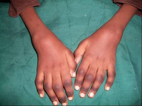
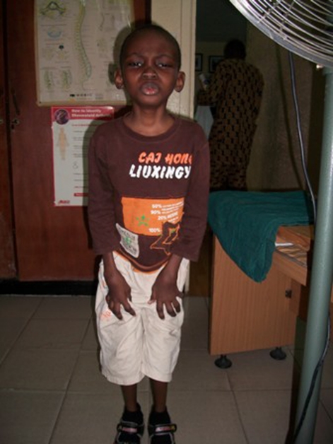
Physical examination showed polyarthritis involving the joints of the wrists, metacarpophalangeal as well as the elbows (figure 1). He was small for age and had arthritis involving the knees as well as the feet (figure 2). He was also pale, slightly jaundiced and had generalised lymphadenopathy. There were no subcutaneous nodules. He also had ankylosis of the elbows and knee joints as well as neck stiffness. Abdominal examination showed hepatosplenomegaly of 6 cm. The chest was clinically clear. There were no symptoms pertaining to the eyes and examination by an ophthalmologist did not show uveitis. The diagnosis was JIA coexisting with SCD.
Figure 1.
Photograph of a 7-year-old boy with coexistent sickle cell disease (SCD) and juvenile idiopathic arthritis (JIA) showing arthritis of the proximal interphalangeal joints and wrist joints.
Figure 2.
Photograph of a 7-year-old boy with coexistent SCD and JIA with stunted growth and arthritis of joints of the lower limbs.
Laboratory investigations – haematocrit – 18%; erythrocyte sedimentation rate (ESR) – 42 mm/h; white blood cell count and platelets within normal limits; serum ferritin – 1120 (normal 20–300 IU/l); rheumatoid factor – 46 IU/l by rheumatoid factor nephelometry (normal 0–20). Antinuclear antibody was negative. X-ray of the hands showed periarticular osteoporosis without erosions. Mantoux skin test for tuberculosis was negative and chest x-ray reported as clear.
He was started initially on subcutaneous methotrexate – 10 mg weekly and prednisolone tablets – 10 mg daily; folic acid – 10 mg weekly and calcium lactate and naproxen tablets – 125 mg prn. After 2 months on the above, he was started on subcutaneous etanercept – 25 mg weekly in addition to oral methotrexate 15 mg weekly.
At the end of 6 months, there were no painful, tender or swollen joints. ESR was down to 23 mm/h. The weight had also increased from 15.9 to 19.1 kg. He is presently on methotrexate tablets 15 mg, folic acid 10 mg once weekly, calcium lactate and non-steroidal anti-inflammatory drugs (NSAIDs) as required.
Case 2 is a 17-year-old Nigerian girl who was initially diagnosed as SCD, HbSS at the age of 5 years. She had presented then with persistent bone pains, abdominal swelling and jaundice. She had been on regular follow-up by her general practitioner, with occasional admissions and blood transfusions. She had been maintained mostly on folic acid supplement, antimalarials and analgesics as required.
She however presented to her general practitioner about 8 years ago with additional complaints of joint pains and swellings, mostly the hands, feet, knees and elbows. She had also noticed recurrent subcutaneous nodules on the elbows. She was managed as arthropathy of SCD, and had been on various NSAIDs without a sustained improvement. There were no eye symptoms.
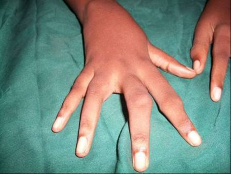
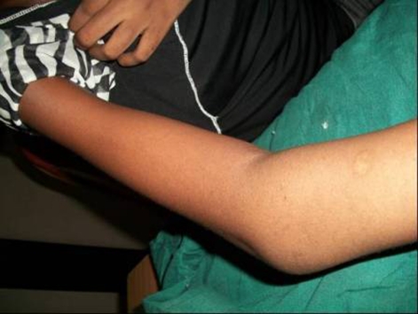
Physical examination showed a small for age girl with swellings and ankylosis of the proximal interphalangeal joints (figure 3) associated with marked tenderness. There was also ankylosis of the left elbow joint (figure 4). There were no subcutaneous nodules. She was neither pale nor jaundiced but she had cervical and axillary lymphadenopathy. The spleen was enlarged to 8 cm but liver was not palpable. Examination of the musculoskeletal system revealed tenderness in most of the joints. Eye examination by the ophthalmologist did not reveal any uveitis.
Figure 3.
Photograph of 17-year-old girl with coexistent SCD and JIA showing arthritis of joints of the hands as well as ankylosis.
Figure 4.
Photograph of the left elbow of a 17-year-old girl with coexistent SCD and JIA showing arthritis and ankylosis of the left elbow.
Diagnosis was JIA associated with SCD.
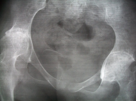
Laboratory investigation and results are as follows – haematocrit – 23%; normal total and differential white blood cells and platelets. ESR was 59 mm/h. Liver and renal function tests were essentially normal. Serology – rheumatoid arthritis nephelometry 24.7 (0–20); anticyclic citrullinated protein (anti CCP) above 300 (0–3 units/ml). Anti nuclear antibody was reported as negative. Radiographs of the hands showed multiple erosions with subluxation while that of pelvis showed narrowed hip joint spaces with sclerosis of the acetabular cups and avascular necrosis of the left femoral head (figure 5). Radiograph of the chest was normal. Mantoux skin test for tuberculosis was negative.
Figure 5.
Radiograph of a 7-year-old boy with SCD and JIA showing sclerosis of both hip joints and avascular necrosis of the left femoral head.
She was treated with subcutaneous methotrexate, 12.5 mg weekly, subcutaneous etanercept – 50 mg weekly, tablet prednisolone – 15 mg daily, folic acid tablet – 10 mg weekly, calcium lactate tablets and naproxen – 250 mg twice daily prn. She however developed exacerbation of an existing ‘chest pain syndrome’ soon after starting treatment. Electrocardiogram showed sinus tachycardia and left atrial abnormalities. Etanercept was given for 24 weeks and discontinued. She is presently on methotrexate tablets 15 mg and folic acid 10 mg weekly, prednisolone 5 mg daily, calcium lactate daily and haematinics. At the end of 6 months there were no painful, tender or swollen joints. ESR is down to 25 mm/h. She is also independent of the mother. The chest pain syndrome has also improved on clopidogrel 75 mg daily.
Investigations
-
▶
Haematocrit
-
▶
Haemoglobin electrophoresis
-
▶
White blood cell count
-
▶
ESR
-
▶
Rheumatoid factor
-
▶
Anti CCP
-
▶
Antinuclear antibody
-
▶
Serum ferritin
-
▶
Liver function tests
-
▶
Chest radiographs
-
▶
Radiographs of the hands and pelvis.
Differential diagnosis
Haemochromatosis arthropathy
Treatment
Subcutaneous etanercept, methotrexate, prednisolone, naproxen, folic acid, clopidogrel.
Outcome and follow-up
After 6 months, the two patients were free of pain, joint tenderness and swellings. ESR is down and the quality of life has improved. They are still being followed up.
Discussion
SCD is a common hereditary disease of the haemoglobin among Nigerians. JIA on the other hand is rarely reported. Because SCD may present with arthropathy, it is not unusual to miss an associated juvenile idiopathic arthritis. This probably accounts for the delay in diagnosis of the two cases presented. The first case did not present to a rheumatologist until 2 years after onset of the arthritis while the second case presented 8 years afterwards. Nistala et al3 had earlier highlighted the delay in presentation of these cases. There is thus resultant destructive arthropathy from the compound effect of the two diseases on the bone and cartilage. They also identified the difficulty of making a diagnosis of an inflammatory arthritis against a background of SCD. The elevated serum ferritin raised the possibility of haemochromatosis. However, this hereditary disease of tissue iron loading due to increased iron absorption is mostly seen among northern and eastern Europeans and has rarely been reported in black Africans. Also, haemochromatosis is seen at the peak age of 40 to 60 years. In addition, the liver enzymes, characteristically elevated in haemochromatosis, were normal in our patients.
The two patients had rheumatoid factor elevation, though marginally in the second case. However, this patient had a significantly elevated anti CCP, a marker of prognosis and joint destruction. The first case had markedly elevated serum ferritin, though this was unavailable in the second case. It is possible that such elevated ferritin while being a marker of JIA may also be attributable to numerous blood transfusions, as is common in SCD patients.
Treatment was with standard medications as in patients with JIA. However, both cases were also placed on subcutaneous etanercept. Nistala et al3 have postulated that there is correlation between increased levels of tumour necrosis factor α in the synovial tissue of joints damaged by arthritis and local sickling. Anti tumour necrosis factor agents may therefore be effective. However the number is too small to make any meaningful conclusion. These cases are presented to highlight the awareness of the possible coexistence of JIA and SCD in patients.
Learning points.
-
▶
SCD is common in African blacks.
-
▶
JIA is uncommonly reported among black Africans.
-
▶
Coexistence of the two conditions is rarely reported in medical literature.
-
▶
Delay in diagnosis and resultant morbidity.
Acknowledgments
Mrs Irene Oduenyi- Clinic Nurse Pathcare Laboratory, Lagos- For serology Dr (Mrs) F Idris, Consultant Opthalmologist. Lagos State University College Hospital.
Footnotes
Competing interests None.
Patient consent Obtained.
References
- 1.Adelowo OO, Umar A. Juvenile idiopathic arthritis among Nigerians: a case study. Clin Rheumatol 2010;29:757–61 [DOI] [PubMed] [Google Scholar]
- 2.Michel M, Habibi A, Godeau B, et al. Characteristics and outcome of connective tissue diseases in patients with sickle-cell disease: report of 30 cases. Semin Arthritis Rheum 2008;38:228–40 [DOI] [PubMed] [Google Scholar]
- 3.Nistala K, Murray KJ. Co-existent sickle cell disease and juvenile rheumatoid arthritis. Two cases with delayed diagnosis and severe destructive arthropathy. J Rheumatol 2001;28:2125–8 [PubMed] [Google Scholar]


